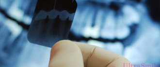
A panoramic photograph of teeth or orthopantomogram (OPTG) is a radiographic examination technique that makes it possible to study in detail the anatomical features of both jaws, the condition of soft and bone tissue, dental roots, maxillary sinuses and joints. The study is carried out using equipment with reduced radiation exposure and does not pose any threat to the health of the body. Today we’ll talk about what this is, why a panoramic shot is needed and how often it can be taken.
What is OPTG and why is it performed?
Unlike a standard targeted photograph, an orthopantomogram allows you to obtain a fairly clear and detailed image of both jaws at once with the ability to study not only the condition of the teeth and their root system, but also the jaw bones, paranasal sinuses and other important anatomical structures. To understand what this diagnostic technique is, you first need to answer the questions: what does it do and why is it carried out. So, here's what the x-ray shows:
- deviations in the formation of occlusion - defects in bite, position, shape and size of teeth;
- early stages of caries and hidden foci of infection;
- periodontal pockets;
- neoplasms: cysts, granulomas, fibromas;
- hidden inflammatory processes and the extent of their damage;
- condition of the temporomandibular joint;
- impacted teeth that have not yet erupted - including wisdom teeth;
- condition of the root canals after treatment;
- diseases of the paranasal sinuses, for example, sinusitis.
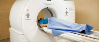
The procedure is mandatory before orthodontic treatment, prosthetics and, of course, implantation. In the latter case, the image gives the doctor the opportunity to carefully examine the condition of the jawbone, select suitable locations for implants, or prescribe a bone tissue augmentation procedure. To understand what the result of OPTG looks like, take a look at the example image below. Important! It should be noted that today, within the framework of one-stage implantation techniques, the preparatory stage involves undergoing a computed tomography scan. This is a more modern X-ray method that allows you to obtain a three-dimensional image of the patient’s jaws with the possibility of layer-by-layer study of tissues, without distorting the actual sizes and shapes of the teeth. OPTG is also performed before treatment or removal of wisdom teeth or “eights”. The image will allow you to identify the exact location and position of the elements, detect hidden carious lesions and make a decision on the need for removal. In general, an orthopantomogram helps to carefully examine the clinical picture, make an accurate diagnosis and prescribe adequate treatment.
What difficulties does radiophobia create during bite correction?
Every sane person who decides to invest substantial funds in correcting a bite with braces wants to get an answer from the dentist to three main questions during preliminary consultations.
- What's wrong with my teeth?
- How can I improve the situation?
- How to save money on braces treatment?
At the same time, the most demanding clients often refuse an X-ray examination: “I will not be exposed to radiation again. It’s enough that they recently forced me to undergo fluorography.”
What should a potential patient of the orthodontist center understand? Locks glued to tooth enamel alone will not correct the bite. The treatment program is developed, activated and controlled by a person. This is a professionally trained specialist who, even before installing metal or aesthetic ceramic braces, must see the goal and imagine the path leading to it.
Teeth can be moved in different ways. The main task that the doctor solves is to choose:
- the correct direction of their movement;
- a device that can do this in the optimal way - effective and most comfortable for the patient.
For this, in-depth diagnostics are performed, and the provider of the most important diagnostic information is precisely the X-ray examination.
What equipment is used as part of the study?
This is not a new method - by now, OPTG has been actively used in dentistry for decades. And if earlier film cameras were used to obtain a panoramic image, today they have been replaced by more modern digital devices - orthopantomographs. The image is generated in a special computer program and, if necessary, can be printed on film. Images taken using digital equipment are more accurate and detailed, which virtually eliminates the risk of making an incorrect diagnosis. Answering the question about how long a panoramic photograph of teeth is valid, it must be said that the information received will remain relevant for several months - a maximum of six months. If more time has passed since the examination, the procedure must be repeated.
Read also
What is dental restoration
If the aesthetic properties of teeth are lost, a person experiences certain discomfort.
What to do if your tooth aches
Sometimes aching pain in a tooth can appear for no apparent reason at first glance.
Indications and restrictions
The procedure is completely painless, carried out as quickly as possible and without any discomfort for the patient. OPTG is prescribed in the following cases:
- as part of implantation – carried out to study bone tissue, select implant models and places for their implantation. Based on the data obtained, the doctor will be able to draw conclusions about the advisability of building up the jawbone;
- to assess the quality of dental canal treatment and the condition of the roots - before prosthetics;
- before starting orthodontic treatment - before installing braces and other corrective structures;
- before an upcoming operation, complex removal, including wisdom teeth;
- to examine the dental system in children, assess the correctness of its formation and identify possible deviations;
- in order to determine the extent of damage to periodontitis and periodontal disease;
- to identify neoplasms in hard and soft tissues.
This procedure may be required in various clinical cases. X-ray diagnostics is not advisable in the first and last trimester of pregnancy - it is carried out only in the presence of emergency indications and with the permission of a gynecologist.
Features of conducting OPTG for children
We figured out what is visible in panoramic photographs and why they are taken, and found out that this is a completely safe diagnostic method. Now it should be noted that an orthopantomogram for children is carried out taking into account some nuances. A radiation exposure of 55 μSv (1 shot) is completely safe for a child, so a similar procedure is allowed from 5-6 years of age. The examination assumes that the patient must stand still for some time, but for young children this task can be quite difficult . When it comes to small patients, the procedure is carried out in exposure mode - the area of exposure is reduced, and the sensor is adjusted to a trajectory of movement corresponding to the small size of the jaws.
“My son also recently had a panoramic photo taken. He's 10, everything went great. Now there are new technologies everywhere, everything is safe. The procedure took less than a minute, and the doctors assured me that there was nothing wrong with it. How else can you start correcting your bite? The orthodontist prescribed OPTG for us. So far I’ve made the plate, but in a couple of years we’ll get braces. There’s no place here without pictures!”
In pediatric dentistry, OPTG is used to detect the rudiments of permanent teeth, assess the correctness of their formation, to diagnose adentia and identify malocclusions. Most often, this procedure is prescribed by an orthodontist before starting to correct defects in bite and teeth position. The procedure is completely safe for the child
Harm and degree of radiation exposure
Let's start with the fact that radiation exposure is measured in micro- or millisieverts - μSv or mSv, respectively. According to the resolution of the Ministry of Health of the Russian Federation, the permissible radiation dose within the framework of OPTG for an adult patient should not exceed 55 μSv, and for children under 15 years of age - 24 μSv 1 . It should be noted right away that only digital X-ray equipment meets these requirements, while film devices provide a slightly higher load. To determine how many times you can take a panoramic photo, you need to take into account that the maximum permissible radiation dose for an adult patient per year corresponds to 1000 μSv. However, it is still not worthwhile to partake in such procedures, even despite their obvious safety and harmlessness . X-ray irradiation has a negative effect on a weakened body, especially with repeated examinations. The children's body is more susceptible to this kind of influence, therefore it is strictly forbidden to exceed the permissible dosages when examining small patients. When contacting the clinic for OPTG, be sure to clarify what type of equipment is used and what the degree of radiation exposure from one image will be. Some private dentistry purchases cheap, outdated equipment, which can produce 2-3 times the recommended dose. The best option where you can take a panoramic photograph of your teeth is a specialized diagnostic center equipped with modern high-precision equipment.
Radiation safety during X-ray dental examinations (Part 2)
M. A. Chibisova Doctor of Medical Sciences, Professor, Head of the Department of Radiology in Dentistry, non-state educational institution "St. Petersburg Institute of Postgraduate Dentistry" (SPbINSTOM), chief physician of the group's radiology diagnostic service
Radiation safety of the population is the state of protection of the present and future generations of people from ionizing radiation harmful to their health (Federal Law “On Radiation Safety of the Population” No. 3-FZ of January 9, 1996, Art. 1). Aspects of radiation safety of personnel and patients, issues of labor protection when working with sources of ionizing radiation are regulated by a number of regulatory documents listed above. Monitoring compliance with radiation safety measures is entrusted to the service of Rospotrebnadzor of the Russian Federation.
In the process of working with sources of ionizing radiation, several principles must be observed: the principle of regulation - not exceeding the permissible limits of individual radiation doses of citizens; the principle of justification is the prohibition of the use of ionizing radiation, in which the benefit obtained for humans and society does not exceed the risk of possible harm from this exposure; optimization principle - limiting the levels of exposure of personnel and patients below dose limits by maintaining radiation doses at such low levels as technically possible to achieve, provided that the required volume and quality of diagnostics are ensured. The principle of justification when conducting x-ray examinations implies the priority use of alternative (non-radiation) methods, x-ray examinations must be carried out according to strict indications, the choice of the most gentle methods of x-ray examinations, the risk of refusing an x-ray examination must obviously exceed the risk from radiation during its conduct. The doses received by the patient from each x-ray examination must be entered into a personal sheet for recording medical radiation doses, which is a mandatory attachment to the outpatient card.
Protection against ionizing radiation is aimed at reducing the physical dose of radiation below the maximum permissible dose.
The following types of radiation protection are distinguished: 1. Protection by screens:
- stationary devices (brick, barite concrete, lead, leaded glass, etc.);
- non-stationary (aprons, gloves, collars, etc.).
2. Protection by distance. 3. Time protection:
- reduction of research time;
- reduction of staff working hours;
- reduction in the number of studies.
To assess the radiation hazard of ionizing radiation, the concept of effective dose is introduced - a value used as a measure of the risk of long-term consequences of irradiation of the entire human body and its individual organs and tissues, taking into account their radiosensitivity. The unit of effective dose is the sievert (Sv), but the derivatives millisievert (mSv) and microsievert (µSv) are commonly used in everyday practice.
In accordance with SanPiN 2.6.1.1192-03 and NRB-99/2009, maximum permissible radiation doses have been introduced for various categories of personnel and patients. For employees directly involved in X-ray diagnostic studies (group A personnel), the permissible effective dose is 20 mSv per year on average for any consecutive 5 years, but not more than 50 mSv per year. For group B personnel (employees who, due to working conditions, are exposed to ionizing radiation) - 5 mSv per year on average for any consecutive 5 years, but not more than 12.5 mSv per year. For the population, i.e. practically healthy individuals for whom X-ray examination is carried out for preventive purposes or for scientific research - 1 mSv on average for any consecutive 5 years, but not more than 5 mSv per year.
For patients, limits on annual radiation doses for diagnostic (as well as therapeutic) purposes are not established. This is due to the voluntary nature of the study, the benefits of which for the patient’s health must exceed the amount of radiation damage. When the accumulated dose of medical diagnostic radiation to a patient reaches 500 mSv, measures must be taken to further limit his exposure if radiation procedures are not dictated by vital indications. Monitoring of patient dose loads is carried out during each x-ray examination.
Dental devices with regular film without an intensifying screen and panoramic devices are allowed to be placed only in the X-ray department (room) of a medical or dental institution.
In addition, for pregnant women, studies are carried out in such a way that the dose received by the fetus does not exceed 1 mSv for two months of undetected pregnancy. If the fetus receives a dose exceeding 100 mSv, the doctor is obliged to warn the patient about the possible consequences and recommend terminating the pregnancy. Studies in pregnant women should be carried out only when clinically indicated, if possible in the second half of pregnancy.
For persons (non-staff) assisting in supporting patients (seriously ill patients, children) during X-ray procedures, a dose limit is set at 5 mSv per year. When performing X-ray dental examinations, the relevant requirements must be met.
Dental devices and pantomographs operating with a highly sensitive image receiver (without a darkroom), and dental devices with digital image processing, the workload of which does not exceed 40 (mA x min.)/week, can be located in the premises of a dental institution located in a residential building , including in premises adjacent to residential premises, subject to the requirements of radiation safety standards within the premises in which X-ray and dental examinations are carried out.
If several devices for X-ray dental examinations are installed in the room, then the anode voltage switching system should provide for the possibility of operating only one device at a time.
Composition and area of premises for X-ray dental examinations: 1. Room for X-ray diagnostics of dental diseases using radiography with a dental apparatus working with ordinary film without an intensifying screen:
- Treatment room - at least 8 m2.
- Photo laboratory - at least 6 m2.
2. Room for X-ray diagnostics of dental diseases using radiography with a dental apparatus working with a highly sensitive film and/or digital image receiver, including a radiovisiograph (without a darkroom):
- Treatment room - at least 6 m2.
3. X-ray diagnostic room using panoramic radiography or panoramic tomography (digital volumetric tomography, 3DCT):
- Treatment room - at least 8 m2.
- Control room (may be absent when using devices equipped with means of protecting personnel workplaces) - at least 6 m2.
- Photo laboratory (may be absent when using devices with digital image processing) - at least 8 m2.
When installing more than one dental X-ray device in a treatment room, the area of the room must increase depending on the type of device, but not less than 4 m2 for each additional device.
X-ray and dental equipment (domestic or imported) is permitted for delivery and operation if there is a registration certificate from the Ministry of Health and Social Development of the Russian Federation and a sanitary-epidemiological conclusion. A dental institution conducts x-ray examinations only if it has a license for the corresponding type of medical activity. An institution using X-ray dental equipment must have the following documentation: a sanitary and epidemiological certificate for the type of activity (operation, storage, testing, etc. of the X-ray machine (devices) in the X-ray room (rooms); design documentation for the X-ray room; technical passport for the X-ray room ; instructions on labor protection, including requirements for radiation safety, for the prevention and elimination of radiation accidents; sanitary rules, other regulatory and instructional documents regulating radiation safety requirements.
The administration of the dental institution determines the list of persons working on dental X-ray machines, provides the necessary training and instruction, and appoints a person responsible for radiation safety, accounting and storage of the X-ray machine, and for radiation control. The room where X-ray examinations are carried out must have a set of mobile and personal protective equipment for staff and patients:
- Large protective screen with a viewing window for devices working with regular film without an intensifying screen, panoramic devices, pantomographs (when the control panel and treatment room are located in the same room, when working with X-ray dental devices with highly sensitive image receivers, it is allowed to use X-ray protective curtains instead of a screen) - 1 PC.
- One-sided lightweight protective apron (for staff) - 1 pc., protective collar (for staff) - 1 pc.
- Protective dental apron (for the patient) or a protective cape (cape) and an apron for protecting the gonads (for the patient) - 2 pcs.
Personnel working on X-ray machines must be trained in the rules of working on this device and trained in ensuring radiation safety of personnel and patients, which must be confirmed by relevant documents. Persons over 18 years of age who do not have medical contraindications are allowed to work on an X-ray dental apparatus, after training, instruction, testing of knowledge of the safety rules for conducting work, instructions in force in the institution and classified by order of the administration of the institution as category A personnel.
The administration of the dental institution ensures continuous individual dosimetric monitoring of employees working on dental and panoramic X-ray machines. In order to protect the patient’s skin during X-ray procedures, the length of the device tube must provide a skin-focal distance of at least 10 cm for a device with a rated voltage of up to 70 kV and 20 cm for higher values of anode voltage. Ways to reduce radiation exposure to staff and patients: replacement of outdated equipment, high-quality maintenance of equipment, availability of a full set of personal protective equipment, use of highly sensitive films, transition to digital technologies, optimization of research protocols, use of specialized programs for reconstruction and image processing.
X-Ray Protection
Radiation protection for X-ray rooms when conducting intraoral examinations and for X-ray dental rooms (the radiovisiograph is located in the dental office):
- For patients - an X-ray protective dental apron FRZS-“R-K” (the attenuation factor of X-ray radiation by X-ray protective material, expressed in lead equivalent value, not less than: at U=100 kV 0.35 RV). This apron is designed to protect the patient's body, including the gonads, pelvic bones and thyroid gland from the radiation beam during dental or cranial examinations. The most reliable form of patient protection when taking dental x-rays.
- For personnel - a protective one-sided lightweight FRZOL-“R-K” apron (the attenuation factor of X-ray radiation by X-ray protective material, expressed in lead equivalent value, not less than: at U=100 kV 0.25 RV). This apron is designed to protect the front of the human body from the throat to the shins during X-ray examinations - with or without a stand. In the second case, it is recommended to wear an X-ray protective collar over the apron.
Radiation protection for X-ray rooms when conducting extraoral panoramic X-ray examinations on an orthopantomograph, cephalostat and digital volumetric tomograph (3DCT):
- For patients - a protective dental apron for panoramic examinations (for an orthopantomograph) FRZS-“R-K” (the attenuation factor of X-ray radiation by X-ray protective material, expressed in lead equivalent, not less than: at U = 100 kV 0.35 Rv). This is a specially designed apron model to protect the patient's body front and back along the spine during extraoral dental examinations.
- For pediatric patients - a protective one-sided heavy children's FRZOT-“R-K” apron (the attenuation factor of X-ray radiation by X-ray protective material, expressed in lead equivalent value, not less than: at U=100 kV 0.35 RV). This apron is designed to protect the front of the body, including the shoulder girdle, from the throat to the shins. Recommended for use when taking photographs of teeth, head and limbs, complete with a collar to protect the thyroid gland. When performing extraoral panoramic examinations, pediatric patients are recommended to additionally use an X-ray protective skirt (0.35 Rv). Unlike an apron, it covers the body area on all sides, and the double wrap adds protection to the front.
Radiation, electrical and fire safety techniques when working with radiovisiographs
Ensuring radiation, electrical and fire safety during X-ray dental examinations and the rules of work on X-ray computerized installations (radiovisiographs) are regulated by Sanitary rules and norms SanPiN 2.6.1.1192-03 “Hygienic requirements for the design and operation of X-ray rooms, devices and the conduct of X-ray examinations, clause 11 and paragraph 12." The placement and permanent protection of premises for X-ray dental examinations are determined by the type of X-ray equipment and the size of the workload of the device.
Dental devices and pantomographs operating with a highly sensitive image receiver (without a darkroom), and dental devices with digital image processing, the workload of which does not exceed 40 (mA-min.)/week. and anode voltage of 70 kV, can be located in the premises of a dental institution located in a residential building, including in those adjacent to residential premises, subject to the requirements of radiation safety standards for the population within the premises in which X-ray dental examinations are carried out.
Composition and area for X-ray dental examinations: a room for X-ray diagnostics of dental diseases using radiography with a dental apparatus working with a highly sensitive digital image receiver, including a videographer (without a darkroom) - a treatment room with an area of 6 sq. m (not less). Ventilation requirements for rooms for X-ray dental examinations must comply with the ventilation requirements for dental departments. In medical practice, domestic X-ray and dental equipment can be used, manufactured according to technical specifications agreed with the Russian Ministry of Health and passed a hygienic assessment. Imported X-ray equipment is allowed for delivery and operation if there is a registration certificate from the Ministry of Health of Russia and a sanitary-epidemiological conclusion. The room where X-ray dental examinations are carried out using a radioviseograph must have a set of personal protective equipment for staff and patients: a protective one-sided apron - light (for medical personnel - a dentist and his assistant) - 2, a protective dental apron (for the patient) - 2, a cape (drape) protective and collar (for the patient) - 1.
In order to protect the patient’s skin during X-ray procedures, the length of the device tube must provide a skin-focal distance of at least 10 cm for a device with a rated voltage of up to 70 kV and 20 cm at higher values of anode voltage
The administration of a dental institution is obliged to determine the list of persons working on dental digital X-ray machines, provide the necessary training and instruction, and appoint by order of the institution a person responsible for radiation safety, accounting and storage of the X-ray machine, and for radiation control. The administration of the institution is responsible for ensuring the radiation safety of staff and patients. Personnel working with radiovisiographs must be trained in the rules of working with this device, trained in ensuring radiation safety of personnel and patients, and have a document from an institution accredited in these matters. Persons over 18 years of age who do not have medical contraindications are allowed to work on an X-ray dental apparatus, after training, instruction, testing of knowledge of safety rules for conducting work, instructions in force in the institution and classified by order of the administration of the institution as category A personnel.
The administration of a dental institution is obliged to ensure constant individual dosimetric monitoring of employees working on dental X-ray machines. In order to protect the patient's skin during X-ray procedures, the length of the device tube must provide a skin-focal distance of at least 10 cm for a device with a rated voltage of up to 70 kV and 20 cm at higher values of the anode voltage. To ensure safe conditions for conducting x-ray examinations, measures must be taken to protect against the effects of electricity, lead and other non-radiation factors, as well as fire and anti-epidemic measures must be taken. Electrical safety of technical equipment, including personal computers of personnel workstations, is ensured by the use of electrical sockets with a grounding contact. Electrical appliances and dental devices can be connected to grounding through plug sockets with an additional grounding contact (European standard). The presence of a grounding strip is not required if the design of the device provides a grounding conductor.
The network resistance must correspond to the rated power of the X-ray power supply with a three-phase rectification circuit. Mobile dental X-ray machines must remain stable when the floor is tilted up to 15°. The moving parts of the device must have a clamping force limiter of up to 300 N. The movement of X-ray devices must be carried out in accordance with the load standards when moving heavy objects. Each X-ray room (including a mobile X-ray unit) must be equipped with carbon dioxide fire extinguishers of the OU-2 type and have free access to fire extinguishing equipment. (A filled transformer tank is not considered a fire hazard). The number and location of fire extinguishers is agreed with the fire safety authorities.
Radiation exposure and protection of patients and personnel during X-ray examinations in dentistry
When radiography of the teeth of the upper jaw on a dental apparatus, the zone of a direct diverging beam of rays includes not only the maxillofacial area, but also the neck, chest and abdominal cavities - organs and tissues located far beyond the boundaries of the area under study. The tube of the 5D-1 device does not sufficiently shield the primary radiation. The cross-sectional area of the primary beam is often 2 times that required for intraoral radiographs. In 5D-2 X-ray machines, aperture of the primary beam ensures the minimum possible divergence angle. When performing intraoral radiographs, the patient's torso is shielded with a standard protective apron made of leaded rubber. However, its large mass, bulkiness, and large cutout around the neck make it difficult to use, especially when examining children. In addition, the thyroid gland remains unprotected. Therefore, specially designed protective screens made of leaded rubber or new models of lightweight protective aprons with a collar for the thyroid gland have been proposed, providing ease of use and sufficient neck protection.
In order to reduce the cumulative effect of ionizing radiation, especially in children, repeated radiographs are taken after 3 weeks, and if several images are taken, then no earlier than after 5 weeks. Taking into account the peculiarities of the centering of the X-ray beam during panoramic radiography and panoramic tomography (zonography) of the dental system, the body is exposed to much less radiation than when performing intraoral contact radiographs of the teeth. During these studies, the thyroid gland is protected by an apron made of leaded rubber, and the patient’s torso is protected by a double-sided apron or drape.
Dosimetric values
Of the dosimetric quantities that can be used to estimate the dose load on a patient, the following are interesting: Absorbed dose of ionizing radiation (dose of ionizing radiation) - D - the energy of ionizing radiation transferred by it to a unit mass of the irradiated substance. Its unit of measurement is the gray (Gy). 1 Gy is equal to the absorbed dose of ionizing radiation, at which the energy of ionizing radiation of 1 J is transferred to a substance weighing 1 kg; 1 Gy = 1 J/kg. 1 Gy = 100 rad (from the English Radiation Absorbed Dose).
Equivalent dose of ionizing radiation (equivalent dose) - H - absorbed dose taking into account the quality factor of ionizing radiation. For X-ray radiation, the absorbed dose (D) and equivalent dose (H) are equal. The unit of equivalent dose is the sievert (Sv). 1 Sv = 100 rem (biological equivalent of an x-ray). Effective dose - Nef - is the equivalent dose of such uniform irradiation of the whole body, which can cause the same long-term (stochastic) effects as non-uniform irradiation. In other words, if there is (for example, in dental practice) uneven irradiation of the body, then it is necessary to use the concept of an effective equivalent dose, which makes it possible to establish an equally effective equivalent dose of uniform irradiation.
Effective dose - Nef - is the equivalent dose of such uniform irradiation of the whole body, which can cause the same long-term (stochastic) effects as non-uniform irradiation
For all types of radiography, radiation exposure to patients is assessed using an effective equivalent dose (EDD), which is measured in microsieverts (µSv) and is determined by measuring the exposure of vital organs that are most sensitive to the effects of ionizing radiation (lens of the eye, cerebral hemispheres, salivary glands, tongue, active bone marrow, thyroid and mammary glands). During dental radiography, leaded rubber aprons are used to protect the mammary glands and gonads. To protect the thyroid gland, it is advisable to use special leaded screens - collars. Protection of medical personnel from x-ray radiation includes strict adherence to operating and safety rules in the x-ray room. When the device is turned on, the X-ray room employees must be behind a protective screen or behind the room wall. Fixation of the film in the mouth when taking intraoral photographs is carried out by the patient himself. The use of X-ray room personnel for this purpose is strictly prohibited.
Radiation safety of patients during X-ray examinations in dentistry
In 70-80% of cases, the clinical diagnosis is established on the basis of X-ray data (M. M. Solovyov, 2000). The share of x-ray examinations in dentistry relative to other types of x-ray procedures is quite large. Often dental x-ray examinations occur at the age of 18-20 years. This implies the need to control doses during x-ray examinations in dental practice. During dental radiography, lead aprons are used to protect the lungs, heart, mammary glands and genital area; an apron to protect the thyroid gland - special screens - in the form of a collar. In this case, equivalent doses are reduced by 10-100 times, so the use of aprons-collars is mandatory. However, the total effective equivalent dose is reduced by only 15-20%, since the cerebral hemispheres, pituitary gland, salivary glands, tongue and lens of the eye are not protected. During orthopantomography, the dose loads are relatively low (load for the tongue and salivary glands) and are comparable to those during intraoral radiography of individual areas of the oral cavity (Fig. 1, 2).
When performing an X-ray examination in dentistry, the following is necessary: 1. Patient protection: In children and adults, it is desirable to perform an orthopantomogram (or digital volumetric tomography - 3DCT) and reduce X-ray examinations to a minimum;
- an apron made of leaded rubber (preferably with a collar - to protect the thyroid gland);
- all extraoral images are performed with intensifying screens, and intraoral images are taken on highly sensitive screenless film;
- radiography is carried out preferably with “hard” (not lower than 60-70 kV) radiation and the shortest possible exposure;
- mandatory use of an aluminum filter (at least 1 mm thick);
- X-ray examinations are performed only according to strict indications (the number of X-ray examinations is reduced to a minimum in children and pregnant women); Repeated X-ray examinations - if possible, no earlier than after 10-15 days.
2. Personnel protection: when working, it is necessary to move away to the side opposite to the working radiation beam, at a distance along the length of the cord to the control button;
- full compliance with the rules of design, operation and safety of work in the X-ray room;
- mandatory use of protective equipment (screens, aprons) by personnel;
- when the X-ray machine is turned on, you must be no closer than 1.5-2 m from the X-ray tube and, of course, behind a protective screen or behind the office wall;
- during intraoral dental radiography, films are fixed in the mouth by the hands of the person being examined or by the patient himself using a positioner;
- systematic dosimetric monitoring is required.
Effective doses for x-ray examinations in dentistry
Effective doses for x-ray examination (for patients according to the Testing Laboratory Center of the Federal Research Center of St. Petersburg Research Institute of Radioactive Geography of the Ministry of Health of Russia dated July 22, 2011 and December 21, 2012, studies were performed on x-ray machines: panoramic x-ray machine “Orthophos XG 3D / Ceph” with cephalostat (Moscow, 79); (Kamennoostrovsky, 42b); (Suvorovsky, 57); three-dimensional dental computed tomograph - targeted 3DX Accuitomo Morita (Nevsky, 82); panoramic 3DKT Galileos "Sirona" (Nevsky, 82); (Komendantsky, 17) and radiovisiographs, "Sirona" using X-ray dental devices such as "Trophy-Kodak", "Sirona" (Fig. 3-6).
During one office visit, the patient is allowed to take 5-6 targeted images
The effective dose for one study when producing a digital orthopantomogram is 0.055 mSv (for a patient under 15 years old - 0.024 mSv); digital teleradiogram - 0.007 mSv. The effective dose when performing one zone on a three-dimensional dental computed tomograph is 0.102 mSv (for a patient under 15 years old - 0.068 mSv).
Effective dose of one digital intraoral x-ray of a tooth on a radiovisiograph “Trophy-Kodak”, “Sirona”: lower jaw - 0.002 mSv (for a patient under 15 years old - 0.001 mSv); upper jaw - 0.005 mSv (patients under 15 years old - 0.003 mSv).
In a year, you can take up to 100 images using a radiovisiograph (for adult patients). During one office visit, the patient is allowed to take 5-6 targeted images.
Maximum effective dose per year for radiologists and x-ray laboratory technicians when working in stationary x-ray rooms; for dentists and medical assistants when working on a radiovisiograph in x-ray dental rooms is 2 rem = traffic limit 20 mSv per year.
The maximum effective dose to the population received from preventive X-ray diagnostic studies is 1 mSv per year. 0.01 rem = 0.1 mSv, 0.1 rem = 1.0 mSv, 1 Sv = 100 rem, 1 rem = 10 mS.
Rice. 1. Digital orthopantomogram. Chronic generalized periodontitis of moderate severity. Rice. 2. Teleroentgenogram of the skull in a lateral projection, hyperplasia of the lymphoid ring of the nasopharyngeal vault. Rice. 3. 3DCT. Chronic osteomyelitis in the area of the angle of the lower jaw on the right after removal of the 48th tooth. Rice. 3. 3DCT. Chronic osteomyelitis in the area of the angle of the lower jaw on the right after removal of the 48th tooth. Rice. 4. 3DCT. Chronic granulomatous periodontitis of teeth 26, 27, 28, odontogenic changes in the mucous membrane of the left maxillary sinus. Rice. 4. 3DCT. Chronic granulomatous periodontitis of teeth 26, 27, 28, odontogenic changes in the mucous membrane of the left maxillary sinus. Rice. 5. 3DCT of the dentition, cyst-like formation of the right maxillary sinus. Rice. 5. 3DCT of the dentition, cyst-like formation of the right maxillary sinus. Rice. 6. 3DCT of the dentition, cyst-like formation of the right maxillary sinus. Rice. 6. 3DCT of the dentition, cyst-like formation of the right maxillary sinus.
Conclusion
In addition to solving the problems of organizing x-ray examinations in dental clinics and observing radiation safety when performing x-ray examinations, it is necessary to widely introduce innovative methods of radiation diagnostics for practical use - three-dimensional dental computed tomography (3DCT) when conducting diagnostic examinations in outpatient dentistry, maxillofacial surgery and otorhinolaryngology. Three-dimensional dental computed tomographs are installed in private and public dental clinics; they allow moving to a new level of three-dimensional diagnostics, which significantly expands the possibilities of visualizing the dental system, maxillofacial area, maxillary sinuses and temporomandibular joints. The use of modern methods of radiation diagnostics makes it possible to optimize the diagnostic and treatment process, reduce the time required for examining dental patients, reduce the individual dose of radiation exposure, and improve the quality of dental care.
- Vasiliev A. Yu., Vorobyov Yu. I., Truten V. P., Chibisova M. A. et al. Radiation diagnostics in dentistry. - M., Medicine, 2007. - 495 p.
- Gerasimova L.P., Zaripova N.R., Verzakova I.N., Rayanova R.A. X-ray diagnostics in therapeutic dentistry. Tutorial. - M., 2011. - 196 p.
- Hygienic requirements for the design and operation of X-ray rooms, devices and the conduct of X-ray examinations. Sanitary rules and regulations. SanPiN 2.6.1.1192.-03. Ministry of Health of Russia / Moscow. - 2003. - 76 p.
- Control of effective radiation doses to patients during medical X-ray examinations / Guidelines MUK 2.6.1.1797-03 Ministry of Health of Russia. - M., 2004. - 23 p.
- Radiation diagnostics in dentistry: national guidelines / Ch. ed. volume Vasiliev A. Yu. - M.: GEOTAR-Media, 2010. - 288 p. (Series “national guide to radiation diagnostics and therapy” / chief editor of the series Ternova S.K.).
- Malakhovsky V. N., Trufanov G. E., Ryazanov V. V. Radiation safety of X-ray studies. - St. Petersburg: ELBI-SPb, 2007. - 104 p.
- Standard instructions on labor protection for personnel of X-ray departments dated January 28, 2002, No. 19. - M., 2002. - 60 p.
- Chibisova M. A. Licensing of dental clinics and offices for activities in the field of using sources of ionizing radiation. - SPb.: MEDI publishing house, 2007. - 40 p.
Digital OPTG - description of the process
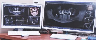
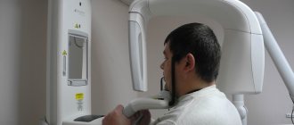
Now let's move on to consider the question of how a panoramic photograph of teeth is taken. To begin with, the patient is asked to remove all metal products from himself so that they do not affect the operation of the equipment and the final result. Next, the patient stands in the place indicated by the specialist, bites down on a special plate that ensures the immobility of the jaws, and after turning on the orthopantomograph, its moving part begins to rotate around the head. The procedure takes literally half a minute - the exact duration will depend on the type and newness of the equipment. Next, the resulting image is formed in digital form, that is, in a special computer program, or on film media (outdated version). A digital photograph can also be printed onto film if necessary.
Is it possible to take photographs of pregnant women?
Let's turn to the regulatory documentation, the same SanPiNam 2.6.1.1192-03.
Thus, paragraph 7.16 clarifies that pregnant women are prescribed for X-ray examinations only for clinical indications. Studies should, if possible, be carried out in the second half of pregnancy, except in cases where the issue of termination of pregnancy or the need for emergency or emergency care must be decided. If pregnancy is suspected, the question of the admissibility and necessity of an x-ray examination is decided based on the assumption that pregnancy exists.
As for the dose, paragraph 7.18 of the current SanPiN says that X-ray examinations of pregnant women are carried out using all possible means and methods of protection so that the dose received by the fetus does not exceed 1 mSv for two months of undetected pregnancy. If the fetus receives a dose exceeding 100 mSv, the doctor is obliged to warn the patient about the possible consequences and recommend terminating the pregnancy. Considering that the fetus is clearly not in the head, and below the head we protect everything we can, the answer to the question of whether it is possible to take dental photographs of pregnant women and men is more than clear:
- Can. but carefully.
Information content of the image and reading (deciphering) technique
If you undergo an examination at a specialized diagnostic institution, the finished image is usually accompanied by an explanation from a specialist with a list of detected problems. Many people are interested in how to decipher an image on their own, how to read it correctly, and whether it can be understood at all. It should be noted here that in order to correctly explain the results of OPTG, you need to have a good understanding of the anatomy of the maxillofacial apparatus, and without the appropriate education, little can be understood. It is better to entrust this task to an experienced, qualified doctor.
Orthopantomogram during implantation
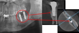
As part of implant treatment, a panoramic image is taken more than once. First, an X-ray examination is carried out at the preparatory stage - the doctor carefully examines the resulting image, assesses the condition of the bone tissue, selects places for implants, and makes a verdict on the need for bone grafting. If we are talking about one-stage immediate loading protocols, then patients are usually sent for a computed tomography scan of the jaw - a 3D image provides more detailed information about the clinical picture.
3D diagnostics makes it possible to study a more detailed three-dimensional image
You will have to undergo a repeat examination after implantation. In this case, OPTG will give the doctor the opportunity to assess the quality of the operation performed, monitor the process of engraftment of artificial roots, and recognize possible complications in the later stages of their development. Several photographs may be required during the osseointegration period of the implants.
Why do you need a panoramic shot of the jaw?
A panoramic photograph of the teeth gives the dentist the opportunity to assess the condition of all teeth, their relationship to each other, and determine changes in the roots of the teeth and in the bone tissue around the teeth. The visible part of the tooth makes up less than 50% of the entire tooth. Your dentist can only assess what is happening inside the gums and bones using x-rays.
Panoramic dental x-rays help dentists of all specialties make an accurate diagnosis, develop the correct treatment plan and monitor the outcome of treatment.
A panoramic photograph (orthopantomogram) is necessary in almost all areas of dentistry. Digital orthopantomography also provides the opportunity for a detailed examination of individual areas of the jaw for a more accurate diagnosis. Without X-rays, it is impossible to imagine diagnosing caries under crowns and fillings, as well as in hard-to-reach places, root canal treatment, dental prosthetics, dental implantation, periodontal disease treatment, bite correction, and wisdom teeth removal. The orthopantomogram is used in both adult and pediatric dentistry.
More information about the use of panoramic photographs of teeth for diagnostics in dentistry can be found here.
What are the advantages and disadvantages of OPTG
An orthopantogram can significantly improve the quality of diagnosis, and today dental treatment is practically not carried out without radiography, except for minor problems. Among the undeniable advantages of the technique, experts highlight the following points:
- speed of the procedure - literally in a couple of minutes the image is ready for study;
- modern equipment makes it possible to adjust the height and trajectory of the emitter, which made diagnostics possible for children and wheelchair users;
- minimal radiation exposure and absolute safety for the body;
- high quality and accuracy of the obtained images;
- the ability to zoom in on the area under study and study it in detail on a computer monitor.
With an orthopantomogram, minimal radiation exposure is used. If we talk about the disadvantages, the main disadvantage of OPTG is obtaining a flat two-dimensional image. As a result, the image may slightly distort the dimensions of teeth, root canals and bone tissue. Such deviations can vary from 15 to 30%. To obtain a more accurate image, it is better to resort to computed tomography.
What determines the radiation dose during CT?
The amount of radiation a person receives during a tomography depends on three main factors:
- Scan area. It's not about whether it will be a targeted shot or a panoramic shot, but about the part of the body that is to be examined. It has been scientifically proven that individual tissues of the body react differently to exposure to x-rays. One of the smallest doses is obtained when examining the dental system.
- Scan time. The time required to obtain CT data is taken into account. The use of cutting-edge equipment makes it possible to reduce the exposure time to X-rays to a few milliseconds.
- Type of equipment. Since tomographs are used in different fields of medicine and for different purposes, they are set to a specific time and intensity of radiation. High-field tomographs, better known as MRI, are considered the most powerful. And small-sized dental (intraoral) portable scanners are gentle.
Manufacturers of medical dental equipment know that CT scanning of teeth and jaws is required more often than tomography of other organs. That is why the devices used in modern clinics provide the minimum possible radiation exposure to the body, equal to approximately 0.039-0.06 mSv. This allows not only to repeat tomographic studies several times a month, but also to carry out diagnostics that are as harmless to health as possible, even for children.
Approximate prices
How much a panoramic dental photograph will cost directly depends on whether the study will be carried out using film or digital equipment - in the latter case, the cost will be an order of magnitude higher. Approximate prices are given below:
- a photograph on a film camera – about 700 rubles;
- digital photo - from 1000 rubles and 150-200 rubles for printing on film.
We have already mentioned above that today, for a more detailed layer-by-layer study of the tissues of the jaw apparatus, computed tomography (CT) is performed. Essentially, this is the same panoramic photo, but much more detailed and in 3D format. An important advantage of this technique is the accurate transmission of the sizes and shapes of all elements of the dental system.