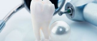
Harm of X-rays for pregnant women
Sometimes, while expecting a child, women need to sanitize their oral cavity. And in order to properly plan therapy, an orthopantomogram is often required. “Is it possible to take dental x-rays during pregnancy? What are the consequences? - expectant mothers often worry.Indeed, irradiation is undesirable during this crucial period. But in reality, not everything is as scary as you might think at first glance.
Dental X-ray during early pregnancy
Directly in the first trimester, the risk of undesirable consequences is the highest, since this is when the baby’s organs are formed. For this reason, doctors do not recommend taking dental x-rays during early pregnancy. After all, cells during active division are very sensitive.
However, many doctors believe that, unlike an X-ray of the back or pelvis, a dental X-ray during pregnancy is not aimed directly at the area where the embryo is located. Therefore, do not worry if you did it before you found out about the joyful event.
In addition, dental x-rays during early pregnancy are allowed in emergency cases when, for example, there is a threat to life. In other situations, it is better to postpone dental x-rays during pregnancy to a more successful period, namely, to the second trimester.
Dental treatment in the I, II and III trimesters
The dentist’s tactics when treating teeth in pregnant women depend on the specific stage of pregnancy - trimester.
Treatment in the first trimester
The first trimester is the time from the 1st to the 13th weeks of pregnancy. This period is characterized by the formation of the rudiments of the organs of the unborn child. At the same time, the placenta does not yet provide adequate protection of the fetus from the influence of factors of the external and internal environment of the mother’s body.
In the first trimester, a pregnant woman is worried about toxicosis, manifested by systematic nausea, short-term vomiting, hypersalivation, slight dizziness, and slight hyperhidrosis. Any, even minor, stress factor can cause a violent reaction in the body, even leading to miscarriage.
In dental treatment, courses of remineralization therapy and professional teeth cleaning without the use of ultrasound are acceptable. Filling, tooth extraction, prosthetics, dental implantation and x-ray diagnostics are contraindicated during this period.
Treatment in the second trimester
The second trimester lasts from the 14th to the 26th week. The placenta in this trimester is fully formed and provides reliable protection for the fetus. Dental treatment carries fewer risks, but still requires consultation with a gynecologist. During this period, you can perform dental fillings, surgical treatment (strictly according to indications), therapy for inflammatory diseases that may worsen by the 3rd trimester or after childbirth, professional removal of dental plaque (hard and soft).
Treatment in the third trimester
The third trimester is the period from the 27th week until birth. During this period, the woman’s body is actively preparing for childbirth, and therefore may be somewhat weakened. A pregnant woman experiences tachycardia, increased breathing, shortness of breath, decreased blood pressure, and increased anxiety. The woman constantly feels tired. Treatment in the 3rd trimester is exclusively emergency. Planned procedures are postponed until the postpartum period.
In all three trimesters, pregnant women are allowed to carry out professional oral hygiene, the method of which will vary depending on the timing of pregnancy and the condition of the body of the expectant mother.
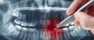
Is it possible to take dental x-rays during pregnancy?
To answer the pressing question “Is it possible to have a dental x-ray during pregnancy?” dentists answer that it is not an absolute contraindication for its implementation. Therefore, if such a study is extremely necessary for the expectant mother, it can be done. The main thing is to follow simple rules:
- So, you can take a dental x-ray during pregnancy, preferably in the second trimester. At earlier or later dates, it is better to refrain from carrying it out.
- To obtain minimal radiation exposure, you can take dental x-rays during pregnancy not with a film fluorograph, but with a computer visiograph.
- During pregnancy, you need to tell the radiologist about it and indicate the due date.
- It is necessary to cover the chest and abdomen with a special protective apron before the examination.
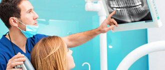
You should also discuss whether it is possible to have a dental x-ray during pregnancy specifically in your case with the gynecologist you are seeing. Perhaps, in order to avoid undesirable consequences, he will have his own opinion.
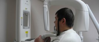
The whole truth about a visiograph in dentistry
Radiovisiographs came into the practice of Russian dentists more than 10 years ago, but still raise questions. The most popular is “Is this the same as an x-ray?” We answer right away: not really. We invite you to understand together the main features of the device and debunk the myths that worry doctors and patients.
How much safer is a visiograph than a regular x-ray?
The device itself is not an emitter, but only receives rays. Consists of a sensor, an analog-to-digital converter and a wire. Nowadays, most models are lightweight and are produced without a separate digitizing unit, that is, they are connected directly to the computer. There are also wireless visiographs that are placed in a special scanner to read the image.
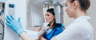
The visiographic complex includes:
- X-ray machine;
- visiograph with software;
- computer.
Yes, the visiograph will not work without an X-ray machine. So why is it even needed then? Isn't it easier to do a regular x-ray?
Firstly, modern equipment is superior to devices of previous generations in terms of safety. If earlier installations emitted wide radiation, now a thin and targeted beam is captured by a sensitive sensor.
Secondly, the sensor recognizes even the smallest particles of radiation in less than half a second or even faster.
I would also like to note the advantage of digital visiography over film devices. Even if you take high-quality film and a good new generation of dental X-rays, it will take approximately 0.6 seconds to take a high-quality image. The same with digital radiography with a sensor is performed in 0.06 seconds. The sensor adequately perceives the signal at exposure from 0.3 tA/sec to 1.8 mA/sec.
So we have the answer to the first question. Radiovisiography is based on the same principles as conventional radiography. At the same time, the level of X-ray radiation during the operation of a radiovisiograph is much lower.
What dose of radiation do patients receive?
We believe you have heard this question from your patients more than once. When X-raying the teeth of the lower jaw using a visiograph, this value is 2 microsieverts, for the upper jaw – 5 µSv.
If film radiography is carried out using highly sensitive film and a low-dose device, the same indicator will be equal to 7 and 13 μSv. When working with old domestic equipment and low-quality film - up to 80 µSv.
How many pictures can you take? It is difficult to regulate the exact number of x-ray examinations, since not all devices are equipped with a dose counter. Accordingly, you will need to calculate it for each type of device and for each tooth. It is logical to focus on the maximum permissible effectively equivalent dose for a person per year, excluding its excess.
According to SanPiN, when carrying out preventive medical x-ray procedures and scientific research, this dose should not exceed 0.001 sievert per year (1 millisievert, 1000 microsievert). The load is equivalent to three plain chest films.
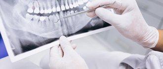
Is it possible to take dental photographs of pregnant and lactating women?
Radiovisiography is allowed only for clinical indications when there is a threat to life. We recommend that private doctors treat this with special care: for example, the patient’s signature on the card has no legal force, and all responsibility lies with the dentist.
Breastfeeding mothers can take photographs, since ionizing radiation is scattered when passing through soft tissues and biological fluids of the body. Thus, it will not affect the child in any way. To make the patient feel calmer, you can skip the next feeding after the photo.
Where can a visiograph be placed?
Since the device only works in conjunction with an X-ray machine, you must have a license that gives you the right to use equipment that generates X-ray radiation. The standard kit for obtaining a document contains a planned diagram of the office for using a visiograph.
Depending on the characteristics of specific equipment, biological protection is calculated by supervisory authorities. They also study the area of the office, the lead equivalent of the wall materials, the location of the office in the building, and the average workload of the imaging unit.
We talked more about organizing an X-ray room in our guide “How to open an X-ray room in a clinic.”
Our resume
A visiograph greatly simplifies the dentist’s work and allows you to quickly obtain digital images. Thanks to the user-friendly software, all images can be edited, archived and compared.
Despite the greater safety of the visiograph compared to standard X-ray machines, do not forget about the protection of personnel. You must stand behind a state-issued protective screen or leave the premises. If your practice has lead aprons, you can stand at least 2.5 meters to the side of the X-ray tube.
Write a comment
Marina
February 21, 2022 at 03:24 pm
Although opinions differ about the harmful effects of x-rays on the fetus, I think the decision should be made by the doctor in a specific case and for a specific pregnant woman. For example, in the seventh month I had a toothache. The dentist removed the nerve and filled the tooth without checking with x-rays. And he said that it would be better to check it later and, if necessary, change it, rather than endanger the child.
Dasha
February 21, 2022 at 10:29 pm
I believe that the dental issue needs to be resolved before pregnancy. Dental treatment in any case involves the use of some medications, drugs, painkillers; all this is not advisable for a pregnant woman. But if this is already necessary, then you need to consume vitamins to the maximum, without them you can’t go anywhere. In general, I think there is nothing wrong with this.
Nastya
February 23, 2022 at 07:22 pm
During pregnancy I was going to get braces. Naturally, it was necessary to take two pictures... I didn’t risk it + I had an ultrasound scan 4 times. Although I remember reading on the Internet, many girls are not afraid - they do it. My friend, a dentist, told me that pregnant women often come with recently taken photographs... I don’t know... here, of course, everyone makes their own choice.
Inna
March 9, 2022 at 03:33 pm
When I was pregnant, I still tried to protect myself from x-rays, the older generation even prohibits the use of cosmetics, and this is an x-ray, no matter how radiation. And as per the law of meanness, it was when I was pregnant that I was diagnosed with a cyst, naturally, that’s why I needed an x-ray. Despite this, the pregnancy went well, the baby was born on time.
Lili
February 8, 2022 at 04:47 pm
This is all nonsense, they are just intimidating naive mothers about how much radiation is needed to affect the fetus... If you listen to the media, you can’t eat meat, drink milk or wash with tap water. They constantly make a big deal out of nothing. I know people who, even if they are not pregnant, are afraid to take an x-ray, and do not yet use a microwave and do not fly on airplanes.
Nastena
February 9, 2022 at 07:41 pm
I’m 5 weeks pregnant, I haven’t gone to the dentist and in fact I only recently found out that I’m pregnant. They sent me to the antenatal clinic to the dentist’s examination room. She scolded me and made me take an x-ray because I had a lot of bad, almost completely destroyed teeth. Well, I’m afraid of dentists; I haven’t been to the dentist for a long time. So I read that x-rays are not allowed, in case there is a miscarriage, what should I do?
Evgeniya A
February 10, 2022 at 09:17 pm
During my second pregnancy, I fell on ice and broke my leg. So at 10 weeks they did an x-ray of my legs! And nothing, ugh, ugh, happened, my son is 8 years old. And here are the teeth... hmm, girls, if you have problems with your teeth, don’t be afraid of this meager radiation exposure, the infection that spreads from a bad tooth throughout the body is much worse. If mommy’s tooth keeps rotting, then the child also comes under the influence of bacteria.
List of abnormalities in the fetus caused by radiation
If a woman had an x-ray in the first 12 weeks of pregnancy , this can cause serious deviations in the development of a number of systems and organs in the fetus:
- spine;
- bronchi;
- hearts;
- faces;
- jaws;
- reduction in the size of the skull and brain.
Radiation contributes to the development of health problems in the unborn child:
- dystrophy;
- blood diseases;
- incurable chronic diarrhea;
- predisposition to cancer.
Also possible:
- frozen pregnancy;
- miscarriage.
But all of the above is unlikely. To cause abnormalities in the fetus, a pregnant woman needs to receive a radiation dose of 3 mSv, and one procedure on modern dental equipment produces a load of 0.02 mSv, and on older devices 0.3 mSv.
The essence of the X-ray machine
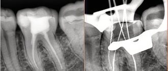
During treatment, prosthetics or implantation, it is necessary to take dental x-rays many times: before, during and after the intervention. The devices familiar to many caused irreparable harm to the patient’s body, because the radiation dose in them is very high. This was necessary so that the reflected X-rays could be imprinted on the film, thereby creating an image.
“Classic” X-ray also required a staff member who knew how to develop images, as well as an entire darkroom. And patients had to wait a long time for examination results.
conclusions
Having studied world practice and research on the use of X-rays in dentistry during pregnancy, the following conclusions can be drawn:
- the harm of x-rays has not been scientifically proven;
- the safety of x-rays has not been scientifically proven;
- conclusions about the effect of x-rays on the egg and sperm during pregnancy planning are not based on anything and have no practical evidence;
- in theory, only a radiation dose of 3 msv affects fetal development;
- a radiation dose of 3 mSv is physically impossible to obtain even during the entire 12 weeks of pregnancy if images are taken every day;
- modern equipment delivers irradiation in a narrow direction, so the beam only affects the tooth specifically;
- It is not recommended to take x-rays in the first trimester;
- The recommended period from which x-rays can be taken is 16 weeks;
- if the degree of neglect of the dental problem is serious and there is a possibility of a negative impact on the health of the woman (and therefore the fetus), we can neglect the theory about the dangers of x-rays;
- The final decision should be made by the patient together with her doctor.
If you find an error, please select a piece of text and press Ctrl+Enter.
Tags: diagnostics during pregnancy
Did you like the article? stay tuned
Previous article
Small lower jaw: etiology and treatment methods
Next article
Infiltration anesthesia in dentistry: what it is and how it is carried out
Alternative Methods
Today in dentistry there is no alternative to x-ray examination. In their quality, we can only consider various types of equipment of this type, which are represented on the medical equipment market by a wide range of assortments.
The best option is a digital visiograph , characterized by a minimum radiation dose for a single exposure. It allows you to immediately receive images and store them on digital media.
The only disadvantage of a visiograph is that it can only be used to take targeted images covering no more than 3 teeth.
© Dan Race/Fotolia
To obtain a three-dimensional image of one or both jaws, an orthopantomograph , which, with a small dose of radiation, produces high-quality digital images.
an apex locator is used as an alternative , allowing one to measure the length and condition of the dental canal. Exposure to x-rays is completely eliminated.
The device is used for intermediate diagnostics, which avoids additional X-ray exposure.
How is a panoramic dental photo taken and is it harmful?
What is a caries marker and how is it used?
What to do if a temporary filling falls out, see the link.
When is the best time to treat teeth for pregnant women?
When the problem is critical, you need to consult a dentist at any time, and as soon as possible. But first, a pregnant woman needs to find out from her gynecologist whether there are any restrictions on dental treatment. If you can be patient, it is better to carry out treatment procedures in the second half of the term, since in the first and third the formation and intensive growth of the baby’s internal organs occurs, so there is a possibility that any intervention and medication will have a detrimental effect on the child’s development.