Indications for tooth extraction during lactation
During the procedure, drugs are used that can penetrate into mother's milk. If it is possible to postpone the date of the operation, then extirpation is carried out after the child is transferred to artificial nutrition. A dental unit is removed urgently during a purulent inflammatory process, as it contributes to the spread of infection throughout the body.
In what cases is surgery necessary:
- suppuration of a cyst, abscess, periostitis;
- unsteadiness due to periodontitis, periodontal disease of the third, fourth degree;
- odontogenic osteomyelitis, sinusitis, phlegmon;
- tooth root fracture;
- pulpitis, which cannot be cured due to the complexity of the canals;
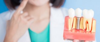
- impacted figure eight, complicated growth of the third molar, pressure on neighboring teeth;
- deep caries that affected the root.
Unscheduled extraction is also performed in cases of severe, persistent pain.
The procedure has contraindications, including acute respiratory infections, chronic diseases in the acute stage, oral infections, sinusitis, severe pathologies of the liver, kidneys, heart, blood vessels, leukemia, bleeding disorders, and mental disorders.
How does radioactive ionizing radiation affect the human body?
Radioactive radiation triggers the production of free radicals. Their excess with a low antioxidant (protective) status of the body leads to the destruction of cellular components, including the destruction and shortening of telomeres - the terminal sections of DNA molecules. Lipids and membrane proteins are also subject to oxidation.
Normally, the human body easily tolerates diagnostic measures and recovers on its own - nothing additional is needed. Following the oxidative processes caused by free radicals, restoration begins, and the body’s resources are sufficient for this.
At the end of the 20th and beginning of the 21st centuries, the telomerase enzyme was discovered (active in germ cells, stem cells and cancer cells). For its discovery, E. Black-Burn, K. Greider and J. Szostak were awarded the Nobel Prize in 2009. Telomerase is responsible for the “lengthening” of telomeres, which means that their destruction cannot be considered irreversible. However, scientists have also noticed another pattern: cancer and the growth of an oncological tumor are possible when DNA molecules are significantly shortened and damaged, while the telomerase enzyme remains in an active state. This is a kind of “failure” of the genetic program, which leads to dangerous consequences.
In general, the average healthy adult body is able to recover from radiation exposure equal to 50-100 mSv per year. With greater systematic exposure to radiation, radiation sickness develops.
Preparing for removal
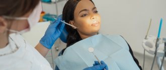
If the tooth begins to ache, the gums are swollen, and the breath smells bad, then this may indicate severe inflammation in the dental system. At the appointment, the dentist collects an anamnesis, conducts a visual examination and sends the woman for an x-ray. It is important for a nursing mother to avoid psycho-emotional stress, so she is allowed to take sedatives in advance.
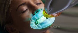
The best option for maintaining mental stability and eliminating fear is oxygen sedation with nitrous oxide (NAS). It is done in the doctor's office before the procedure. The patient inhales the gas and falls asleep. Additionally, a local anesthetic is injected into the gum.
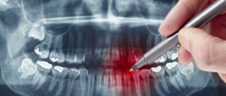
What signs indicate the presence of pathology?
The peculiarity of an x-ray image is expressed in the fact that the result obtained on the film is displayed in the form of a negative image: the denser the tissue, the lighter it will be in the image. That is, in place of bones there will be white areas, in place of soft tissues there will be gray areas, and layers of air will be black.
In this regard, the doctor calls the bright areas in the image darkening; they indicate the presence of pathological formations. For example, if such areas are found in the lungs, they will indicate pneumonia, in other organs - the presence of a tumor, stones and other processes.
The process of tooth extraction in a nursing mother
The procedure for a woman during lactation is no different from that performed for ordinary patients.
Extirpation of a dental unit is carried out in stages:
- antiseptic treatment of mucous membranes;
- anesthesia with local antiseptics or general anesthesia;
- applying forceps and disconnecting the ligament;
- rocking or dislocation;
- extraction from the hole;
- stop bleeding.
If the third molar from below is removed, the operation becomes more complicated. In order to pull it out, the doctor cuts the gum, removes part of the bone, divides the roots into fragments and removes them separately. To prevent infection, the wound is treated with antibacterial drugs. After all manipulations, the gums are sutured with absorbable or non-absorbable suture material.
How often can I have x-rays?
A diagnostic x-ray is prescribed by the attending physician in case of suspicion of the presence of any disease, in order to confirm or refute them. SanPiNom limits are not established in such cases; radiation examination in this case is dictated by vital indications.
For preventive purposes, it is recommended to undergo a chest x-ray (fluorography) once a year, and a breast examination (mammography) once every two years. In medicine, the maximum permissible radiation dose is 1 mSv/year.
X-ray and anesthesia during lactation
Before extirpation of a dental unit, an x-ray or computed tomography is almost always done. This is necessary to assess the structure, position and condition of the root system. To protect the patient from x-ray radiation, she is wearing a lead apron. It blocks dangerous waves, but after the procedure it is better to express the first batch of milk and pour it out.
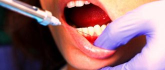
For local anesthesia, private clinics use drugs approved during pregnancy: Ultracain, Ubistezin, Artifrin, Alfacain and similar medications. They do not contain toxic components that can cause allergies and other side effects. Primacaine has a short half-life, so feedings may not be missed.
Anesthesia is used only in extreme cases, if a complex and lengthy extraction is being performed. If general anesthesia is necessary, the child is temporarily transferred to artificial nutrition. During this period, the mother must express milk regularly to ensure milk production continues.
If antibiotic therapy is necessary after a complex removal, then breastfeeding will have to be stopped for several days. During long-term withdrawal, doctors recommend giving artificial formula not through a pacifier, but through a spoon or even a syringe without a needle. Otherwise, the baby may refuse to breastfeed, since the process of sucking milk from a bottle with a nipple is much easier.
Benefits of radiography
The radiation research method is widely used both for diagnostic purposes and for assessing the results of treatment. The advantages of this research method are obvious:
- high information content and a wide range of indications;
- simplicity and speed of obtaining results;
- affordable cost of research;
- lack of special preparation on the part of the patient (in most cases);
- The images can be saved and used for consultation with a related specialist.
Recommendations for the recovery period
In order to avoid complications after surgery, you need to follow all the doctor's advice. After the procedure, the doctor applies a cotton swab to stop the bleeding. It should be held for no more than 10 minutes, otherwise the blood clot will dry out and come off along with the cotton wool. With high blood pressure, bleeding continues for a longer time, so the tampon can be kept in place for 15 - 20 minutes.
In the first days you cannot:
- overheat the body, apply warming compresses;
- engage in heavy physical labor and sports;
- lick a blood clot;
- eat hot, spicy, rough food;
- drink through a straw;
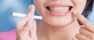
- smoke;
- touch the socket with a toothbrush and other objects.
You are allowed to eat after 3-4 hours. The issue of feeding a child should be discussed with a doctor. Local anesthetics practically do not enter the blood, and components penetrate into mother's milk in minimal quantities. If the doctor has not prescribed other medications, then you can feed the baby within a few hours.
After the anesthetic wears off, pain appears. Possible increase in body temperature. To relieve acute symptoms, you can take Paracetamol, Ibuprofen, Naproxen. You can reduce pain and swelling by using cold compresses.
Rinsing your mouth and brushing your teeth is allowed on the second day. For rinsing, pharmaceutical preparations are used: Romazulan, Chlorhexidine, Miramestin. They will prevent the development of infection and inflammation. You can rinse your mouth with an aqueous solution of baking soda and salt. To improve the healing of damaged tissue, decoctions of oak bark, chamomile, and calendula are used.
What device do you use for radiographic examination?
The Clinic on Komarova uses the latest generation universal X-ray complex - AGFA DX-D 300 (2016), the only one in the Far East. Modern digital technologies make it possible to obtain images of the highest quality with minimal radiation exposure, which have undeniable diagnostic effectiveness.
For comparison, when performing digital chest radiography AGFA DX-D 300, the radiation exposure is only 0.03 mSv (millisievert) per procedure, which is 17 times lower than with conventional fluorography (0.5 mSv), 10 times lower than with conventional radiography (0.3 mSv) and 2 times lower than with digital fluorography of the chest organs (0.05 mSv).
One of the unique features of the AGFA DX-D 300 is an individual approach to research. The German company has thought through everything to improve the level of patient comfort. The maximum flexibility and motorization of the AGFA DX-D 300 allow the equipment to independently take a position in space relative to the patient during the examination. This unique feature makes it easy to carry out a wide variety of examinations on any population, be it children, the elderly or patients with limited mobility.
Prevention measures
Extraction of a dental unit is carried out in cases where the disease is advanced or therapeutic treatment does not have an effect. Since teeth decay faster during pregnancy, it is necessary to carry out proper oral hygiene and undergo a preventive examination by a dentist while carrying a child. If pathology is detected in the early stages, removal will not be required.
Doctors' recommendations during lactation:
- brush your teeth twice a day;
- rinse your mouth after every meal, use dental floss;
- eat well;
- limit consumption of sweet foods.
During lactation, teeth can and should be treated. Even if anesthesia is performed with lidocaine, there is no need to worry. You just need to check with your doctor for how long you should refrain from feeding. The components of the drug enter milk in minimal concentrations, so when used in small dosages they will not harm the baby’s health.
Alternative Research Methods
In some cases, minimal radiation doses can lead to negative consequences. Therefore, if it is possible to make a diagnosis using alternative research methods, then it is better to resort to ultrasound. And if the results obtained are not enough, then a more expensive research method is used - MRI.
Women at risk and those whose babies are susceptible to allergies should approach such diagnostic methods with caution. But you shouldn’t neglect your health, so if the procedure is recommended and justified, then it is better to take an x-ray and, if possible, protect the baby as much as possible. After all, if the mother is healthy, then the baby will grow strong and healthy.