How does the infection manifest and why is it dangerous?
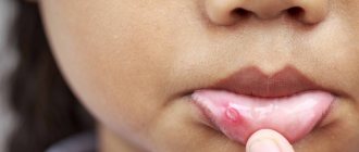
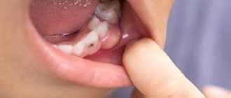
The most striking symptom of oral candidiasis is a white, loose coating on the mucous membrane. It mainly covers the tongue, cheeks, and may affect the gums and palate. Plaque can be easily scraped out; the tissue underneath is prone to redness and may bleed. Other signs of the disease include:
- unpleasant taste;
- dry mouth;
- burning;
- the appearance of cracks in the corners of the lips;
- difficulty or painful swallowing;
- unpleasant sensations with habitual movements of the tongue.
Candidiasis can be acute or chronic. In most cases, it is the acute form that manifests itself; the chronic form is typical for carriers of HIV infections and smokers. Depending on the degree and form of the disease, other symptoms may be observed, so even if one or two appear, you should immediately contact your dentist.
Diagnosis and treatment of superficial candidiasis
T
The term “superficial candidiasis” combines lesions of visible mucous membranes, skin and its appendages caused by
Candida fungi.
Usually these are those forms of candida infection that (not counting pediatrics) require contacting a dermatologist. Superficial forms of candida infection are also candida keratitis, blepharitis, and external otitis. Unlike deep forms of candidiasis, which have much in common in pathogenesis, forms of superficial candidiasis are pathogenetically noticeably different from each other. Thus, with oral candidiasis, the influence of nonspecific and immunological protective factors is clearly visible, which determines the inclusion of this nosological entity in the group of HIV-associated infections. With candidiasis of the skin and nails, the main pathogenesis is a violation of the barrier function of the skin.
Like most forms of candidiasis, superficial candidiasis is classified as an endogenous infection, with the source of the pathogen in the body of the patient himself. Nosocomial and HIV-associated infection is, as a rule, only oral candidiasis. The main approach to the treatment of superficial candidiasis is etiological, i.e. removal of the pathogen mass. According to our data, 84% of the etiology of modern candidiasis is due to the species C. albicans
, 9% – for
C. parapsilosis
;
2% each for C. tropicalis
and
C. krusei
, 1% for
C. glabrata
.
C. albicans, C. parapsilosis
and
C. tropicalis,
sensitive to modern antimycotics, accounted for more than 95% of the etiology of candidiasis of all localizations.
Oral candidiasis
The most common acute pseudomembranous form
, or thrush. It is characterized by a coating of films resembling curdled milk. Thrush takes a chronic form in patients with AIDS and other forms of immunodeficiency. Characterized by a long, persistent course, resistance to therapy. Clinical features are the frequent involvement of all parts of the mouth, sometimes difficult to separate films with an erosive, bleeding base.
Acute atrophic form
may occur after acute pseudomembranous form or independently. It is often a complication of antibacterial therapy or a consequence of the use of local (including inhaled) or systemic corticosteroids. The lesions are represented by spots of erythema with a smooth, as if varnished surface. The acute atrophic form is often accompanied by pain, burning and dryness in the mouth. The mucous membrane becomes very sensitive to tactile, chemical and temperature stimuli, which makes ingestion of rough food, cold and hot liquids painful.
Acute stomatitis from dentures also refers to the acute erythematous (desquamative) form. A clearly defined area of bright erythema and edema is found under the prosthesis.
Chronic atrophic form
Oral candidiasis is more common in older people who use dentures, and therefore its synonym is denture
stomatitis
. This form has very sparse symptoms, so it is usually detected only when changing dentures. Erythema and swelling of the mucosal area adjacent to the prosthesis are characteristic.
Median rhomboid glossitis is characterized by the appearance of a diamond-shaped or oval lesion of papillary atrophy in the middle of the dorsum of the tongue.
Chronic hyperplastic form
(hypertrophic, plaque, or candida leukoplakia) is more often observed in smokers, sometimes in those who use dentures. With it, white spots and plaques of varying sizes appear on the mucous membrane of the cheeks (less often on the tongue). They differ from lesions caused by pseudomembranous candidiasis in that they are difficult to separate from the underlying epithelium. The chronic hyperplastic form of oral candidiasis requires special attention due to the fact that in 15–20% of cases it becomes malignant. Particular attention should be paid to those lesions where elements of erythema and leukoplakia are combined, since there is a high probability of malignancy that has already occurred. It is still not clear, however, whether malignancy is a consequence of candida infection or a secondary infection of an already altered epithelium occurs. The hyperplastic form must be differentiated from leukoplakia and lichen planus.
Chronic granulomatous
, or nodular, form of oral candidiasis is rare. Small nodules appear on the mucous membrane of the tongue, so that it sometimes resembles a cobblestone pavement.
Erosive-ulcerative
, or locally invasive form of oral candidiasis is very rare. Similar forms were described in diabetic ketoacidosis and were observed by us in chronic candidiasis associated with autoimmune polyendocrinopathy (APECED). These forms should be distinguished from secondary candida colonization or infection of existing erosive and ulcerative lesions (for example, from trauma to a prosthesis).
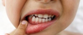
Candidiasis of the corners of the mouth
(angular, or angular, stomatitis, angular cheilitis, candida infection), can accompany any of the listed forms of oral candidiasis or develop independently. The disease can be found in people who have deep folds in the corners of the mouth, which usually occur with a low bite, in older people who lose teeth with age, and when using removable dentures. Other causes of seizures can be bacteria (especially in children), vitamin deficiency, and iron deficiency.
Usually the jam is noted in both folds of the corners of the mouth. The clinical picture is represented by erythema and cracks in the corners of the mouth; an easily removable whitish coating (a layer of macerated epithelium) can be seen. The lesions may be painful when opening the mouth and moving the lips.
The disease is prone to chronic relapsing or persistent course. Over time, infiltration develops around the crack, the crack itself deepens, and its edges thicken.
Candida lesions are differentiated from bacterial (staphylococcal) lesions, which are more characterized by bright widespread hyperemia, slit-like erosion in the center, eczematous skin lesions and crusts. Similar conditions can also be observed with vitamin deficiencies.
In the area of the pharynx and tonsils
A pseudomembranous form of candidiasis may be observed. As a rule, rashes are not accompanied by any additional subjective sensations. Isolated candida lesions of the tonsils and pharynx are very rare; a combination with oral candidiasis is usually observed.
Candidiasis of skin folds
Candidiasis of large folds, also called “candida intertrigo” or “intertriginous candidiasis,” in adults affects the folds of the perineum, gluteal, inguinal and axillary folds, folds on the abdomen and neck in obese people; Women often experience lesions of the vulva and the skin under the mammary glands.
Candidiasis of large folds
At the beginning of the disease, a whitish strip of macerated stratum corneum appears in the depths of the fold. Here surface cracks and erosions form.
The formed erosion has polycyclic edges, sharply delimited from the surrounding skin and bordered by a white rim of exfoliating epidermis.
Around the main focus – erosion – “dropouts” appear, represented by small surface bubbles and pustules. Opening up, these elements turn into erosions, also prone to growth and fusion. The lesions are accompanied by itching. Inguinal athlete's foot and erythrasma often have the same localization as candidiasis of large folds.
Candidiasis of interdigital folds
affects the skin between the fingers, most often on the hands. A synonym for candidiasis of the interdigital folds is interdigital yeast erosion (erosio blastomycetica interdigitalis). The first elements of the rash appear on the skin of the lateral surfaces of the proximal phalanges of the fingers. As a result of maceration, a white stripe of detached epidermis is formed, and then erosion, usually located linearly along the fold. The boundaries of the erosion are clear and bordered by an overhanging whitish fringe of macerated skin. The lesion usually does not extend beyond the main phalanx and does not extend to the back of the hand.
Most often, interdigital erosion is observed in the narrowest third and fourth interdigital folds. As a rule, bilateral lesions or lesions of the right hand predominate.
Subjective sensations include itching and burning, and in advanced cases complicated by bacterial infection – pain. With secondary pyococcal infection, regional lymphadenitis and lymphangitis occur. The disease often takes on a chronic, relapsing course.
Yeast interdigital erosions should be distinguished, first of all, from dyshidrotic eczema on the hands, and on the feet – from lesions caused by dermatophytes.
Candidiasis of smooth skin
Candida lesions of smooth skin can be a consequence of the spread of the process from the area of the folds, or independently appear anywhere under compresses, wet bandages, or during prolonged bathing. In women breastfeeding infants with oral thrush, lesions appear in the peripapillary area. In these cases, the clinical picture of the lesions resembles that of candida intertrigo of the folds: small blisters and erythematous papules at the onset of the disease, the formation of point erosions, growth and fusion of foci, large erosions with a whitish fringe along the edge. The course of the disease is acute, the lesions tend to resolve after eliminating the factors provoking maceration. The rash is accompanied by itching. Differential diagnosis of such lesions is usually carried out with microbial eczema.
Candidiasis of the skin of the palms
can occur in two forms.
One of them, vesiculopustular
, occurs in children and resembles dyshidrotic eczema, manifesting itself as small superficial blisters and pustules on the palms and palmar surface of the fingers, resolved by peeling.
The other, hyperkeratotic
form, resembles tylotic eczema, manifests itself with moderate hyperemia and an accentuated pattern of palmar furrows, the skin of which acquires a brown or brownish tint.
The course of the disease is chronic; patients are occasionally bothered by itching. As a rule, with candidiasis of the palms, candida paronychia with onychomycosis or interdigital yeast erosions are also observed.
Dermatophytic lesions of the palms, with which this form of candidiasis has to be distinguished, are characterized not by brown, but by white color of the palmar grooves and ring-shaped or lamellar peeling, as well as damage to the nails of the hands and feet.
Candida balanoposthitis
Burning and itching in the area of the glans penis may occur within a few hours after sexual intercourse. In mild cases, they last no more than 1–2 days and pass, resuming after the next intercourse. There is moderate hyperemia and mild superficial peeling. Discharges are rare.
Papules may appear on the skin of the head and foreskin, turning into superficial pustules and vesicles, and then into characteristic round erosions, bordered by a white strip of macerated epidermis, which, when fused, have a polycyclic outline. A whitish coating may be observed on the surface of these elements. The formation of erosions occurs most often in the contacting areas of the skin of the head and the inner layer of the preputial sac. A whitish pseudomembranous plaque can be observed on the inner layer of the foreskin.
Over time, the process can spread to the skin of the penis and inguinal folds. In severe cases, persistent, long-lasting erosions, ulcerations, infiltration and cracks of the foreskin, and the development of phimosis are observed. In patients with diabetes mellitus, acute (fulminant) forms of candida balanoposthitis have been described, occurring with severe swelling of the head, cracks and ulcerations.
Candida allergy with superficial candidiasis
To the so-called Candida
–mykids, or “candidida” include cases of skin rash caused by hypersensitivity reactions to
Candida spp
.
Unlike true skin candidiasis, fungal elements are not found in rashes with Candida
mycids.
In this regard, the diagnostic criteria for Candida
mykids seem unclear, since candida colonization and even candidiasis of the skin or internal organs, observed simultaneously with mykids, do not exclude the possibility of developing allergic reactions to other allergens.
A reliable criterion can be considered the resolution of the rash upon eradication of Candida spp
., cure of candidiasis of another localization.
Manifestations of candida mycids included rashes resembling seborrheic, microbial and dyshidrotic eczema, urticaria, ring-shaped erythema, and dermatitis.
One of the forms of allergic reactions to candida colonization is considered itching in the area of the outlet openings of the intestinal tube, genitals (i.e. in the mouth, anus, vagina). Itching in the anus ( pruritis ani)
) when associated with candida colonization of the intestine, it can be combined with asymptomatic or manifest candidiasis of the rectum, fissures and maceration at the anus.
Currently, the predominantly candida etiology of pruritis ani
is disputed; some authors point to the association of itching with other microbes, in particular with dermatophytes.
Sensitization to Candida
(regardless of the skin manifestations of candida allergy) often accompanies chronic forms of candidiasis.
C. albicans
antigen in these patients revealed a significantly more frequent occurrence of reactions of types I, II and IV of hypersensitivity compared to control groups. At the same time, changes in type I sensitization parameters did not correlate with the presence of atopic history. This indicator directly correlated with the presence of itching, and inversely with a positive NBT test and the presence of a bacterial infection. This can be regarded as a feature of the reaction of the macroorganism and, to a large extent, as evidence of the Th2 profile of immunoreactivity and the ineffective fungicidal response of the macroorganism. Type II hypersensitivity (antibody-dependent cellular cytotoxicity reactions) directly correlated with the presence of paronychia, chronic dermatophytosis, increased levels of B-lymphocytes, early activation of T-lymphocytes in patients with chronic candidiasis against the background of APECED.
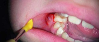
Candida paronychia
Candida paronychia is generally characterized by a chronic undulating course, moderate inflammation, absence of pain or mild soreness. Cases of acute candida paronychia have been described, but it remains unclear what is most often the cause of acute phenomena - Candida spp.
or secondarily attached bacteria.
Candida paronychia most often affects the middle and ring fingers of the right hand and the middle finger of the left hand. The little finger and index finger are affected less frequently, and the thumb is even less frequently affected. Involvement of all fingers or both hands is less common, and paronychia on the feet is extremely rare.
At the onset of the disease, the skin of the nail fold or surrounding skin turns red, becomes thinner, and its pattern is smoothed out. The roller swells, its edge becomes rounded or undermined, and the nail skin disappears. Cracks can be observed along the edges of the roller, erosion can be observed on the inner (becomes visible when the adjacent nail plate is removed) or outer side. Soreness or mild pain of a pulsating nature appears. An abscess forms in the groove under the nail fold, causing pain when pressed. When pressing on the cushion, a scant whitish-yellow purulent discharge may appear from under it.
Over time, the inflammatory phenomena subside, pain disappears, swelling and hyperemia disappear or become less noticeable. Fine-plate peeling is observed on the skin of the roller, especially pronounced at the edges. The absence of nail skin also indicates a disease that continues in the chronic stage.
The duration and wave-like nature of the course of paronychia is evidenced by changes in the surface of the nail plate. Characteristic is the appearance of alternating transverse grooves and elevations parallel to the lunula and the free edge of the nail, sometimes called Beau lines. The number of furrows and their depth generally correspond to the number and intensity of exacerbations of paronychia.
Candida onychomycosis
Onychomycosis caused by Candida spp.
, usually presented in a proximal form, less often in a distal form.
Proximal form
Candida onychomycosis, as a consequence of paronychia, begins with dullness and clouding of the nail plate at the proximal edge, usually at one of its corners. Soon the nail plate in this area becomes brittle, crumbles, and acquires a yellowish-brown or brown color. As the nail grows, a strip of modified nail plate forms at its lateral edge. The nail looks like it has been cut off from the side. In addition to the proximal-lateral type of lesion, there is also a proximal type involving the entire width of the plate.
In distal form
Candida onychomycosis causes a change in the color of the nail plate at its free edge. Over time, the plate becomes brittle, loose, and rises due to subungual hyperkeratosis. The distal form of candida onychomycosis is observed more often in patients with peripheral circulatory disorders, Raynaud's syndrome, and existing skin diseases (eczema, psoriasis, etc.).
The clinical picture of the distal form of nail candidiasis resembles that of a more likely dermatophyte lesion. The latter must always be excluded using laboratory diagnostics. The rare proximal form of onychomycosis caused by dermatophytes is not characterized by paronychia, but the appearance of white spots under the nail plate is typical.
Total dystrophic onychomycosis and paronychia affecting all fingers, previously described for chronic skin candidiasis, accompanied by changes in the terminal phalanges in the form of drumsticks, are rare these days. According to our observations, in the modern picture of nail lesions in chronic skin candidiasis, paronychia of the 1st–3rd fingers of the hand, preceded by a nail injury, predominate, and onychomycosis on the legs is most often caused by dermatophytes.
Treatment of superficial candidiasis
Oropharyngeal candidiasis
Drugs for local etiotropic therapy of candidiasis are divided into antiseptics
and
antimycotics.
Antimycotics - polyene antibiotics and imidazoles - are prescribed in the form of solutions, aerosols, gels, drops, regular and chewable tablets. Previously, our country also produced caramels with decamine 0.15 and levorin cheek tablets 500,000 units. Caramels with imidazole derivatives are now produced abroad.
To local polyene antimycotics
Drugs used in the treatment of oropharyngeal candidiasis include nystatin, levorin, natamycin and amphotericin.
Imidazole derivatives
include miconazole, econazole, clotrimazole and others. In Russia, local forms of imidazoles include a solution of clotrimazole (Candide).
If the doctor has at his disposal only drugs in forms that are not specifically adapted for use in oral candidiasis, then they should be adapted independently. Otherwise (for example, if nystatin tablets are swallowed), the drug is wasted.
It is necessary to explain to the patient that any drug for local treatment should remain in the oral cavity as long as possible. Nystatin tablets should be chewed and kept in the mouth for a long time, but it is better to prepare a suspension from them. The unpleasant taste of the suspension can be weakened by adding up to 50% sucrose. Solutions and suspensions linger longer in the oral cavity if compresses with cotton wool soaked in them are prescribed rather than irrigation. We also recommend using any antifungal ointment (preferably with 2% active substance), applied between two layers of cotton wool and placed behind the cheek (sandwich application).
The duration of treatment of acute forms with local antimycotics is usually 2–3 weeks, with antiseptics – somewhat longer. It is recommended to carry out treatment until complaints and clinical manifestations disappear, and then for another 1 week (in any case, at least 2 days).
Antiseptics
with antifungal action are usually prescribed in the form of lubricants or rinses. It is more effective to use modern antiseptics - 0.12% chlorhexidine bigluconate or 0.1% hexetidine solution (Hexoral, also available in aerosol form). Rinse for 30–60 seconds with 10–15 ml of any of these solutions twice a day after meals. The aerosol is applied for 1–2 seconds. Unlike rinses with antifungals, antiseptic solutions should not be swallowed.
Preference is also given to antiseptics in chronic forms of candidiasis, stomatitis from dentures, and seizures, since in these forms of the disease the role of mixed infection and microbial associations is assumed.
For candida infections, the treatment of choice is ointments containing an antifungal (preferably also an antibacterial) agent and at the same time corticosteroid hormones. These include Candida-B, Triderm, Lorinden S, Lotriderm, Pimafucort, Mycozolon, Travocort.
Systemic therapy
it is prescribed, as a rule, only for certain indications (chronic and severe forms, predisposing conditions, combination with candidiasis of other localizations, ineffectiveness of local therapy, etc.). This is determined by the fact that despite the highest efficiency, systemic therapy also has disadvantages, primarily the possibility of developing resistance and high treatment costs. At doses usually prescribed for oral candidiasis, side effects and toxicities are rare, except in the continuous long-term treatment of chronic forms of the disease. Let us repeat that with the exception of fluconazole, itraconazole and ketoconazole, all other oral antimycotics are classified as local treatments for candidiasis.
is considered the drug of choice in systemic treatment of oral candidiasis .
It is prescribed for candidiasis (in the absence of immunodeficiency) at 50–100 mg/day: on the first day – 100 mg, then 50 mg for 10 days. Itraconazole is prescribed at 100–200 mg/day for 7–10 days, ketoconazole at 200–400 mg/day.
When C. albicans
the dose of the systemic drug is increased (in the case of fluconazole to 400–800 mg/day). In some cases, in particular, with cross-resistance to azoles, parenteral amphotericin is prescribed at 0.5–0.7 mg/kg/day for 1 week.
With frequent relapses, it is possible to prescribe pulse therapy with fluconazole, from 150 mg once a week. Intermittent schemes can reduce stability or significantly delay its development.
To prevent relapses for any of the indications for systemic therapy, you can use various antimycotics and antiseptics, preferably in the form of rinses or lozenges/caramels.
Basic preventive recommendations for patients with chronic forms of oral candidiasis: rinse your mouth thoroughly after each meal (for rinsing you can use water, a 2-3% solution of baking soda, a weak solution of potassium permanganate); brush your teeth with toothpastes containing antimicrobial additives; follow recommendations for wearing dentures; treat caries, periodontitis, and other oral diseases.
Skin candidiasis
There are three main areas in the treatment of skin candidiasis. The first and most popular is the destruction of pathogens in the affected area. Doctors often forget about the other two areas necessary for cure. One of them is the destruction of the endogenous source of the pathogen in the intestines or genitourinary tract. Another is the correction of conditions predisposing to candida intertrigo.
The first task is to eliminate the pathogen in the skin lesion
- achieved by prescribing local agents - antimycotics or antiseptics. Time-tested, simple and affordable means - alcohol or water (the latter is better for large folds) solutions of aniline dyes: methylene blue (2-3%), brilliant green (1%), Castellani liquid, ointments and pastes containing 10% boric acid . A 2% aqueous solution of resorcinol, a 0.25% solution of silver nitrate, and a 0.1% solution of rivanol are also used. Almost any local antimycotic can be used, regardless of the class of compound (including morpholines and allylamines), in the form of 1–2% creams, ointments, and solutions. Both local antiseptics and some antimycotics, in particular imidazole derivatives, are attractive due to their broad spectrum of action, including bacteria. Combined products containing an antimycotic along with an antiseptic or antibiotic and, as a rule, a steroid hormone, should be used in the presence of inflammatory phenomena in the lesion, often caused by the addition of bacterial flora.
External agents are used until the skin lesions are completely resolved, and then for another 1 week.
The second task is the elimination of the intestinal or genitourinary reservoir of Candida spp.
– usually achieved by prescribing special local remedies: orally for intestinal candida dysbiosis and in the form of suppositories or douching for vaginal candidiasis. For intestinal dysbiosis, the drugs of choice are: nystatin, prescribed in tablets at 1–2 million units/day, or natamycin at 0.4 g/day (children’s doses are half of those indicated). Duration of treatment is 1–2 weeks.
The administration of systemic antifungal drugs leads to widespread destruction of the pathogen both in skin lesions and in all its reservoirs outside the skin. Fluconazole is prescribed 150 mg once, for torpid course - 150 mg/day once a week for 2-3 weeks. Itraconazole is prescribed at 100 mg/day for 2 weeks or 400 mg/day for 7 days. Ketoconazole is prescribed 200 mg/day for 1–2 weeks. The advisability of prescribing systemic antimycotics is determined by the effectiveness of previous therapy, the patient’s condition, including concomitant diseases, contraindications to the use of specific drugs and drug compatibility, the motivation of the patient who wants to get rid of the manifestations of the disease as soon as possible, as well as the availability of drugs. In most cases, treatment of skin candidiasis does not begin with systemic medications.
The third task is the correction of predisposing conditions
– consists of treating diseases (for example, diabetes), a complication of which is candidiasis of the folds. It is necessary to eliminate local factors that provoke intertrigo: for interdigital erosions, protect the skin of the hands with gloves while working; hygiene of body folds using drying powders, lotions, pastes - for candidiasis of large folds. The first and third tasks can be combined by adding any antifungal agent in the form of an ointment or cream, or nystatin powder (100,000 units per 1 g) to the zinc paste.
Candida balanoposthitis
Candida balanitis and balanoposthitis in general can be treated according to the same principles as vulvovaginal candidiasis. Local treatment of balanoposthitis includes toileting the head and preputial sac, baths with weak antiseptic solutions: potassium permanganate, 2% soda solution, 1% boric acid solution once a day. Local antimycotics in the form of a cream are used 1–2 times a day for 2 weeks.
Among systemic agents, preference should be given to a single dose of fluconazole at a dose of 150 mg; for chronic recurrent and persistent forms, repeated courses of systemic therapy are recommended.
Candida paronychia
The treatment tactics for candida paronychia depend on the stage of the process (acute or chronic), the severity of inflammatory phenomena, changes in the nail plate, the presence of candidiasis of the skin or mucous membranes, and the success of previous therapy.
The question of prescribing systemic therapy is fundamental.
. We recommend prescribing systemic drugs (itraconazole, ketoconazole and fluconazole) for onychomycosis, namely for permanent changes in the nail in the form of onycholysis and hyperkeratosis. Since the proximal form of onychomycosis develops with paronychia, the combination of paronychia and onychomycosis is always an indication for systemic therapy.
The need to prescribe systemic agents is explained not only by the involvement of the nail plate, because Candida spp.
, as a rule, are incapable of aggressive growth in it and independent maintenance of the process. Onychomycosis that persists as the plate grows indicates the preservation and persistence of the source of infection under the nail fold.
For the same reason, constant changes in the plate in the form of wavy transverse grooves, which indicate a chronic recurrent form of the disease, regardless of onychomycosis, can be considered an indication for systemic therapy. We also consider the combination of paronychia with candidiasis of the skin or mucous membranes to be an indication for systemic therapy. In this case, only local therapy will be labor-intensive and does not guarantee either clinical cure or elimination of the source of infection.
In case of fresh paronychia or its exacerbation, or the presence of inflammatory phenomena, local therapy
. Hot baths with soda, a 3% solution of boric acid, and potassium permanganate help soften parts of the nail for their subsequent removal and cleaning, promote better implementation of antimycotics, and the antiseptics included in their composition act on the attached bacterial flora.
After the baths, you can prescribe any antifungal agent. Foreign authors suggest long-term (up to six months) prescription of local antimycotics - azole agents, allylamines, ciclopirox or amorolfine. We prefer a short course of systemic antimycotics to long-term local therapy. Fluconazole
Prescribe 150 mg once a week for 2–6 weeks.
Itraconazole
is prescribed at 200–400 mg/day for 1 week, or two three-day courses of 200 mg/day with an interval of 1 week.
ketoconazole
200 mg/day for 2 weeks. Systemic therapy can be combined with treatment with local antiseptics or antifungals.
Simple and affordable local remedies for the treatment of paronychia are solutions of aniline dyes (1% brilliant green is the most widely used), Castellani liquid, iodine tincture. In the acute stage of paronychia, the nail folds are lubricated with antiseptic solutions 2-3 times a day for 1-3 weeks.
For severe inflammatory phenomena, applications of pure ichthyol and corticosteroid ointments, preferably combined with antiseptics or antimycotics (for example, Lorinden S, Triderm), can be prescribed. In addition, in some cases, nonsteroidal anti-inflammatory drugs can be prescribed orally.
In case of paronychia combined with onychomycosis, when a decision is made to prescribe only local therapy, after the baths the softened affected parts of the nail plate are removed. For the same purpose, they use Arievich detachment, ureaplast, and a combined antifungal patch of mycospores, applying them to the nail for 2 days, after which they clean, exposing the nail bed.
Candida onychomycosis
For proximal onychomycosis, fluconazole is prescribed at a dose of 150 mg/day for 4–6 months. Itraconazole is prescribed at 400 mg/day (in two doses) in two one-week courses of pulse therapy with an intercourse interval of 3 weeks. Ketoconazole is prescribed at a dose of 200 mg/day for 2–4 months, the duration of therapy is calculated using the KIOTOS index.
In the proximal form
In distal form
Candida onychomycosis, the therapeutic decision is also made based on the KIOTOS index.
For clinical index values less than 2, or for KIOTOS less than 3, local therapy is chosen, including removal of damaged parts of the nail plate and the prescription of local antimycotics. For values exceeding those indicated, systemic therapy is prescribed. Its doses are appropriate, and the timing usually does not exceed those for the proximal form. Literature:
1. Antonyev A. A., Bulwakhter A. A., Glazkova L. K., et al. Candidiasis of the skin and mucous membranes. – M.: Medicine. – 1985.
2. Arievich A. M., Stepanishcheva Z. G. Candidiasis. M.: Medicine. – 1965.
3. Marchenko A.I., Rudenko M.M. Candidiasis of the oral mucosa. – Kyiv: Health. – 1978.
4. Potapova S. N. Yeast paronychia and onychia. – L.: Medgiz. – 1962.
5. Sergeev A. Yu., Sergeev Yu. V. Candidiasis: nature of infection, mechanisms of aggression and defense, diagnosis and treatment. – M.: Triad-Kh. – 2000.
6. Sergeev Yu. V., Sergeev A. Yu. Onychomycosis: fungal infections of the nails. M.: Geotar. – 1998.
7. Stepanova Zh.V. Fungal diseases. – M.: Kron-Press. – 1996.
Causes of oral candidiasis
How does the disease occur? The fungus Candida lives in the body from birth and, under certain circumstances, provokes illness. What becomes the trigger for infection:
- weakened immunity;
- taking antibiotics;
- long-term use of removable dentures, especially if they do not fit tightly;
- insufficient oral hygiene;
- smoking;
- oncology;
- use of inhalers for asthma;
- excess simple carbohydrates in the diet;
- pregnancy period;
- infancy or old age.
It can be seen that there are many reasons, often they act together, less often - individually. In general, oral candidiasis is rare in adults. It mainly affects children in the first years of life and older people. Among these categories, the prevalence of the disease reaches 10%.
How is oral candidiasis treated?
To begin with, let's say that treatment is necessary, otherwise the infection spreads to the pharynx, tonsils, and gums. Then the disease descends into the lungs, gastrointestinal tract, liver and causes irreparable harm to them.
When the first signs of candidiasis of the oral mucosa appear, you should contact your dentist or therapist to confirm the diagnosis. When you see your doctor, describe all your symptoms, even those that are not on the list. For example, you have noticed increased anxiety or a tendency to stress lately. Information about the medications you are taking, recent illnesses, and being around sick people is also important. To confirm the diagnosis, it is often enough to take a swab from the oral cavity and carry out the appropriate analysis. If the disease has already spread to other organs, a more thorough examination may be required, for example, endoscopy - diagnosing the condition of the esophagus and stomach using a flexible tube with a camera at the end.
How to treat oral candidiasis? Treatment can be local or systemic depending on the form and stage of the disease. When local, antifungal agents and antiseptics are prescribed. These can be sprays, rinses, gels, lozenges and more. Treatment on average takes up to 3 weeks, as a general rule - until symptoms disappear and another week. Systemic treatment is used for chronic forms or when the infection spreads to other organs. It usually involves the use of more medications that are intended to treat the directly affected areas.
Under no circumstances resort to self-medication! A fungal infectious disease has nothing to do with inflammation, so your actions are more likely to harm your body. In doing so, you can reduce the harmful effects of the infection and make you feel better. To do this, eat natural yogurt without sugar and take probiotics as recommended by your doctor. But these actions are not a complete treatment for candidiasis in the mouth!
Patients and methods
In the Department of ENT Pathology of Children, State Budgetary Healthcare Institution “NIKIO named after. L.I. Sverzhevsky" DZM on the basis of the State Budgetary Institution "DGKB No. 9 named after. G.N. Speransky" DZM examined and treated 306 children with chronic tonsillitis and 402 children with chronic adenoiditis aged 2 to 16 years.
The diagnostic algorithm for examining children with fungal infections of the pharynx included: collection of complaints and medical history, otorhinolaryngological examination, endoscopic examination of the nose, nasopharynx and oropharynx, microbiological diagnostics (bacteriological and mycological).
When collecting an anamnesis of the disease, attention was paid to the child’s complaints, previous viral infections, and the presence of chronic diseases, in particular diabetes mellitus; features of the course and frequency of recurrence of the inflammatory process of the pharyngeal and palatine tonsils. The collection of medical history included anamnesis of antibacterial, corticosteroid and immunosuppressive therapy, since with long-term use of these drugs immunosuppression occurs, leading to the activation of a fungal infection.
During an objective examination, attention was paid to the condition of the nasal mucosa; for the presence and nature of pathological discharge in the common nasal passage; for the presence or absence of changes in the mucous membrane of the oropharynx, palatine tonsils, regional lymph nodes (submandibular, anterior cervical, posterior cervical).
When conducting an endoscopic examination of the nasopharynx, we assessed not only the shape, size and location of the pharyngeal tonsil in relation to the paranasal structures, but also the absence or presence of signs of inflammation of the pharyngeal tonsil (swelling, hyperemia, smoothness of the grooves, the presence of secretions, the nature and extent of pathological overlays).
When collecting pathological material from the surface of the pharyngeal tonsil in children, we used a method developed by us, which allows us to take material directly from the pharyngeal tonsil, excluding contamination from adjacent paranasal structures (priority certificate for the issuance of a RF patent for an invention No. 2011107242). The use of endoscopic control during the selection of pathological material made it possible to visualize and select the exact localization of the pharyngeal tonsil site. When carrying out this method, a rigid endoscope with 0° optics was used.
To determine the prevalence of fungal inflammation, samples were taken from different loci (from the palatine tonsils, from the nasopharynx and from the surface of the mucous membrane of the posterior pharyngeal wall).
Laboratory microbiological diagnostics were carried out in two directions: microscopy of pathological discharge from various loci of the pharynx and nasopharynx, as well as cultural research methods - inoculation of pathological discharge on liquid and solid nutrient media, followed by counting fungal colonies and determining the growth rate. The preparations were stained using the Gram method, and the calcofluor white staining method was also used. When conducting a cultural study, Sabouraud's medium was used to identify the fungus. Species identification was carried out using standard API 20 test systems (bioMerieux, France).
The sensitivity of fungi to antifungal drugs was determined using the disk diffusion method to modern antifungal drugs: fluconazole, ketoconazole, clotrimazole, itraconazole, voriconazole and amphotericin.
To identify the bacterial flora, 5% blood agar, 10% yolk-salt agar, and Endo medium were used as nutrient media. After isolating a pure culture, the sensitivity of bacteria to antibiotics was determined using the disk diffusion method.
To exclude side effects of systemic antifungal drugs, on the 5th day of taking antifungal therapy, a biochemical blood test was performed (monitoring liver enzymes: ALT, AST, alkaline phosphatase).
How to avoid candidiasis?
To prevent the disease, you must follow a few simple rules:
- maintain oral hygiene;
- consult a dentist at least twice a year, even if there are no complaints;
- control nutrition, avoid excess carbohydrates in food;
- refrain from smoking, which can cause a chronic form of the disease.
Treatment of candidiasis of the oral mucosa can take a long time and also cause irreparable harm to health, so we recommend that you take care of yourself in advance by following preventive measures. If symptoms have already begun to appear, consult a doctor as soon as possible so that the infection does not have time to spread to other organs. You can undergo oral cavity diagnostics at Comfort Dentistry. Our specialists will tell you in detail about the prevention of common diseases of teeth and gums, and also, if necessary, provide high-quality treatment.