general information
Streptodermia refers to a pathology caused by streptococci. If we talk about the external manifestations of the disease, blisters and purulent rashes that itch appear on the skin.
Streptoderma in children can occur in two different forms: acute and chronic. Acute is characterized by pronounced symptoms, and chronic - by periodicity.
According to the degree of damage, the disease is classified into superficial, deep and intertriginous. Each form has a specific feature.
Molluscum contagiosum in children
Molluscum contagiosum is more common in children than in adults. Children up to one year old practically do not get sick with molluscum contagiosum. This is due to the fact that in the first year of life the child’s circle of contacts is small: the child moves in a limited space, often specially prepared for him and under the strict supervision of adults, without trying to come into contact with other children. But as soon as a child begins to actively communicate and independently explore the world, the threat of becoming infected with molluscum contagiosum increases sharply.
The peak incidence of molluscum contagiosum in children occurs between the ages of 2 and 6 years. Immunity at this age is still weak. Children become infected through toys or dirty hands. The virus penetrates the skin in the place where the integrity of the skin is damaged - through wounds, abrasions, cracks. Children's skin is delicate and sensitive, and the activity of a preschool child is great. As a result, numerous microtraumas occur, opening the way for infection. Cases of infection with molluscum contagiosum while swimming in the pool have also been described.
From 6 to 10 years, the incidence of molluscum contagiosum decreases. Instilling household hygiene skills is of great importance. The sooner your child starts taking care of clean hands, the better.
Symptoms
If streptoderma occurs, what is the treatment? Before we know the treatment options, let's find out the symptoms. The disease in children occurs in extremely severe forms:
- temperature up to 39C,
- poisoning,
- large lymph nodes.
The texture of the skin may vary depending on the form of the pathology:
- Superficial. Initially, red spots form on the skin. After a couple of days they turn into blisters with a certain liquid inside. The blisters grow up to two cm in diameter, after which they rupture. Yellow crusts form in this area. After this, the skin in this area heals, and the pathology develops further.
- Dry streptoderma . This form usually occurs in boys. Light and pink spots up to 5 cm in diameter form on the skin. They are found everywhere on the body. After they disappear, scars may remain.
- Streptococcal infection. The pathology extends specifically to the corners of the lips; it is encountered when there is a deficiency of vitamin B. Small cracks form on the skin, causing discomfort. After some time, they form into pustules that become crusty. The child has difficulty consuming food, as it causes pain.
- Felon. In a situation where streptococcus spreads to the area around the nails, panaritium develops. Oddly enough, this disease is more often encountered by children at an early age who bite their nails.
- Streptococcal diaper rash. This form of pathology is typical for breastfed children. Blisters form in the folds of the skin.
Understanding how streptoderma begins to develop in children, treatment which requires timely contact with a dermatologist and the implementation of therapeutic actions, not a minute can be lost. If you constantly put off going to the doctor, the pathology can develop into a chronic form, and it will be almost impossible to achieve a final cure.
streptoderma, treatment
What to do if a newborn has a callus on the lip
- First of all, determine whether it is really a milk callus and not a symptom of any disease;
- As a rule, a milk callus is located on the upper lip of a newborn. It can be observed much less frequently on the lower one;
- A real milk callus does not cause discomfort to the baby, he continues to eat and suck the breast or pacifier, behaves calmly, does not cry or be capricious;
- A true milk callus is white or light in color and has a clear liquid inside;
- Milk callus does not require treatment. It disappears on its own, may appear again and be regular;
- Do not open the bladder or release the liquid, otherwise you will injure the tissues and increase the risk of infection!;
- If the callus bursts on its own, treat the area with hydrogen peroxide;
- If the callus causes discomfort during feeding and provokes anxiety, then an infection has occurred and the disease begins;
- A watery blister with yellowish liquid and bright redness around indicates the presence of stomatitis or thrush;
- An elevated temperature and the appearance of several watery formations are signs of herpes;
- Herpes, stomatitis and thrush must be treated.
Causes
The causative agent of streptoderma, the photo of which you will find below, is group A streptococcus. However, it is worth noting that infection can only develop in a situation where certain factors are present:
- skin damage,
- lack of following basic hygiene rules,
- weak immune system,
- disruption of the endocrine system,
- dermatological diseases,
- depressive mood
- vitamin deficiency,
- constant washing of the skin, which leads to damage to the protective layer,
- strong influence of different temperature ranges,
- poisoning,
- circulatory disorders.
Therapy
Streptoderma has occurred ? Even in cases where the initial examination was carried out by a pediatrician, only a dermatologist can select therapy. Doctors in this field have an understanding of drugs with a narrow scope of action.
The first step is to limit the child’s diet. The course of treatment also involves avoiding water procedures, which can cause the spread of the disease. Healthy areas of the skin should be washed with a solution of chamomile, and damaged areas should not be touched at all.
The sick person needs to choose the right wardrobe, from which items of clothing made from synthetics and wool must be removed.
Doctors advise popping blisters that form on the skin with a disinfected needle, after which the abscess should be drained twice a day. Healthy areas of the skin are washed with a boron solution.
If crusts appear on the skin, they should be treated with antibacterial gels or ointments.
In more complex situations, many other medications may be prescribed:
- antibiotics for streptoderma of the tetracycline or chloramphenicol series,
- means that prevent the occurrence of an allergic reaction,
- means aimed at improving the body's immune system,
- vitamin preparations,
- antipyretic.
The set of medications must be discussed with the doctor. With the right treatment, the symptoms disappear within a week, but after the severe form is eliminated, scars may remain. This is what streptoderma looks like .
what does streptoderma look like?
Types of the disease, their symptoms
There are a large number of types of disease, each of which has its own symptoms. Cheilitis happens:
- catarrhal, resulting from injury. Its characteristic symptoms are redness, peeling, swelling and soreness of the lips.
- eczematous, developing due to the occurrence of eczematous processes. The reason is an allergy to a certain type of food, cosmetics (lipstick, cream).
- glandular, associated with an abnormality of the salivary glands. It happens that cheilitis on the lips accompanies periodontal disease, lupus, etc. The lower lip often suffers and begins to peel.
- meteorological. It is caused, as the name suggests, by weather conditions (high air humidity, strong wind, decreased or increased temperature, exposure to ultraviolet radiation, etc.).
- exfoliative, characterized by a chronic course. It is divided into two forms - dry (the blood supply to the lips is disrupted and, as a result, dry scales are formed, which can be easily removed, but after a few days they appear again) and exudative (the border swells and becomes inflamed, crusts appear).
- angular, called jam. Due to streptococci, inflammation begins in the corners of the lips. Vitamin deficiency and frequent licking of lips can also provoke it.
- atopic, accompanying atopic dermatitis. The red border of the lips flakes and itches, the lips crack.
- hypovitaminosis, which occurs due to a lack of B vitamins. Dryness and a pronounced burning sensation affect not only the lip area, but also the entire mucous membrane of the mouth, including the tongue. In this case, the mucous membrane turns red, and vertical cracks form in the area of the red border.
Regardless of the type of cheilitis, common symptoms will be:
- peeling of the lips, observed both only near the border and over the entire surface;
- swelling and swelling, burning and itching in the lips, the appearance of cracks and crusts on them.
Causes of complications and re-infection
The reason for re-infection may be incorrect therapy when, for example, if you have primary criteria for improvement, you decide to stop therapy. The most important thing is to remember that you need to unquestioningly follow the advice and instructions of your doctor.
streptoderma complications
Are we going to treat?
So, if you and your baby have had to deal with such an unpleasant phenomenon as a callus on the lip of a newborn, first of all, do not panic. If in doubt and if you want not to leave everything to chance, contact your child’s attending physician; after assessing the situation, he will suggest the correct algorithm of action.
In the vast majority of cases, no treatment will be needed, but delicate skin will not be harmed at all by light moisturizing and nutrition; it is best to use natural olive oil, it will soften the callus and prevent a rough and rough crust from forming in its place.
Some doctors recommend cauterizing a callus on a newborn’s lip with an antiseptic (iodine, brilliant green, furatsilin). Treatment with such means is a controversial and controversial method, because they are quite aggressive in their effects and can cause burns, especially on such a delicate area of the body as the lips. If a blister has burst, the best treatment for the wound is hydrogen peroxide; it will disinfect and dry the hole without further injuring the skin.
What can streptoderma be confused with?
Pyoderma
With pyoderma, the sebaceous and sweat glands are damaged. The rash appears in certain areas of the skin - parts of the body, scalp.
Herpetic rash
Herpes is characterized by the presence of blisters with constant inflammatory processes. They burst and provoke development.
Thrush or candidiasis
Candidiasis is characterized by erythematous papules or reddened, wet plaques. In the pathology under consideration, the lesion, as a rule, occurs in the mucous membranes or fold areas.
Manifestations of allergies
An allergic rash continues to occur as long as the influence is the causative agent of the allergy.
Will a bottle save you from calluses?
This, however, does not mean that it is worth limiting the baby’s sucking; moreover, pediatricians do not recommend weaning the baby or switching to mixed feeding before he is 6 months old. A callus on the lip of a newborn with artificial feeding occurs as often as in those babies who eat mother's milk.
If this problem greatly worries parents or the child experiences certain difficulties while eating, you can try changing the nipple on the bottle. The silicone nozzle is more durable and hygienic, but traditional latex is softer and more delicate to the touch, and many children like the tactile sensations after touching it more.
Types of disease
- Streptococcal impetigo. The most common standard form of the disease. A characteristic rash forms on the face, arms and legs. This form is also the most limited, since the pathogen does not move beyond the upper layer of the skin, which has certain protective mechanisms. The appearance of the integument does not change, only individual blisters with liquid appear on the reddened areas. Initially, the patient will experience itching, after which the blisters become darker, which helps the blister to crust over. The duration of this process reaches one week. However, it is worth remembering that if the rash resolves, bacteria are further transferred to healthy areas of the integument, which has its negative consequences for the sick person.
- Bullous impetigo. The rashes are located on the arms and legs and had a fairly impressive diameter. Once the blisters open, these areas of skin develop ulcers that can spread quickly.
- Streptococcal diaper rash. Most of the patients are young children and overweight people. The location of the rash is under the mammary glands, in the axillary folds, intergluteal or inguinal-femoral folds. If you are overweight, the abdominal area is affected.
- Ringworm common. Appears in the form of bright pink rashes. The place of appearance is the face. The rash may become smaller due to exposure to direct sunlight. Those areas of the skin that are affected will not take on a tan like before.
- Streptococcal infection is characterized by the formation of blisters in the corners of the mouth. After the blisters disappear, a crack appears in their place, which in a matter of minutes becomes covered with a yellow crust. It also happens that children tear off the crust, but it forms again.
- Streptococcal ecthyma is the most complex form of the disease. The disease is accompanied by the appearance of characteristic lesions and scars on the arms and legs. In case of development of ecthyma, as a rule, there is a deterioration in health.
Unusual oral mucocele in a child
An oral mucocele is one of the most common benign tumors of the oral mucosa, and is a cavity filled with mucus, a secretory product of the salivary glands (muco means mucus, coele means cavity).
The mechanism of development of such pathologies usually comes down to two options: extravasation of mucus associated with a traumatic lesion, and retention of mucus as a result of obstruction of the minor salivary gland. When mucoceles are localized at the bottom of the mouth, they are called ranulae, since the inflammation resembles the cheeks of a frog. The most common location of mucocele is on the lower lip, there is no prevalence by gender and age of patients, the peak incidence occurs in the second and third decades, rarely occurring in children, which makes the diagnosis and treatment of pathology in this group quite difficult. Mucocele has common clinical features with many other tumors and ulcerations of the oral cavity, which requires a careful differential diagnosis. In this article, we describe an interesting and unusual clinical case of mucocele of the lower lip in a child, focusing on the etiopathogenesis, clinical presentation and various treatment options.
Clinical case
An 11-month-old boy was referred to the clinic with complaints of a “small ball” on his lower lip and associated difficulty sucking for more than 3 months.
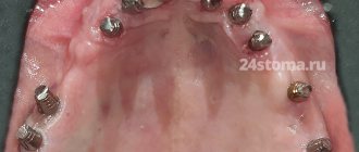
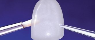
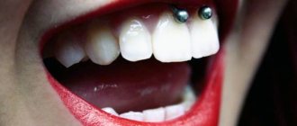
The child was in good general health and showed no other symptoms. No bad habits or local trauma were detected. Clinical examination revealed the presence of a soft tissue nodule on the mucous membrane of the lower lip (Figure 1), which was similar in color to the mucous membrane and measured 5 cm at its widest diameter, was set on a wide base, had a soft consistency, clear boundaries and a smooth surface. Based on the medical history and clinical examination, a preliminary diagnosis of mucocele was made. After a thorough examination and obtaining written consent from the parents, an excisional biopsy was performed under local anesthesia. Due to the small age of the child, parents and an assistant took part in fixing the hands. Since the baby was constantly crying, this ensured that the mouth was always open. Infiltration anesthesia was administered around the formation (2% lidocaine with epinephrine 1:80,000, one carpule). Before the injection, anesthetic gel is applied topically for 2 minutes. The lip was then retracted using finger pressure to bring the mass into greater convexity. A thick silk suture is passed through the mass along the widest possible diameter, then a surgical knot is formed, followed by excisional biopsy using an electrocoagulator (Photos 2 and 3), thus minimizing pain and postoperative bleeding. On the first day after surgery, an analgesic was prescribed to eliminate possible pain after the intervention.
Photo 1: Mucocele on the lower lip of an 11-month-old child.
Photo 2: Excision of the formation using an electrocoagulator.
Photo 3: View immediately after surgery.
The tissue sample was sent for histopathological analysis, which revealed a large area of mucous secretion containing mucinophages, mucin containing cells, surrounded by a compressed connective tissue wall and developing granulation tissue (Figure 4). The diagnosis of mucocele was confirmed. After 2 hours, the patient began breastfeeding as usual. Recovery was uneventful with a return to a normal diet within a week.
Photo 4: Hematoxylin-eosin stain revealed keratinizing epithelium with underlying connective tissue consisting of a large accumulation of mucin surrounded by granulation tissue and chronic inflammatory cells.
The child was re-examined after 15 days, 6 and 12 months. No recurrence was observed after 12 months (Figure 5).
Photo 5: View of the surgical site 12 months after the intervention, without relapse.
Discussion
Yamasoba identified two main etiological factors that can lead to mucocele:
- Injury
- Salivary gland duct obstruction
Basically, it is physical trauma that can cause the secretion of the salivary glands to spill into the surrounding submucosal tissue. Then, due to stagnation of mucus, inflammation occurs. The habit of biting your lip and sticking your tongue are also aggravating factors for this pathology.
The extravasal type goes through three stages of development:
- At the first stage, mucous secretion is poured out of the duct into the surrounding tissues, and leukocytes and histiocytes are also detected.
- At the second stage, due to the presence of histiocytes, macrophages and giant multinucleated cells as a result of a reaction to a foreign body, granuloma develops. This stage is called the resorption phase.
- At the third stage, a pseudocapsule is formed from connective tissue without epithelium around the mucous formation.
The retention type of mucocele is often observed in large salivary glands. This happens due to stretching of the duct and block by a stone or dense mucosa. There is a relationship between the severity of the pathology and the degree of duct obstruction.
Clinical characteristics
Clinically, they are characterized as single or multiple, round, fluctuating nodules, varying in color from normal pink to dark blue, often asymptomatic. Tissue cyanosis and vascular block are associated with distension of the underlying tissue and the clarity of the collected fluid, which may produce a bluish color. Sometimes they can rupture, leaving a slightly painful erosion that heals within a few days. Parafunctions such as lip biting, lip sucking, and trauma explain why the lower lip is the most common location for extravasal mucoceles. They are mainly found in children and young patients with no gender predominance, and are also rarely observed in children under 1 year of age.
Diagnostics
The medical history and clinical studies allow us to make a diagnosis of superficial mucocele. The appearance of the mucocele is pathognomic, but a correctly collected history of the formation is also important: localization of the formation, history of trauma, frequent recurrence, variations in size, bluish tint and consistency. Typically, mucoceles are mobile formations of soft or elastic consistency, depending on the number of tissues involved in the formation of the pathology. Despite this symptom of fluctuation, a drained mucocele does not fluctuate, and a chronic mucocele fluctuates much less due to well-developed fibrosis. In the retention type of mucocele, the cystic cavity with well-defined epithelial walls is lined with cubic cells. This type shows less inflammatory response. The extravasal type is a pseudocyst without an epithelial wall and contains inflammatory cells and granulation tissue. But even in the absence of epithelial cover around the mucosa, good encapsulation is found.
Radiography is an important contribution to the diagnosis of ranula. Localization of such formations is carried out using CT and MRI. High levels of amylase and proteins can be determined by chemical analysis. Histopathological examination is critical to confirm the diagnosis and usually shows the presence of ductal epithelium, granulation tissue, mucin accumulation and inflammatory cells.
Mucocele clinically resembles many other tumor-like and ulcerative formations of the oral cavity, so differential diagnosis requires a careful approach. Palpation is also important to differentiate the formation from other pathologies. Lipomas and tumors of the minor salivary glands do not show fluctuations, while cysts, mucoceles, abscesses and hemangiomas do. A simple technique known as fine needle aspiration biopsy (FNAB) is very useful and informative, especially when angiomatous lesions are involved in the differential diagnosis.
Treatment
The most common treatment for this pathology is simple surgical excision. Other treatment options include CO2 laser ablation, cryosurgery, intralesional corticosteroids, micromarsupialization, marsupialization, and electrocoagulation.
There is no difference in the treatment of retention and extravasal mucocele. Small mucoceles are removed along with the marginal tissue of the gland, and in the case of large formations, marsupialization will help avoid damage to the labial branch of the mental nerve. Lacrimal catheters are used to dilate the duct and resolve retention-type mucocele obstruction. To prevent recurrence during mucocele excision, it is also necessary to remove the surrounding glandular acini, remove the formation below the muscular layer, and avoid damage to the adjacent gland and duct. If the fibrous wall of the mucocele is thick, the removed tissue is sent for histological analysis to exclude neoplasm of the gland. Micromarsupialization can be accepted as an alternative treatment method in pediatric practice, since this technique is relatively simple, painless and rarely leads to relapse. This technique (after disinfection and anesthesia) consists of pulling a thick silk thread through the formation along its largest diameter and then making a surgical knot. The suture is removed after 7-10 days, which is sufficient time for the mucocele to disappear. The advantage of the CO2 laser technique is to minimize recurrence and complications, as well as to quickly and easily ablate the formation. This method can also be used in patients for whom long-term interventions are contraindicated. The remaining alternative options have not objectively proven to be highly effective. The administration of corticosteroids and gamma-linolenic acid usually becomes necessary only with multiple mucoceles, when surgical excision of each formation is difficult to perform.
Conclusion
Mucocele is the most common benign lesion of the oral cavity. Since this pathology is painless, dentists often detect it during the next examination of the oral cavity. Treatment of mucocele is quite problematic, since the risk of relapse is very high. However, surgical excision with dissection of surrounding tissue and minor salivary glands can lead to positive and durable results.
Authors: Neha Bhargava, Prateek Agarwal, Nitin Sharma, Mayank Agrawal, Mohsin Sidiq, Pooja Narain
Therapy and drugs
Throughout the spread of the disease, basic hygiene rules should be observed: in the first days, it is forbidden to take baths and wet impetigo, and it is forbidden to comb the affected areas.
Therapy for this disease is aimed at destroying the pathogen and strengthening the protective functions of the immune system. As a complex treatment, antibacterial drugs of the category of cephalosporins, macrolides and penicillins are prescribed. If you have bullous streptoderma, you should stop taking medications. As an immunocorrective treatment, the drugs “Likopida”, “Amiksin” and their analogues are effective. Treatment with drugs for . Restoration of microflora in the intestines occurs with the help of probiotics and prebiotics. Appropriate antihistamines will help get rid of scabies.
Means that increase the body’s resistance to infections in the form of solutions - “Eleutherococcus”, “Echinacea”, “Leuzea”, etc.
Sets of vitamin nutrients should also be taken in accordance with the instructions.
Local treatment includes antiseptic drugs that prevent the spread of putrefactive bacteria. Such medications include various alcohols, brilliant green, as well as their analogues - “Fukartsin”, “Chlorhexidine”, “Miramistin”, “Rivanol” or the cauterizing agent “Resorcinol”.
The use of a huge number of zinc-based pastes, preparations and ointments for streptoderma in children with intense scabies.
Treatment
The main method that allows you to remove rashes and cure skin pathologies is hygienic care. It is important to change the diaper immediately after a bowel movement and every three hours, washing the baby with running water. Between shifts you need to give air baths and leave the baby without clothes for a few minutes. At the same time, it is necessary to abandon synthetic clothing and low-quality children's cosmetics.
Bathing is carried out in water with a decoction of medicinal herbs. You can add chamomile, yarrow, and string. After bathing the child, dry thoroughly and powder the skin with powder.
In some cases, the doctor also prescribes treatment of rashes with drying antiseptics (“Furacilin”, “Chlorphilipt”), and creams containing panthenol (“Bepanten”, “Dexpanthenol”). White, red and deep miliaria are treated with antifungal and antibacterial ointments. However, it is not recommended to use any medications without prior consultation with a doctor. Source: Modern dermatological and cosmetic products for caring for the skin of children. Kotlukov V.K., Kuzmenko L.G., Antipova N.V. Medical Council, 2013. p. 8-12.
Doctors recommend setting the air temperature in the nursery at 20-22 degrees and monitoring the humidity, which should not exceed 50-70%. A good solution would be to purchase an air humidifier that will help create the right microclimate. These simple steps can help you get rid of the conditions that increase your risk of developing heat rash.
Preventive measures and prognosis
Due to the high risk of infection, sick children should keep their distance for some time and not contact other people. Quarantine is provided for up to 10 days. For the entire period of therapy, it is necessary to follow the rules of hygiene.
To prevent the development of the disease, it is necessary to disinfect the child’s personal belongings. Sick children should have a proper diet rich in nutrients. To improve the quality of the immune system, certain measures should be taken.
streptoderma treatment
As a rule, streptoderma ICD 10 in children is cured without any problems. Exacerbation of the disease and re-infection are observed mainly in children from disadvantaged families or in children with weak immunity. With timely treatment, symptoms disappear within one week. If you do not attach any importance to this, the disease can develop into a more severe form. In the worst case, the disease can lead to blood poisoning. For streptoderma, treatment with ointments and antibacterial agents is mandatory.
Thrush in a newborn
With thrush, a white coating with a red rim forms on the baby’s gums, tongue and cheeks. In addition, a rash and redness may appear on the body, and yellowish blisters or blisters on the lips. At the same time, the baby is capricious, refuses to eat, sleeps poorly, and feels discomfort.
In this case, it is also important to improve hygiene, disinfect things and objects that can get into the baby’s mouth. Products are boiled or sterilized for at least twenty minutes. To cure thrush, you can also treat your mouth with a soda solution up to eight times a day. To prepare this remedy, dilute a teaspoon of soda in a glass of boiled warm water.
Stomatitis and thrush can be caused by poor hygiene, too early or incorrect complementary feeding, fungus and thrush on the breasts of a nursing woman. To prevent the development of infection, maintain your hygiene and the hygiene of the mine. Be sure to clean your mouth after each feeding and burping, and remove any remaining milk or food from your baby’s lips. Thoroughly clean and sterilize bottles, nipples, baby dishes, toys and other objects with which the baby has constant contact.
Brush your mouth, tongue and teeth regularly. Up to six months, use special finger wipes or a sterile gauze bandage, after six months they switch to silicone finger brushes, and after a year you can already use a children’s toothbrush with soft bristles. Read more about oral and dental hygiene for a newborn in the article “How to properly brush your child’s teeth.”
Prevention
To reduce the risk of infection, you should follow the following rules:
- follow the rules of hygiene,
- disinfect damage to the skin,
- strengthen the name system with useful substances,
- do not carry out frequent water procedures, as this may pose a certain danger for the child,
- In case of primary symptoms, you should immediately consult a doctor.
Streptoderma on the face can be treated, but only if you consult a doctor in a timely manner. The earlier therapy was carried out, the easier it is to exclude complications and transition to the chronic stage. You can undergo a full examination in a clinical clinic.
Diagnostics
You should not self-medicate or try to diagnose your child yourself. Before you begin to treat prickly heat in a child, when the first rash appears, you should contact a pediatric dermatologist or pediatrician. The specialist will conduct an examination and distinguish the pathology from other diseases that are accompanied by a skin rash. For an experienced pediatric specialist, this is not difficult even with an initial visual examination.
In some cases, additional examination is necessary for a comprehensive diagnosis. The doctor may prescribe scraping for pathogenic fungal infections, as well as bacterial culture for microflora.