Tooth canal treatment: when is it necessary and how is it carried out?
Tooth canal treatment is a procedure that is carried out for certain indications and allows you to stop active inflammation in the internal tissues of the tooth and avoid the removal of a dental unit. Treatment of tooth canals has its own specifics and is complex; only an experienced and competent specialist can carry out the procedure efficiently. In the article we will look in detail at the process of root canal treatment: we will find out under what circumstances the procedure is carried out, how it goes, what methods are used for treating tooth canals.
How can root canals cause pain?
- deep caries, which affects the tooth enamel and forms a carious cavity, which becomes the cause of serious dental disease;
- infection entering the pulp can inflame it. As a result, the pulp loses its vitality, so pulp disease must be treated. Inflammation of the pulp leads to inflammation of the soft tissues of the gums - periodontitis. Signs of pulp disease: acute pain, swelling of the gums, the tooth reacts to cold and hot food.
Where is the tooth canal located, how is it structured
Human teeth have almost the same anatomical structure, including three main parts: crown, neck and root. Inside the tooth there is a pulp chamber, from which canals originate and extend all the way to the root. The pulp chamber contains the pulp or nerve of the tooth, which is a bundle of nerve fibers and blood vessels. Nerve fibers and vessels also pass through the entire internal space of the dental canal. The shape of the tooth canals can be either straight or curved, and their number varies depending on the type of tooth. Thus, the canines and incisors of the upper jaw have one canal, the canines and incisors of the lower jaw have two. There may be two or three canals in molars, and the maximum number of canals is observed in wisdom teeth - from three to five. The exact number of dental canals is determined using radiographic examination.
As mentioned above, the shape of the canals can have bends and turns, which complicates the process of their high-quality cleaning and treatment. Meanwhile, when treating canals, it is important to thoroughly clean their internal space - otherwise it will not be possible to stop the inflammatory process and save the diseased tooth.
Anatomy of a dental canal
In addition to hard tissues (enamel and dentin), the structure of all teeth contains quite large cavities. They are called pulp chambers and are filled with soft connective tissues, vessels, and nerves. Smaller branches extending along the length of the root extend from the pulp cavity; they are called dental canals. Their number depends on the number of roots. In the front teeth there is only 1 canal, in the lateral teeth their number can reach up to 5.
The canal begins at the root apex, at which it opens with the apical foramen. A nerve and several vessels pass through it into the tooth. Further, the channel is a narrow tube with many branches. Thanks to the presence of these structures, the tooth receives nutrition and metabolic processes take place in it. And sensitive nerve fibers allow the body to monitor the condition of dental tissue.
Indications for dental canal treatment
Tooth canal treatment is a specific dental procedure that is advisable to perform in the following clinical cases:
When an active inflammatory process is detected inside the root of a dental unit. The inflammatory process in this part of the tooth can lead to gradual tissue necrosis. If tissue necrosis begins, the diseased tooth will have to be removed.
The condition is diagnosed by radiography;
Treatment of tooth canals is required for pulpitis - inflammation affecting the pulp bundle;
Endodontic treatment is carried out for periodontitis, a disease affecting the apical part of the tooth root;
In some cases, cleaning and treatment of dental canals is required for advanced forms of caries.
Dental canal treatment is also indicated for abscesses, the onset of an inflammatory process under an old filling, or a fractured tooth root. The need for measures to treat tooth canals may be indicated by such symptoms as: severe, excruciating pain in the tooth, occurring mainly at night, swelling of soft tissues, discoloration of the gums and tooth enamel.
In some cases, tooth canal treatment is carried out before prosthetics. The nerve is removed from the tooth, and the canal cavities are cleaned, treated with antiseptic drugs and filled.
Classical treatment methods
Dentistry has long been dealing with the problem of root canal inflammation. Treatment was carried out using two methods:
- opening the tooth chamber, placing arsenic paste, removing it after the tissue has died, and filling;
- opening the dental chamber, placing a resorcinol-formalin mixture, which blocked the inflammatory process, filling.
There are classical and modern treatment methods. When comparing, it can be clearly noted that classical therapy has a number of disadvantages:
- both compositions have a toxic effect on the body;
- risk of incomplete pulp death;
- risk of residual infection and relapse;
- painful procedure.
These methods are now practically not used, since modern means and drugs make it possible to completely clean the inflamed cavity and destroy the infection painlessly, with minimal trauma and without the risk of relapse.
Main stages of root canal treatment
Treatment of tooth canals is a complex, specific process that includes several successive stages. Below we will consider all stages of the treatment process in detail.
Diagnostics
During the initial visit to the dentist, the doctor will conduct a thorough examination of the patient’s oral cavity and will also prescribe x-rays. An image of the tooth will allow you to assess the condition of the dental canals, see their number, determine the stage of development of the inflammatory process, and also select adequate treatment for a specific clinical case.
Anesthesia
To gain access to the canals of a tooth, the dentist will drill out the coronal part of the tooth to reach the pulp chamber, behind which the canals are located. Both the pulp and the canals are penetrated by nerve fibers and therefore the specialist’s actions can cause some discomfort to the patient. To relieve a person of discomfort, local anesthesia is performed before starting root canal treatment. The drug is selected individually for each patient.
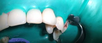
Moisture insulation of the area of the diseased tooth
To prevent pathogenic microorganisms that may be contained in saliva from entering the internal space of the tooth, the area of the diseased tooth is first protected from moisture by covering with a special latex bandage - a rubber dam.
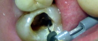
Reaming a tooth
To gain access to the dental canals, the doctor will drill the tooth, removing all tissue affected by caries. The access hole is made either on the chewing surface of the tooth (when treating molars) or on the inside (when treating incisors and canines). After drilling, the doctor will remove either the entire pulp bundle or only part of the tissue affected by the inflammatory process.
Treatment of dental canal cavities
For high-quality treatment of dental canals, a specialized tool is used - files. Before the cleaning process, it is important to obtain detailed information on the structure of the dental canals, as well as their length. To do this, a photograph of the diseased tooth is taken, and a special type of device is used - an apex locator. The dentist will use the files to clean the canal cavities, and then rinse them with an antiseptic solution and treat them with antibacterial agents. During cleaning and antiseptic treatment, the dentist will expand the cavity of the canals, clean and smooth their internal surfaces. Then the cavities of the dental canals are thoroughly dried with paper points.
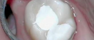
Next, the doctor puts medicine into the canal cavity, places a temporary filling on the tooth and sends the patient home. After a few days, you should come back to the dentist to evaluate the results of the treatment. If the image shows that the inflammation has been stopped, the canals are filled.
Sealing
Gutta-percha pins are used to fill dental canals. It is important that the dental canal is sealed correctly and efficiently, otherwise a recurrence of inflammation is possible and the tooth cannot be saved. After filling the canals, the crown part of the tooth is restored with a photopolymer filling or orthopedic structures. It will be useful to know that mild pain in the tooth within 14 days after the procedure is considered normal by dentists. Taking painkillers prescribed by your doctor will help eliminate discomfort. However, if the pain does not go away, its character is sharp, and does not subside even after taking anesthetics, you should immediately contact the dentist!
Recommendations
Endodontic treatment at the Dentpremium clinic has high success rates. However, to prevent complications arising due to improper dental care after therapy, the following recommendations should be followed:
- exclude spicy, hot and cold foods from the diet for 3–5 days;
- maintain oral hygiene (brush your teeth, use an irrigator and dental floss);
- do not chew gum and hard foods, do not chew candies, nuts, etc.
In order to prevent pathological processes in the roots of the tooth, it is necessary to visit the dentist twice a year for preventive purposes, as well as to promptly treat diseases of the oral cavity.
Note! Increased sensitivity and minor pain in the first days are normal. If the pain is severe, you should contact your dentist.
How long does root canal treatment take?
The duration of tooth canal treatment depends on the complexity and characteristics of the clinical case, as well as the number of canals that the specialist will have to treat. On average, the duration of the procedure can range from 20 minutes to 1.5 hours.
It is also worth considering that for high-quality root canal treatment, you will have to visit the dentist’s office several times.
Filling with hot gutta-percha
Sealing the root passages with heated gutta-percha can be done in several ways. The most common of them are:
- injection method (filling the channel with material heated to 200 degrees);
- method of vertical condensation (laying filling material into the canal, distributing it with instruments along the side canals, installing a softened pin on top of it);
- continuous wave method (introducing a basic central pin into the tooth using a heated instrument, placing side pins around it, completely sealing the canal).
Is it painful to treat root canals?
Thanks to the use of modern anesthetics, root canal treatment has become a painless procedure. The patient may feel mild discomfort only during the injection of the anesthetic.
Therefore, there is no need to be afraid of root canal treatment; moreover, you must remember that you should never postpone the procedure due to fear of the dentist! Inflammation in a tooth that has progressed to a certain stage of development cannot be cured, which means that the diseased tooth will have to be removed and a prosthesis put in its place. In addition, more serious complications may occur.
Obturation using the Thermofil system
“Thermophile” is a titanium, plastic or steel cone-shaped rod coated with gutta-percha, which is in the alpha phase state. This coating is characterized by a lower melting point, increased fluidity and the ability to penetrate into the microtubules of the root passage. When introducing thermophile into the root canal, the base rod fills the main space, and gutta-percha fills only a small part of it. This is what makes it possible to solve the problem of significant shrinkage of the thermoplastic gutta-percha filler after cooling and to prevent the formation of microspaces between the walls of the root passage and the filler.
The procedure for sealing root passages, which involves the use of the Thermofil system, consists of the following stages:
- selection of suitable thermophile diameter;
- antiseptic treatment of the rod and its heating;
- introduction of sealant into the root canal;
- inserting the rod into the root canal until it stops;
- removal of excess gutta-percha from the dental cavity;
- restoration of a damaged tooth crown.
The use of the described technique allows for effective obturation of the main canal and its branches.
Modern method of treating tooth canals: treatment of the canal cavity under a microscope
The quality of dental canal treatment depends on the correctness and accuracy of cleaning and processing of their internal space. This is a very painstaking work, since the channels themselves are quite narrow (no more than one millimeter), and in addition, they can have bends and turns. If a specialist makes a mistake when treating the canals and does not remove particles of the affected dentin, a relapse of the inflammatory process is guaranteed. The use of a special tool in treatment - a dental microscope - will help eliminate possible errors and inaccuracies in the treatment of dental canals.
Using a microscope, the dentist will perform a high-quality removal of all infected tissues, while preventing injury to healthy tooth tissues. The use of the device simplifies the process of cleaning and processing channels with significant lengths, anomalous structure,
with curvatures and branches. In addition, the use of a dental microscope during the treatment of tooth canals improves the quality of tooth restoration as a whole, and also contributes to the timely detection of hidden caries.
At our dental clinic “Uni Dent” in St. Petersburg, you can receive services for dental canal treatment under a microscope. The procedure is carried out by highly qualified doctors using modern instruments and materials, and therefore when you contact us, you can rest assured that the treatment result will be of high quality! Dentistry "Uni Dent" - come to us for a beautiful smile and painless dental treatment!
Possible complications after canal filling
In the absence of complications, the process can take about one to one and a half hours. But in severe and advanced cases, it may require several visits to the dentist.
The most common type of complications after filling a tooth canal is the neglect of the inflammatory process, accompanied by focal tissue destruction. In this situation, a temporary filling is installed from a non-hardening paste containing medications. The time of wearing it and the composition of medications is determined by the doctor during an individual consultation. This treatment shows high effectiveness for diagnoses of cystogranuloma and periodontitis.
Materials for canal filling
Unfortunately, situations are common in which pain continues to torment the patient even after all medical procedures have been completed, and after the period adequate for rehabilitation has expired. The reason for this reaction of the body may be an error in choosing the filling material. There is still no universal substance that would suit everyone. Consequently, dentists, based on their knowledge and the results of examining patients, each time make a choice in favor of one material or another.
Today the following substances are used for canal filling:
- fillers. They are represented by gutta-percha, silver and titanium pins;
- sealers. These are different types of cements, including polymer, natural, glass ionomer or containing calcium hydroxide, as well as polydimethylsiloxanes.
Gutta-percha can be called perhaps the most popular material due to the complex of its properties:
- leaves the tooth color unchanged;
- copes with absolute sealing;
- not subject to dissolution;
- not subject to deformation.
What a miracle is BeeFill Pack technology?
To completely seal the root canals of teeth, we use the BeeFill Pack system from the German company VDW GmbH, which completely eliminates the possibility of re-infection.
Indications for root canal filling with BeeFill technology:
- caries, pulpitis, periodontitis;
- depulpation;
- purulent abscess;
- refilling of canals as a result of poor quality treatment;
- severe dental injury;
- preparation for prosthetics.
Not all dental clinics use the technology of three-dimensional filling of the root canal system with thermoplastic gutta-percha, which meets established European standards.
BeeFill Pack system capabilities
Filling root canals with gutta-percha pins is considered a successful and safe method of strengthening and preserving a tooth. With the usual filling method, it is not possible to achieve complete adherence of the material to the walls of the crown. And only the use of the unique BeeFill Pack technology ensures:
- Constant and uniform heating of gutta-percha.
- Precise delivery with optimal pressure, which allows the material to penetrate into all branches of the root canal.
- Possibility of filling difficult root canals.
- Accuracy of channel length determination.
Comprehensive control of the filling procedure eliminates the penetration of gutta-percha into the periodontal tissues. The use of the BeeFill Pack device not only makes the dentist’s work easier, but also allows the doctors of our Research Center clinics to carry out effective treatment and high-quality root canal filling.