Patients with chronic periodontitis often encounter a situation where a cyst forms at the apex of the root - a cavity in the bone tissue filled with pus. Most often, a cyst develops due to infection in the root canals; sometimes the cause of a cyst is trauma, inflammatory processes in the periodontium (gums) and adjacent areas. It can also occur due to undetected caries, pulpitis or periodontitis, as well as due to poorly filled root canals.
Such a neoplasm may not bother a person for a long time. As a rule, a cyst makes itself felt during periods of weakened immunity, against the background of a cold. In such cases, the infectious process in the cyst cavity worsens, which leads to abundant pus formation. The patient feels acute pain, swelling and swelling of the gums appears, and a fever may appear. The main thing is to seek dental care on time. In these cases, resection of the apex of the tooth root and dental surgery to remove the cyst are used. However, you can often get by with therapeutic treatment.
Why does a dental cyst form?
The cause of cyst formation is an untreated root canal infection.
The reasons for infection in the root of a tooth can be different:
- As a complication of insufficiently treated or completely neglected
- caries/pulpitis,
- periodontitis,
- periodontal diseases,
- inflammation under the dental crown,
- Error and infection during endodontic treatment (root canal treatment).
- Tooth injury.
- Immune disorders in the body.
- Chronic diseases of the ENT organs: runny nose, sinusitis, tonsillitis.
Children in primary and mixed dentition can also suffer from dental cysts. Even newborns may experience teething with a purulent cyst - the so-called. Bona knots.
What are dental cysts?
Types of cysts
Root cyst
Develops from granuloma as a continuation of pulpal necrosis (death of pulp tissue) and as a result of periapical inflammation. It is located in the apical (apical) third of the tooth root and sometimes has a transverse orientation due to additional canals, the initial paths for the spread of pulpous necrosis.
Root cysts are radiolucent capsules with well-defined boundaries. They can vary greatly in size from 2-3 mm to 2-3 cm. In the absence of secondary inflammation, the roots develop asymptomatically. Even the largest neoplasms do not displace teeth or expand bone tissue.
Once diagnosed, treatment requires eliminating the underlying cause. The most common treatment for pulp necrosis is endodontic treatment. If it is unsuccessful, a decision is made on surgical curettage or enucleation.
Residual
Remains after removal of the pathology. It can form in the granuloma left after tooth extraction or around the left apex of the root.
The residual cyst is clearly visible on x-ray. Its dimensions range from a few millimeters to several centimeters.
Follicular cyst on the gum
A cyst in the gum develops from a follicular sac that encloses each unerupted tooth. This is a capsule clearly visible on an x-ray, most often surrounding the crown of an unerupted tooth. May cause extensive tooth damage. It has a rapid growth rate, can move to adjacent teeth and lead to expansion of the cortical plate.
Clinically, a follicular cyst always occurs in the area where the enamel meets the cementum of the tooth (the place of attachment of the follicle). Due to its growth, adjacent teeth may tilt, and in rare cases, resorption (destruction) of tooth roots occurs. Unerupted teeth can also become displaced due to aggressive growth. If a follicular neoplasm has been identified, its treatment may include surgical curettage (curettage).
Eruption cyst
This is a fluid-filled follicular sac that usually occurs during teething. It covers the erupting tooth and often contains blood. Outwardly it looks like pronounced swelling of soft tissues of a bluish color (due to the presence of blood). Most often it occurs due to slow tooth eruption and can rupture spontaneously. However, in some cases where it is found, treatment may include surgical resection to help the tooth erupt faster.
Primordial (keratocyst)
Develops from the tooth germ as a result of degeneration of the stellate reticulum during tooth development before the onset of calcification of its tissues. Keratocysts can arise from any developing tooth germ, but most often they are localized in the area of premolars and the third molar of the mandible.
Clinically very prone to relapse. If the patient is diagnosed, treatment consists of careful curettage and close follow-up to ensure early detection of recurrence of pathology.
Lateral periodontal
The exact reasons for their occurrence are not fully known. They are rarely large. They are clearly visible on an x-ray and are most often closely associated with the lateral surface of the tooth root. Typically, teeth associated with these cysts have normal pulp. When a lateral periodontal neoplasm (cyst) is detected on the root of a tooth, treatment consists of surgical curettage without compromising the integrity of the tooth. It is important that the removed tissue is sent for histological analysis to rule out more serious pathologies such as early adamantinoma.
Calcifying odontogenic
Their etiology is also completely unknown. They can be found in any area of the supporting surface of the jaws, most often in the lower part. Morphologically they are quite diverse. Clearly visible on x-ray and may contain various opacities depending on the degree of calcification of the tumor. Once diagnosed, treatment consists of surgical curettage or resection.
Why do you need to remove the cyst?
If a dental cyst is not removed in time, even with an asymptomatic course of the disease, the consequences can be very serious - this is a real “time bomb”.
- The main danger of a cyst is that sooner or later it will destroy the affected tooth, and then “get” to the neighboring teeth.
- Cysts are reborn. Slowly but surely, so in 15-20 years it may already be a malignant formation.
- An infection is raging in the cavity of the cyst, so with a general illness, hypothermia, or a decrease in immunity as a result of simple stress, acute purulent inflammation is possible - flux, abscess, phlegmon.
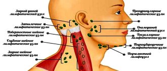
- The infection can spread to adjacent lymph nodes and cause inflammation - lymphadenitis.
- Gradual thinning of the jaw bone in the affected area, due to purulent melting, turns into osteomyelitis. Even a spontaneous fracture of the jaw is possible.
- The cyst enlarges and can grow into the nasal cavity or maxillary sinus.
- Infection in the cyst can cause sepsis, a blood poisoning.
If treatment is not started on time, the tooth will have to be removed. And this is the minimum “evil”.
What are the symptoms of a dental cyst?
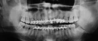
For a long time, the cyst may not manifest itself in any way until it becomes large enough in size, so it is very rare that a cyst is diagnosed at an early stage of the disease. This mostly occurs accidentally when the patient is having an x-ray for another reason.
At a later stage, the patient begins to worry about:
- Aching pain that gets worse over time. Such pain is difficult to relieve with regular painkillers.
- Pain when biting.
- Swelling of the gums and/or cheeks.
- An abscess, gumboil, or fistula may form on the gum.
- General malaise, fever.
Symptoms of anterior tooth cyst
This disease is especially dangerous due to its practically asymptomatic course at the initial stage - few people will worry if they notice a slight change in the color of the enamel or a slight displacement of the tooth. But a front tooth cyst is much more likely to be noticed than other types, since the frontal units have to “work” more often, because they are located only on one side of the jaw. However, other types of formations can go unnoticed for years, for example, if the patient is used to chewing on only one side. A cyst on the front upper or lower tooth is noticed much earlier, because pain is immediately felt.
Dangerous symptoms!
When the size of the granule with pus begins to exceed 10 mm, pronounced symptoms of a front tooth cyst arise:
- constant nagging pain with a feeling of fullness
- high fever and painful condition, which sometimes continue after the procedure for removing the cyst of the front tooth has already been completed
- swelling of the gingival tissue at the location of the granule, in severe pathologies it spreads to the face
- protrusion of a tubercle in the affected area
- enlarged lymph nodes - occurs if bacteria from the granule with pus begin to spread through the blood
How is a dental cyst treated?
The first thing that should be said is that today a dental cyst is successfully treated with a high percentage of preservation of the affected tooth, although in the past the diagnosis of a “cyst” meant 100% removal.
There are 2 main approaches to treatment:
- Therapeutic. Therapeutic treatment of dental cysts is used when
- There is access to the dental canal through the dental crown
- The canals are not sealed or can be opened
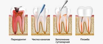
This method of treatment is very similar to the treatment of periodontitis, with the only difference being that the doctor additionally removes pus from the cyst cavity and injects special medicinal preparations into the root canal.
This treatment takes at least 1-2 visits, is carried out under sterile conditions, using a rubber dam and a dental microscope, because requires special care from the doctor to prevent relapses.
Treatment of cysts with laser with access through the coronal part of the tooth is another type of therapeutic treatment. Its main difference is that the laser immediately completely disinfects the area of the removed cyst, the treatment is quick and bloodless, the disadvantages include the risk of burns.
There are several methods for surgical removal of cysts, but cystectomy - complete removal of the cyst with all its contents - is the most modern and most effective of them.
Cystectomy is indicated for:
- A sealed root canal that is difficult to open.
- The affected tooth is replaced with a crown
- Pain, swelling of the cheeks/gums.
What do patients say about apex resection? Reviews are usually only positive!
“Resection of the top of the tooth took about an hour, the operation was performed under pain relief. It didn’t hurt very much near the tooth for about a month. A few months later I had to take an x-ray, the doctor showed that all the tissues had been restored."
“The cyst grew, they told me to remove it, it was a little unpleasant, but quite tolerable - the doctor quickly and accurately carried out the procedure. Everything was restored soon, now it’s completely unnoticeable that there was anything there.”
At Ritsa Dentistry, cyst removal operations are performed by experienced surgeons with extensive experience in performing such procedures. Therefore, you can rely on our professionalism and entrust the treatment of the cyst to our doctors.
How is surgery to remove a cyst (cystectomy) performed?
Removal of the cyst is carried out by a dental surgeon in a surgical office, on an outpatient basis, the operation usually lasts about half an hour, an hour after the manipulation the patient can go home. Improvement in the patient's condition should occur 6-8 hours after the operation. A sick leave certificate is issued for the recovery period - until the stitches are removed.
To relieve all pain, the patient is given local conduction or infiltration anesthesia; we also numb the injection site with an application gel. After surgery, painkillers in tablets are prescribed for the first time.
I Preparatory stage
- The patient is diagnosed by a surgeon, and a consultation with the attending dentist-therapist is required.
- It is advisable, if the operation is not urgent, to provide the patient with professional hygiene and sanitation of the oral cavity before surgery - this reduces the presence of infection in the oral cavity and healing proceeds faster.
II Carrying out the operation
- The patient is given anesthesia.
- The surgical field is treated with an antiseptic.
- The dentist-surgeon separates the mucoperiosteal flap from the bone.
- Access to the affected area is provided, the cyst along with the affected part of the apex of the tooth is removed.
- The cavity is thoroughly cleaned and washed with an antiseptic.
- An antiseptic is injected into the cavity and the wound is sutured.
- During the recovery process, the cavity heals on its own and the bone tissue is restored.
III Postoperative period
- After the operation, you should not eat for 4 hours, then the food should not be hot, liquid and gentle.
- The patient is prescribed antibiotics to fight the infection and painkillers.
- Hygiene can be carried out according to the standard scheme one day after surgery, according to the recommendations of your doctor.
- Antiseptic rinses and healing gels are prescribed.
- Swelling of the gums and face may occur for several days.
- NOT RECOMMENDED:
- Warm the surgical site, because re-development of the infection is possible.
- Take medications without a doctor's prescription.
How is tooth root apex resection and cyst removal performed?
Typically, the entire procedure takes about an hour. In this case, the position of the tooth is important: surgery on distant teeth requires more skill and time.
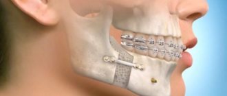
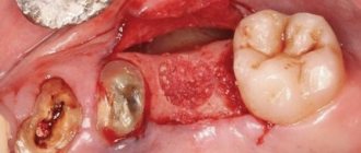
During the operation, which is performed under local anesthesia, an incision is made in the gum near the diseased tooth to peel it away to expose the bone tissue. A small hole is then made in the bone using a special bur. Through it, the doctor should see the apex of the tooth root and the cyst attached to it.
Using a drill, the tip of the root is cut off and then removed from the wound along with the cyst attached to it. After the cyst is removed, synthetic bone is placed into the empty space to restore jaw bone tissue. At the end of the procedure, the mucous membrane of the gums at the incision site is sutured, sometimes a drainage is inserted between the sutures to drain the ichor from the operation site for several days.
When performing a tooth resection, only the inflamed and infected part of the root is removed, healthy tissue is not affected.
Prevention of dental cysts
Taking standard care of your dental and general health will help prevent the development of cysts.
To prevent dental cysts from bothering you, you must:
- Undergo regular preventive examinations with your dentist every 6 months and a professional hygiene procedure. Do not refuse if the doctor offers you an X-ray examination - he has reasons for this.
- Treat caries and pulpitis in a timely manner.
- To identify hidden caries at an early stage of the disease, for this in our clinic we use digital (not x-ray) diagnostics using the Diagnocam apparatus. We recommend undergoing such diagnostics at least once every six months.
- Brush your teeth well yourself using dental floss, irrigator, and mouthwash. Pay attention to cleaning your tongue.
- Strengthen immunity.
Treatment of a cyst without tooth extraction
The success of treatment directly depends on how early it was detected. Therefore, dentists recommend that patients undergo regular preventive examinations and seek help at the first signs of illness. More recently, therapy necessarily involved tooth extraction. Of course, this approach did not suit either the doctors or especially their patients. Today, dentists' methods have changed significantly, and it is possible to get rid of a cyst without losing the beauty of your smile.
Treatment of the cyst can be surgical or conservative. The choice of therapy method depends on: the type, age of the patient, personal wishes and is determined for each patient.