Quick navigation
- Causes of pathologies
- Development of cheilitis
- The course of Fordyce's disease
- Lip cancer
- Herpes
- Development jammed
In the modern world, diseases on the lips are quite common. They can occur against the background of external and internal provoking factors.
When the first symptoms of a disease appear, the patient should seek help from a doctor who can correctly diagnose and prescribe rational treatment.
Causes of pathologies
- Diseases of the lips in humans in most cases are observed with a weakened human immune system, which cannot cope with viruses and bacteria that attack the body.
- If the diet lacks vitamins, this can lead to a pathological process.
- Human lips are full of nerve endings. With nervous overstrain, they become overfilled with blood, which creates an unaesthetic appearance.
- Lip diseases are observed with frequent hypothermia or overheating.
- If the patient has allergic reactions to cosmetics or food, then damage may occur against this background.
- Improper installation of braces or veneers creates skin irritation, which leads to a pathological condition.
- Lip diseases can also be caused by bacteria and fungi.
To ensure adequate treatment of diseases on the lips, it is necessary to establish their cause.
Development of cheilitis
Cheilitis is a benign inflammatory disease that develops on the lips. The pathological condition has pronounced signs, which makes it possible to detect it in a timely manner.
Features of exfoliative cheilitis
A decrease in the defenses of the immune system, a stressful situation, as well as the presence of a hereditary predisposition and mental disorders can cause lip disease.
If the form of the disease is dry, then white crusts appear on the edges of the lips, which are easily removed by hand. Lips often become red and flaky.
The exudative form of the disease is accompanied by redness, burning and swelling of the lips. Most patients complain of pain. The crusts in this form of the disease are gray-yellow in color.
Treatment of the disease should be carried out with the use of sedatives. It is recommended to use interference agents to lubricate the lips. To remove crusts - boric acid. Most patients are prescribed drugs to increase the body's reactivity, for example, Pyrogenal.
Features of actinic cheilitis
This form of the disease is observed with excessive sensitivity to ultraviolet radiation. If a person spends a long time in the sun, this leads to an exacerbation of the disease. Symptoms are pronounced:
- When this pathological condition occurs, the patient feels dry lips. Their surface turns red.
- Scales and cracks appear on the lips.
- If treatment of the disease is not carried out in a timely manner, this leads to the appearance of erosions that do not heal for a long time.
- Some patients were diagnosed with the appearance of ulcers and hardened areas.
- If the disease is chronic, it can lead to a precancerous condition.
To eliminate cheilitis, it is imperative to use photoprotective creams when going outside. Patients are also prescribed corticosteroid ointments. Prednisolone ointment and Flucinar are quite effective in this case.
To slow down the pathological process, it is necessary to take vitamins and nicotinic acid. If necessary, it is recommended to use antimalarial drugs, for example, Delagil.
The course of atopic cheilitis
Atopic cheilitis is a symptom of diseases such as neurodermatitis and atopic dermatitis. It occurs when the human body is exposed to a variety of allergens - plant pollen, household dust, medications, food products, bacteria and microorganisms.
With atopic cheilitis, the border of the lips acquires a bright red tint, dryness and flaking of the skin is observed. Most patients are diagnosed with cracks. Some patients complain of a burning and itching sensation, which may be accompanied by pain.
To eliminate the disease, it is mandatory to prescribe anti-allergy medications. Usually:
- Claritina
- Suprastina
- Fencalora.
To strengthen the immune system, it is recommended to take vitamins that belong to group B. If the disease lasts for a long period, then it is necessary to take corticosteroid ointments. It is recommended to apply them to rashes 4 to 6 times a day.
In some cases, patients are prescribed borderline Bucca rays, which are characterized by a high level of effectiveness.
During the period of therapy, all possible allergens must be removed from food. Carbohydrates should be taken by patients in limited quantities.
Why do cold sores appear on the lips?
Those who suffer from herpes have noticed that exacerbations begin along with ARVI. Herpes occurs when immunity decreases, the protective forces of which are used to suppress ARVI. Colds on the lips also appear when the body’s defenses are weakened: with chronic fatigue, lack of sleep, frequent stress, poor diet, alcohol abuse and active smoking, in women at the beginning of pregnancy and in the first 5-7 days after childbirth.
Photo: Colds on the lips can only appear if the immune system is weakened
The course of Fordyce's disease
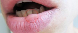
When Fordyce's disease appears, the sebaceous glands on the lips become enlarged. In appearance they look like pustules. The occurrence of a pathological process is quite often observed against the background of hyperplasia of the sebaceous glands.
To treat cysts, it is recommended to use retinol-containing ointments. To hide cosmetic defects, permanent makeup is used.
To remove old rashes, a laser should be used, sometimes cryodestruction or electrocoagulation is used. Unfortunately, not all treatments are highly effective, so relapses occur in 80% of the population.
Lip cancer
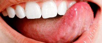
Cancer on the lips are neoplasms that are malignant in nature. In the early stages, the disease manifests itself in the form of ulcers that constantly bleed. In appearance they are similar to herpes. Lumps and increased salivation may also occur.
Attention ! If the ulcers do not go away when using medications, the patient should seek help from a doctor.
The treatment regimen directly depends on the degree of its development, as well as on the clinical picture. In most cases, treatment is carried out using cryotherapy, radiation or surgery. Patients are also recommended to undergo chemotherapy using special drugs.
Lip tumors
Of all the anatomical parts of the oral cavity, the red border of the lower lip is most often affected. Lower lip cancer accounts for approximately 12 - 25% of the total number of tumors in this location. For reasons that are not entirely clear, tumor lesions of the red border of the upper lip are an extremely rare occurrence. The disease most often occurs in people over 60 years of age. Men are more often affected, the male/female ratio approaches 13/1. Predisposing factors are solar radiation, smoking, and alcohol abuse.
Clinically, the disease manifests itself as an exophytic (i.e., protruding) tumor or, much more often, an ulcerative bleeding surface. Since it is very difficult not to notice this lesion, the vast majority of patients seek medical help at an early stage of the disease. This circumstance (treatment of the early stages of any malignant tumors always brings better results) allows us to count on high effectiveness of treatment. In addition, lower lip cancer rarely metastasizes to regional (cervical) lymph nodes. At the time of initial treatment, involvement of the cervical lymph nodes is diagnosed in only 2–12% of patients. Damage to regional lymph nodes significantly worsens the prognosis of the disease. Thus, the 5-year survival rate in the presence of this lesion drops from 90 to 50% and forces one to resort to more aggressive methods of treatment.
Treatment for lower lip cancer is determined by the stage of the disease. Early stages can be cured with surgery or radiation treatment. There are 2 types of radiation treatment: remote and contact therapy (so-called brachytherapy).
The lip has a rather complex anatomical structure, which includes muscles with different directions of muscle fibers, skin, salivary glands, and subcutaneous tissue. Naturally, the goal of surgical treatment is not only radical removal of the tumor, but also restoration of the structure and function of the lower lip. Modern methods and techniques of plastic surgery can achieve good cosmetic and functional results. This is illustrated by the photographs below of patients who underwent surgical treatment for cancer of the lower lip.
Develops against the background of obligate (warty precancer, limited dyskeratosis, Manganotti cheilitis, Bowen's disease) and facultative (leukoplakia verrucous, keratoacanthoma, cutaneous horn, papilloma with malignancy, erosive-ulcerative and hyperkeratotic form of lupus erythematosus and lichen planus, post-radiation cheilitis) precancerous processes .
Warty precancer is characterized by the appearance on the red border of the lip of a sharply demarcated hemispherical lesion with a diameter of 4 mm to 1 cm, which protrudes above the surrounding tissues and has a dense consistency. Its color varies from an almost normal red border to a stagnant red. The nodule may be covered with thin scales and resemble a wart or keratinizing papilloma. Malignancy can occur 1-2 months after the onset of the disease.
The productive form of focal dyskeratosis is characterized by excessive keratinization of the epithelium and looks like a white spot with flat protrusions on a red border or an area of hyperkeratosis with stylized horny protrusions. The destructive form (erythroplakia) is distinguished by the fact that limited defects of the epithelial cover appear on the red border of the lip - erosions, ulcers, cracks. Malignancy can occur 6 months after the onset of the disease.
Abrasive pre-cancer cheilitis Manga-Notty is characterized by the formation of an oval or irregularly shaped erosion on the lip, often with a smooth, polished surface of a deep red color. Crusts may form on the surface of the erosion, which may cause slight bleeding when removed. There is no tissue compaction at the base and around the erosion. Spontaneous epithelization with subsequent recurrence is possible. Malignancy occurs 3 months to 30 years after the onset of the disease.
Lip papilloma is a clearly circumscribed tumor from 1-2 mm to 2 cm in diameter on a wide base with a villous or rough surface. As keratinization increases, the tumor acquires a whitish tint. Lip papilloma is considered a progressive precancerous disease. Compaction of the base of the papilloma and the appearance of pain are signs of its degeneration.
Treatment of diffuse lesions of the lower lip (cheilitis, leukoplakia, etc.) is predominantly conservative. Focal lesions are treated surgically, laser and cryodestruction.
Lip cancer
The term “lip cancer” refers to malignant tumors that arise in the mucous membrane of the red border of the lip. Neoplasms that have developed on the skin near the lip or mucous membrane of the vestibule of the mouth are not included in this group of tumors.
In the structure of cancer diseases in Russia (1997), lower lip cancer accounted for 1.4% of the total number of malignant neoplasms with an incidence of 2.8 per 100,000 population. The disease is most often observed in men over 70 years of age. In rural areas, lip cancer occurs 1.5-2 times more often than in urban populations. In recent decades, there has been a gradual decline in incidence. Approximately 90% of tumors of the lower lip develop due to exposure to known carcinogens, the main of which are tobacco and tobacco-containing substances. Alcohol is comparable in action to tobacco and is part of a group of agents that can potentiate tobacco-induced carcinogenesis. Other important etiological factors are chronic injuries and inflammation of the lip, ultraviolet radiation, changes in weather conditions, and occupation (work in the oil refining, coal, textile industries).
International classification according to the TNM system
Anatomical areas and parts:
1.Upper lip, red border. 2.Lower lip, red border. 3. Angles of the lips (commissures). T - primary tumor: Tx - insufficient data to evaluate the primary tumor, T0 - primary tumor not determined, Tis - preinvasive carcinoma (carcinoma in situ), T1 - tumor up to 2 cm in greatest dimension, T2 - tumor up to 4 cm in greatest dimension , T3 - tumor more than 4 cm in greatest dimension, T4 - tumor spreads to adjacent structures - bone, tongue, facial skin.
N/pN - regional lymph nodes. Regional lymph nodes for all parts of the head and neck, with the exception of the nasopharynx and thyroid gland, are the lymph nodes of the neck, including: submental, submandibular, nodes at the base of the skull near the great vessels (deep cervical), in the bifurcation zone of the common carotid artery (deep cervical) , posterior cervical (superficial cervical), located along the accessory nerve, supraclavicular, preglottic and paratracheal, retropharyngeal, parotid, buccal, postauricular and occipital.
N/pNx - there is insufficient data to assess the condition of the regional lymph nodes, N/pN0 - there are no signs of metastatic lesions of the regional lymph nodes. pN0 - histological examination of material from a selected area of neck tissue includes 6 or more lymph nodes; histological examination of the material obtained using cervical lymphadenectomy includes 10 or more lymph nodes, N/pN1 - metastases in one lymph node on the affected side, up to 3 cm or less in the greatest dimension, N/pN2 - metastases in one or more lymph nodes on the affected side, up to 6 cm in greatest dimension; or metastases in the lymph nodes of the neck on both sides or the opposite side, up to 6 cm in greatest dimension, N/pN2a - metastases in one lymph node on the affected side, up to 6 cm in greatest dimension, N/pN2b - metastases in several lymph nodes on the affected side, up to 6 cm in the greatest dimension, N/pN2c - metastases in the lymph nodes on both sides or on the opposite side, up to 6 cm in the greatest dimension, N/pN3 - metastasis in the lymph node more than 6 cm in the greatest dimension.
Note. The lymph nodes on the affected side include the lymph nodes located in the midline.
M - distant metastases: Mx - the presence of distant metastases cannot be assessed, M0 - no distant metastases, M1 - distant metastases.
The requirements for determining the рТ category correspond to those for determining the T category.
Grouping by stages
Stage 0 TisN0М0 Stage I Т1N0М0 Stage II Т2 N0М0 Stage III Т3N0М0 Т1-3N1М0 Stage IVA Т4N0-1М0 Any ТN2М0 Stage IVB Any ТN3М0 Stage IVC Any Т Any NM1
Clinic . Cancer most often affects the lower lip midway between the midline and the commissure. The main morphological form is squamous cell carcinoma (80-95%). Metastasis later, mainly to regional lymph nodes (5-9.2%). In the clinical course of cancer of the lower lip, two groups are distinguished - exophytic and endophytic. There are transitional forms between them. Exophytic cancers of the lower lip include papillary and warty forms. The papillary form develops from papilloma or against the background of focal productive dyskeratosis. Exophytic cancer visually represents a tumor of irregular shape (round, lumpy, flat), rising above the surface of the red border. There is infiltration of the underlying tissues without clear boundaries. The warty (fungous) form develops against the background of productive dyskeratosis and is characterized by the fusion of multiple small growths on the lip (“cauliflower appearance”), a gradual increase in infiltration of the underlying tissues and tumor disintegration. Endophytic cancer of the lower lip is represented by ulcerative and ulcerative-infiltrative forms. The ulcerative form is characterized by the formation of an ulcerative defect on the lip with a gradual deepening of the ulcerative surface, its bottom becomes uneven, its shape is irregular, the edges rise above the level of the red border, everted. The ulcerative-infiltrative form is characterized by infiltrative growth, which makes it difficult to determine the boundaries of the tumor. Endophytic forms of cancer are more malignant.
Diagnosis is based on anamnesis, examination, palpation of regional lymph nodes (submental and submandibular lymph nodes are examined bimanually), cytological and/or histological examination data. When making a differential diagnosis, it is necessary to keep in mind tuberculosis and syphilitic lesions.
Treatment . In stages I and II, the standard treatment method is radiation therapy (short-focus radiotherapy, electron therapy, contact radiation, interstitial or combined radiation therapy) with a total focal dose of 60 Gy.
The treatment of choice for stage I lip cancer is cryodestruction. Block resection of the lip with simultaneous plastic surgery according to Bruns is rarely used due to the poor cosmetic effect. Surgery is more indicated in the presence of residual tumor after completion of a radical course of radiation therapy; it is performed within 3-6 weeks after the end of irradiation.
For stage II tumors, 2-3 weeks after completion of treatment of the primary lesion, prophylactic upper fascial-sheath excision of the neck tissue on both sides is performed. The block of tissue removed includes the tissue of the submental and submandibular region and the lymph nodes of the fork of the common carotid artery, the lower pole of the parotid salivary gland.
For stage III lower lip cancer, combined radiation therapy is used (external gamma or electron therapy + interstitial or short-focus radiotherapy). The presence of clinically detectable metastases in the lymph nodes is an indication for preoperative irradiation of the cervical lymphatic collector on the affected side. Preventive or therapeutic fascial-sheath removal of cervical tissue (on both sides) is carried out after complete subsidence of radiation reactions and completion of regression of the primary tumor focus. The method of choice for treatment of stage III lower lip cancer, especially when the tumor has spread to the corner of the mouth, is a combined one. In this case, lymphadenectomy is performed simultaneously with removal of the primary tumor. To eliminate lip tissue defects formed after treatment, primary reconstruction is performed using local or regional flaps.
Treatment for stage IV lower lip cancer is combined. For multiple metastases in the lymph nodes of the neck, treatment begins with preoperative external beam radiation therapy. 2-3 weeks after completion of the first stage of treatment, surgery is performed on the primary lesion with simultaneous cervical lymphadenectomy. Fascial-sheath excision of the cervical tissue is performed for single, dislocated metastatic lymph nodes that are not fused with adjacent anatomical structures of the neck. The presence of multiple displaced metastases in regional lymph nodes or single, but limitedly displaced, fused with the internal jugular vein and the sternocleidomastoid muscle is an indication for performing the Crail operation. In case of bilateral metastatic lesions of the lymph nodes, cervical lymphadenectomy is performed simultaneously on both sides. In elderly and weakened patients, this operation can be done first on one side, and after 2-3 weeks on the other.
In case of unresectable tumors and the presence of contraindications to combined treatment, a course of palliative remote gamma therapy is carried out according to a radical program (60-70 Gy). Chemotherapy is used as part of palliative chemoradiation treatment (see Cancer of the mucous membrane of the floor of the mouth).
Recurrent lower lip cancer is treated surgically and in combination. For late limited relapses (developing 1 year or more after completion of short-focus radiotherapy), a course of radiation therapy (60-70 Gy) can be repeated.
Forecast . According to the summary data of various authors, the five-year survival rate for stage I is 91.2-96.9%, for stage II - 82.8-83.6%, for stage III - 63.5-75.4%, for stage IV - no more thirty%.
Prevention of lip cancer consists of timely treatment of precancerous diseases, quitting smoking, eliminating unfavorable external factors (increased insolation, etc.), and using protective lip creams.
SIGN UP FOR A CONSULTATION by phone +7 (921) 951 - 7 - 951
Share link:
Herpes
One of the most common diseases on the lips. There is a constant presence of the herpes virus in the human body. If the immune system's defenses are sharply reduced, the disease manifests itself as blisters on the lips.
If the pathology is not treated in a timely manner, there is a regular increase in their number, then the blisters burst and ulcers appear in their place. In some cases, patients experience increased body temperature and chills.
Treatment of the disease is carried out using antiviral therapy. For this purpose, patients are recommended to use a special ointment, for example, Acyclovir. Traditional medicine also helps - essential oils, chamomile infusion, sea buckthorn oil.
How can herpes be dangerous?
Herpes can cause many complications. Some of them are life-threatening.
- Bacterial complications. Most often, bacteria simply attach to the virus and the blisters fill with pus. If the blisters have already burst, then the ulcers become covered with pus, the pain in the lip becomes unbearable and a crust does not form.
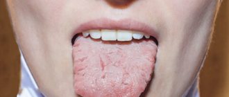
- Herpetic stomatitis is a lesion of the oral mucosa. The disease begins with a sharp increase in temperature, salivation and sharp pain when chewing food. There are many blisters in the mouth, similar to herpes on the lips.
- Herpetic eye damage begins with pain in the eyes and lacrimation. A person cannot open his eyes due to severe pain. A dangerous condition that can result in blindness.
- Herpes of the esophagus. Occurs when herpes spreads from the lips. There are difficulties when swallowing, severe pain, refusal to eat and weight loss.
- Herpetic pneumonia and herpetic hepatitis occur in people with immunodeficiency and occur together with bacterial and fungal processes.
- Herpetic damage to the nervous system occurs in the form of meningoencephalitis and is the most severe form of herpes. It begins with a sharp headache, nausea and vomiting, high fever, movement disorders and convulsions. The disease is unfavorable.
Photo: To avoid complications, you need to start treating herpes in a timely manner.