Safety of X-ray diagnostics
The standard of safe radiation exposure is regulated by the federal law “On Radiation Safety of the Population”. Article 9 of this law “State regulation in the field of radiation safety” states that the average effective dose for the population per year is 0.001 sievert (1 millisievert). In this case, the dose received during one cone-beam computed tomography is 0.09 millisieverts. Accordingly, at least 10 CT scans of teeth can be safely performed per year.
For comparison, during air travel the radiation dose is on average 0.08 millisieverts. The dose of so-called natural radiation is 2.4 mSv. It consists of the effects of cosmic and solar rays, the influence of atmospheric radioactive atoms (radon), radiation from soil and building materials, and food radionuclides. Thus, performing a dental CT scan is absolutely safe for your health.
Contraindications and prohibitions
When compared with traditional radiography, the quantitative exposure of the body to X-rays during multispiral immersion increases by 20 times. If you exceed the permissible number of studies, there is a risk of developing oncopathological processes. This is especially true for young patients, whose cellular structures are in a stage of constant growth and development.
The use of radiographic diagnostic methods during pregnancy in women is absolutely prohibited. The decision to prescribe this type of screening can only be made in the case of a life-threatening anomaly, when the risk of death is higher than the risk of harm from hardware diagnostics. The conditional threshold that removes this contraindication is the minimum age of the patient, which must exceed 14 years. Regarding the frequency of access to multispiral scanning, medical experts recommend not to exceed the schedule, which includes a one-time examination per year. An additional condition is monitoring of excess of permissible exposure over the past five-year period. During vascular tomography, color enhancement in the form of contrast is often used. The difference between MSCT and CT without contrast, the enhancing effect is achieved by intravenous administration of iodine solution. A contraindication is immune rejection to iodine-containing drugs. A true allergic reaction to the product is extremely rare. Associated unpleasant consequences of intravenous administration of the solution are:
- pressure drop;
- increased heart rate;
- fainting state;
- urge to vomit.
With minor manifestations of the reaction, these symptoms are considered to be the norm. The drug is administered under the careful supervision of a medical specialist. If negative consequences occur, urgent restorative manipulations are carried out.
The procedure for performing CT scans of teeth in Moscow at Dental Guru clinics
Computed tomography of the jaw, Moscow. Any Dental Guru clinic has a full range of X-ray diagnostics, including a computer tomography machine. No special preparation required. Before the examination, all metal jewelry and items of clothing (earrings, chains, piercings in the dentofacial area, hairpins) are removed. A lead vest or apron is worn to protect against X-ray radiation. To fix the head, the chin rests on the stand and the forehead on the bracket. During the examination, the sensor scans the dentofacial area. At this time, you should refrain from any movements, including swallowing. The patient sits or stands and the examination takes 30 seconds. At Dental Guru clinics, cone beam computed tomography is performed using a multifunctional Rayscan device.
Execution steps
- The patient puts on a protective apron and takes a comfortable position - sitting or standing.
- The patient's head is securely fixed in one position using immobilization means. For maximum image clarity, the patient must remain motionless during the entire procedure.
- The specialist launches the program. The cone tomograph makes rotational movements around the axis of the head.
- The information received is sent from the tomograph to a special computer program.
- A three-dimensional 3D image of the jaw is formed.
- The entire examination takes 30-60 seconds
Implant planning using CT
A CBCT scan is necessary before dental implantation. Three-dimensional examination reduces the risk of the procedure, significantly improves the quality of implant installation, and shortens the patient’s rehabilitation period. CT scan shows the condition of the bone tissue, possible inflammatory processes, the location of the maxillary sinuses, nerves and blood vessels, helps to create a model of the implant, calculate the depth of its immersion in the bone and the angle of inclination, and the optimal physiological load on the implant.
Why is 3D examination important before implant placement? An artificial root will stand long and firmly if it is surrounded on all sides by bone tissue. And here an individual approach is needed. The structure of bone tissue varies in shape, width, height, density, and possible defects. For longevity, it is important to choose a site where bone volume and density are sufficient. You need to choose the angle of inclination for acceptable aesthetics. In order to determine the required installation depth, you need to know the location of the nerves and blood vessels. Thus, it is possible to obtain a surgical navigation template for implantation for the most accurate virtual planning.
Performing a CT scan with a radiopaque tray when planning a surgical template
Most patients who come for implants already have fillings, crowns, veneers or other dental restorations. All these materials can produce additional glare during CT scanning, so-called artifacts. To minimize them and ensure the accuracy of the CT examination, this procedure is performed using special radiopaque trays. They can be standard (if there are supporting teeth) or individual (when there are very few or no teeth and guidelines are needed for further work).
In these cases, special radiopaque trays are placed in the oral cavity, a CBCT (cone beam computed tomography of the jaws) is performed, then the trays are removed and sent to a dental laboratory. It is thanks to this type of research that it is possible to achieve accurate results and guarantee that the implant is installed in the area in which its location will be best. If implantation using surgical templates is planned, a CT scan with a radiopaque tray must be done. Before implantation with immediate loading, this study allows the fabrication of a temporary prosthesis to be worn before surgery.
Limitations of the use of CBCT devices
The use of CBCT in dentistry has its limitations. Thus, equipment for cone beam tomography is more expensive than orthopantomographs. In addition, the use of a dental tomograph will require new knowledge and competencies from doctors, since diagnostic information in each case must be correctly interpreted.
Another difficulty: the quality of CBCT images and, as a result, diagnostics can be affected by image artifacts, primarily metals. In modern devices, their negative impact can be reduced by fixing elements for the head and special programs, for example, the SMARF algorithm in Papaya 3D Genoray devices (South Korea).
The main disadvantage of CBCT is the inability to study the state of deep soft tissues in the study area due to limited contrast resolution. To study them, the doctor will have to supplement the diagnostic protocol with an examination on an adapted CT or MRI machine.
If you do not do a CT scan before implantation, there may be:
- Loss of sensitivity and pain in the teeth and jaw.
- Injury to the gums due to incorrect calculation of the permissible load.
- Bite defects.
- Loosening and loss of the implant.
- Increased costs to correct problems, including installing a new structure.
This cannot happen in Dental Guru clinics. We care about the interests of our patients. All clinic specialists have extensive experience and high qualifications, confirmed by diplomas and certificates. CT scans of the upper and lower jaw are equally important.
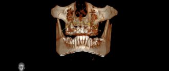
Computed tomography of the upper jaw
To diagnose the condition of the maxillary sinuses before implantation, it is important to do a CT scan of the upper jaw. After tooth extraction, thickening of the mucous membrane in the maxillary sinuses often occurs; there may also be manifestations of sinusitis, a tumor process, the presence of a cyst or polyp. A CT scan of the jaw will show whether there is perforation of the maxillary sinus due to the protrusion of the root or crown of the tooth. The term “odontogenic sinusitis” means that the cause of the disease is in the tooth. The bony septum between the oral cavity and the maxillary sinus is quite thin, so infection from dental tissue can cause sinusitis. Therefore, for implantation to be successful, it is necessary to promptly identify and treat possible diseases and eliminate the inflammatory process.
Computed tomography of the lower jaw
A CT scan of the lower jaw, as well as the upper jaw, shows the condition of the teeth and bone tissue: possible diseases, inflammatory processes, granulomas and cysts. A CT scan of the jaw is important to identify hidden processes and timely initiation of treatment. A CT scan of the upper and lower jaw is usually done at the same time, but some clinics can do a CT scan of the upper and lower jaw teeth separately. The lower jaw bears the main load when chewing and speaking; chronic subluxation of the lower jaw is quite common. CBCT of the temporomandibular joint helps to make a diagnosis and distinguish subluxation from dislocation and fracture.
Benefits of purchasing a dental tomograph
Increased income for the clinic
A CBCT device is a profitable investment for a clinic. The purchase of the device allows the clinic to expand its range of services with more popular and expensive diagnostic procedures. So, if an orthopantomogram in Moscow costs from 1,000 to 3,000 rubles, then 3D tomography costs from 2,000 to 10,000 rubles. At the same time, a CBCT device pays for itself as quickly as an orthopantomograph - due to the high price of the procedures and the large number of indications for the procedure (see above).
If the device performs 5 procedures per day (at an average price of 4,500 rubles for examining two jaws), you will pay for a mid-price dental tomograph costing 6-8 million rubles. in just 1 year. The second year of operation will bring you 8,212,500 rubles. (4500*5*365) – with the same frequency and cost of procedures and excluding expenses.
Own diagnostic center at your disposal
If you purchase a dental tomograph with a cephalostat and an orthopantomography function, then you have a full-fledged diagnostic machine at your disposal. You close the cycle of treating and diagnosing patients within your clinic. Thus, you not only attract new clients, but also eliminate the possibility of losing a patient by sending him to another dental center for diagnostic procedures.
More patient trust
CBCT images display pathological areas and anatomical features of the dental-facial system so clearly that they become understandable even to non-specialists. The patient should no longer take the doctor's word for it. For example, a doctor can clearly demonstrate to a patient the number of canals in a tooth that require filling, and thereby justify the cost of the procedure.
After canal treatment, implant installation or surgical treatment, the doctor can take an image showing the positive results of his work, which increases the patient’s confidence and motivation to continue treatment or rehabilitation procedures in your clinic.
Increasing the prestige of the clinic and increasing the professionalism of specialists
By installing the device, you will dramatically increase the status of the clinic and the level of your staff: when such equipment appears in the clinic, dentists, learning how to use it, improve their professional level.
Protection from lawsuits
During forensic medical proceedings, visual CBCT images can play an important role, namely, confirming the absence of a medical error. Moreover, all information obtained during examinations is stored in the database of the CBCT computer system: the doctor can access it at any time.
Panoramic photo, CT scan of teeth in Moscow, price
For a CT scan of the jaw, the price in Moscow starts from 3,500 rubles. If you search online for “computed tomography of teeth price Moscow” or “computed tomography of jaw price”, then the average price of this examination in the search will be approximately 4,500 rubles. Dental Guru is currently running a unique promotion - a dental CT scan costs only 1,900 rubles. If the question of where to get a CT scan of a tooth is relevant to you, come to Dental Guru. When referred by a doctor, we guarantee non-interference in the patient’s treatment.
The doctor may also order two-dimensional examinations, such as spot and/or panoramic images. The targeted photograph shows the condition of 3-4 adjacent teeth. An OPTG (orthopantomogram) shows the condition of both jaws, including soft and hard tissues, malocclusions, impacted teeth and tumors, but you need to remember that this is a two-dimensional (single-view) image.
Types of modern CBCT machines
Modern dental tomographs differ in scan volume (field of view or FOV), the presence of a cephalostat and patient positioning.
Field of View (FOV)
CBCT machines are classified by field of view or scanning height as follows:
- For a localized area (dentoalveolar complex): about 5 cm
- For one jaw, upper or lower: 5-10 cm
- For both jaws: 7-10 cm
- For the maxillofacial area: 10-15 cm
- For the craniofacial region: more than 15 cm
Field of view (FOV) is one of the most important parameters of a CBCT scan and depends on the size and shape of the detector, as well as the features of the collimator (a device that blocks X-rays that do not pass through the area of interest).
The larger the maximum field of view, the higher the installation cost will be. For this reason, before purchasing a device, you need to decide what shooting modes you will need so as not to overpay for equipment. For example, for implantology, an FOV of 8×8 (in some cases – 10×10) is sufficient; for otolaryngology and maxillofacial surgery – from 10×15. For devices with modular architecture, the choice of FOV can be expanded in the future.
Equipment with cephalostat
The cephalostat is a separate “arm” of the dental tomograph. It is used by orthodontists to create teleroentgenograms—images of frontal and lateral projections of the skull. The photographs allow you to study the position of the jaws relative to the skull and each other, as well as the inclination of the teeth and are mandatory when planning the installation of braces.
There are the following models on the market: with cephalostat in the basic configuration, with the possibility of retrofitting, and without the possibility of retrofitting.
Clinics where an orthodontist works require a device with a cephalostat. If orthodontic services are not provided, but are planned in the future, the medical center requires a dental tomograph with the ability to retrofit with a cephalostat.
Patient position
There are three different types of CBCT platforms, depending on the patient's position:
- Sitting position
- Standing position
- Patient supine position
Devices that scan the patient while standing are more suitable for wheelchairs and take up no more space than orthopantomographs. Sitting units are considered more comfortable, but they take up more space and make the examination of patients with disabilities more difficult. CBCT devices with the patient in the supine position occupy the largest area and volume and are not adapted for examining all patients with physical disabilities.
Commentary by the chief physician of the Dental Guru clinics
To understand the clinical picture, it is necessary to make an accurate diagnosis. Dental Guru dental clinics have all the necessary diagnostic equipment for x-ray diagnostics. These are multifunctional devices with the ability to take pictures of the temporomandibular joint, cephalometry, and a digital radiovisiograph for targeted imaging. We take a panoramic photo of the teeth, CT scan of the upper and lower jaw, the price is now unprecedentedly low. We are always glad to see you in our clinics. Call and make an appointment with specialists!
Recommendations for purchasing CBCT dental tomographs
The choice of a CBCT device is made based on the needs of the clinic, software capabilities and equipment costs.
Thus, before purchasing a device, ask yourself the following questions:
- How much money are you willing to spend on a dental tomograph?
- Which specialists will operate the CBCT machine and what procedures will be performed?
- What are the dimensions of your office and are you limited in space?
- Why do I need this, and what benefits will I get from purchasing a CBCT?