Tooth root caries is a common disease when the neck of the tooth is exposed. It is based on damage to the enamel by microorganisms that feed on food debris and produce acid that can destroy the surface of the teeth. Root caries is often secondary: the infectious process gradually penetrates into deeply located tissues. Inflammation spreads to the nervous tissue of the tooth - the pulp.
With gum recession and other disorders, root caries occurs without visible changes to the enamel. The disease is dangerously asymptomatic for several months or even years. The pathological process occurs inside the tooth and is invisible to the patient.
Make an appointment with a therapist by phone+7(985)532-21-01
Types of caries localizations
Caries (lat. caries rotting) is a slow and hidden pathological process occurring in the hard tissues of the tooth. It is characterized first by focal demineralization of the enamel, then by the destruction of hard tooth tissues with the appearance of a cavity in the dentin. If left untreated, the tooth is lost or complications arise - pulpitis and periodontitis.
Cervical (circular)
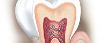
The structure of a tooth is divided into a root, a neck and a crown - the latter is located above the gum. The neck and root are located in the gum and are protected by soft mucosal tissue.
When the carious process begins to destroy the tooth in the area of contact of the crown with the mucous membrane, we can talk about the cervical form of caries.
This category is the most difficult to diagnose and treat. It most often affects the incisors. When biting food with incisors, the food is not only “cut off”, but also gets clogged into the gum pockets, moving towards the gums.
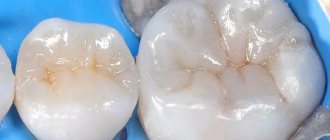
Crown caries
Caries has the ability to affect not only healthy, but also already treated teeth - under a filling or crown. If there is a crown, it is difficult to detect, because the tooth does not have a nerve and pain is not felt. There are no external signs for a long time.
The causes of this type of caries are:
- defect of the dental crown with the appearance of access to the tooth tissue underneath it
- poorly treated tooth before prosthetics;
- gum disease;
- poor oral hygiene.
The problem is that the process spreads into the deep layers and spreads to the bone structures, as a result of which the tooth is often completely removed. That is why preventive examinations and an orthopantomogram - a panoramic image of the entire jaw - are so important.
Radical caries
Basal and cervical caries are similar. Both incisors and chewing teeth are affected. Destruction begins with the enamel, gradually moving to the nerve in the root canal. When the process touches bone tissue, it threatens with osteomyelitis.
The main reason for the occurrence is a cariogenic situation in the oral cavity, when plaque accumulates especially quickly. The enamel in the root part of the tooth is much thinner and more easily destroyed. It is most often diagnosed between the ages of 30 and 60 years.
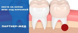
Root caries
Root or subgingival caries occurs hidden. It can be with or without a cavity. Along the flow - active, suspended, secondary. It most often affects molars.
Main types of caries
How caries is treated in dentistry largely depends on which type of spread is typical for a particular person. There are several classification criteria:
- Spread intensity. This category includes single and systemic caries. The difference is that in the first case, the patient is not diagnosed with any additional symptoms. In the second case, the entire row or most of it is also affected. The constant spread of new lesions not only stimulates damage to the enamel and soft tissues, but also impairs the functioning of the human immune system.
- Complexity of development. Uncomplicated caries is the easiest to treat. In this case, there are no additional disorders that lead to the need for additional surgical intervention. Complicated caries is a situation that is accompanied by periodontitis and pulpitis. In this case, treatment must be much longer and more difficult.
In our clinic, doctors treat caries regardless of the stage of the process. We are ready to deal with a problem when there is one of the following four stages:
- Spot. In this case, only the enamel is affected. At the same time, there are spots on the surface with a dark or pale tint. Some people develop stripes instead of spots that are a different color from the rest of the surface.
- Initial surface position. The spot begins to gradually grow, and a hole develops. Sensitivity also increases.
- Middle stage of caries. Dentin begins to deteriorate and pain worsens.
- Deep defeat. Active necrotic damage to hard and soft tissues begins, nerve endings and blood vessels are affected. Treatment in this case will be much more complex and lengthy.
There is also another classification factor - the location and depth of tooth damage. There are five main classes:
- First. It is characterized by damage to the enamel. At the same time, at the first stage it will be very difficult to understand whether there is a lesion, because visually caries shows little of itself.
- Second. Most often it appears on chewing molars, as well as premolars, regardless of the jaw.
- Third. Found on canines and incisors. Most often, the problem is due to the fact that a person eats a lot of sweets.
- Fourth. Characterized by significant destruction of enamel.
- Fifth. The most severe type of flow option. In this case, caries can affect the entire row of teeth, manifesting itself closer to the gums, near the root - where it is very difficult to cure.
What is the danger of this disease?
Caries is dangerous because there are no symptoms for a number of years. It is detected more often in an advanced stage, when tooth extraction becomes the solution.
Visual diagnosis is difficult because all destructive processes occur inside the tooth. Pronounced plaque or tartar hides any stains on the enamel.
The root is hidden under the gum and external irritants do not affect it until a certain time. On the other hand, the root walls are thin, so they are destroyed quickly and with complications.
Diagnosis and symptoms of tooth root caries
Diagnosis of root caries is complicated by the fact that the disease can be asymptomatic for quite a long period of time. A visual examination of the oral cavity only in 11 - 13% of cases helps to identify the presence of the disease. Therefore, additional examination methods are required for diagnosis:
1) probing;
2) X-ray examination (orthopantomogram, radiography, etc.);
3) electrical odontometry (assessment of nerve response);
4) thermometry (checking the reaction to hot and cold).
It is impossible to identify the presence of dental root caries on your own, but basal or cervical caries, which in most cases accompany root damage, is quite possible. This can be done with a toothpick and a mirror. With clean hands, take a toothpick and pass it between the gum and tooth, pressing lightly. If pain occurs or roughness is found on the enamel, there is a possibility of developing caries of the cementum of the tooth root. This means it's time for you to visit the dentist.
Symptoms of root caries, which, as we noted above, may not always appear:
- pain when exposed to high/low temperatures;
- pain when eating sour and sweet foods;
- increased sensitivity of the periodontium;
- discomfort when chewing;
- aesthetic defect at the border with the gum.
Causes of the disease
There are 3 main provoking factors that must act in a complex manner; none of them acts independently:
- Cariesogenic microflora - this includes Mutans streptococci, actinomycetes and certain types of lactobacilli. They should dominate the oral cavity. Bacteria produce organic acids from food carbohydrates, which cause demineralization of cement.
- Consumption of simple carbohydrates is the most cariogenic. Their breakdown produces glucan, which contributes to the appearance of plaque.
- Reduced caries resistance is a deterioration in the resistance of tooth tissue and the body as a whole. This is facilitated by a decrease in calcium content in hard root tissues, bad habits, and lack of saliva.
In addition, the anatomical features of the mouth can also have an effect - a small vestibule, a short frenulum, and bite pathology.
Gum disease, pocket formation
Gum pocket or otherwise periodontitis is a common occurrence in many patients. Many people do not treat it because it does not bother anyone for a long time.
The first manifestation of pathology is bleeding when brushing teeth, although outwardly they may look healthy. Dental pockets or depressions are normal, but their anatomical size should not exceed 3 mm.
Then the pocket is able to self-clean from food debris and epithelial particles. Reservoirs of bacterial accumulations form in deep gaps. This is accompanied by bad breath. Conventional treatment with antibiotics and brushing teeth with toothpaste have no effect.
Causes of periodontitis:
- insufficient hygiene;
- poor quality fillings;
- abnormal position of the dentition, malocclusion;
- hereditary predisposition;
- deficiency of vitamins and minerals;
- immunosuppression.
Basic methods of caries treatment
A specific method of caries treatment is selected depending on the stage of the disease and how severely the tooth tissue is affected.
Let's consider the treatment method using the example of deep caries. It is quite common due to the fact that the patient delays visiting the dentist’s office for as long as possible. Among the central stages are the following:
- Examination of the affected tooth to determine the current condition. The doctor takes an x-ray, which will clearly show the extent of the lesion. This will allow you to create a plan for subsequent treatment.
- Anesthesia. Allows you to work with affected tissues painlessly. Today, high-quality, harmless drugs are used that can be used even in the treatment of children and pregnant women.
- Tooth isolation. It is important that the tooth is protected from saliva accumulating in the mouth.
- Removal of dentin affected by caries. This can be done using different methods, either using a standard drill or a laser. The dentist needs to properly form the cavity and also clean the space well.
- Treatment with antiseptics. This eliminates tissue damage, because there are a lot of blood vessels in the soft tissues of the tooth.
- Drying. If saliva does get into the cavity, the drying process allows it to be completely removed. Also, the air flow blows out any remaining tooth dust that remains after drilling.
- Placement of a temporary filling. Before this, a special calcium pad is also placed. The practice of installing a special temporary filling is necessary in order to reduce the risk of pulpitis.
- Choosing a specific filling color. This is especially important if you need to fill the visible part of the tooth that opens when you smile. Our dentists use many options of materials that can be harmoniously matched to the shade of the enamel.
- Filling. We use high-quality, durable fillings. They are created without the use of harmful substances and are not deposited in the body over time.
The whole process ends with the filling being ground and ground down. The dentist also determines whether it is comfortable for a person to close his jaws, and whether there is a feeling that something is in the way.
The doctor also comes up with a list of measures to ensure protection from most potential dangers and factors that could negatively affect the health of the tooth.
If the damage to hard tissue is small, only the superficial layer is affected, other means can be used:
- Infiltration. Involves a combination of chemical and mechanical means for treatment. This allows you to remove a small spot of caries. This approach is common if a person has been wearing braces for a long time.
- Ozone therapy. The technique allows you to achieve high-quality remineralization, as well as eliminate harmful bacteria that multiply in the body. In this case, drilling is excluded, and anesthesia is not required.
- Processing by air-abrasive method. In this case, a special mixture of air and abrasive material directly affects the carious cavity. At the same time, providing additional protection helps prevent particles from getting on the mucous membranes.
- Laser treatment. Modern lasers do not allow tissue to warm up. Using this technique, even children and pregnant women can be treated.
Our dentists carefully check the patient's current condition in order to understand which remedy will be most effective and safe.
Lifestyle
If you have learned about the risk group for root caries (older age), this does not mean that you can relax. Caries has many causes and can occur at any age if basic precautions are not followed.
Those at risk include smokers, diabetics, pregnant women and even children. The provoking factors in this case are:
- smoking and lack of oral hygiene;
- infrequent brushing of teeth and lack of fluoride;
- poor nutrition with preference for desserts;
- frequent stress;
- alternating hot and cold;
- structural features of the oral cavity;
- alcohol abuse - alcohol breaks down into sugars and acids;
- abuse of coffee and strong tea, which creates an acidic environment in the oral cavity;
- lack of water intake, and therefore lack of saliva;
- gum injuries.
Symptoms of the disease
The process usually occurs without symptoms, but pain may occur when brushing a toothbrush, eating sour, salty, sweet, cold or hot foods.
After eliminating the irritant, everything goes away. The patient can see a doctor if a stain is found on the front surface of the incisors, but often it is hidden under plaque or tartar.
The ongoing carious process gradually reaches the dentin junction, penetrating first into its surface layers and then further. The cavity deepens with bacteria and food debris. There is a smell from the mouth. Irritants cause pain more and more frequently.
With cement caries, teeth become mobile and lose their support, and bleeding gums occur. These are already symptoms of periodontitis. Now, even when chewing food or brushing teeth, severe discomfort occurs.
Digestion begins to suffer. Teeth become hypersensitive to hot or cold.
Next, the process follows the Leus classification:
- Active lesion - the edges of the cavity are undermined. The cavity is filled with softened tissues and tends to grow.
- Suspended caries - no increase in the affected area is observed. The cavity is clean, the bottom is shiny and smooth, the edges are even and dense.
- Secondary caries - occurs under a filling.
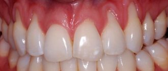
Stages of cervical caries
Cervical caries develops gradually, the process of disease development includes the following stages:
- Superficial caries. Characterized by damage only to tooth enamel. The disease begins with the stage of a “chalk” stain - the appearance of a white mark on the surface of the tooth. In the affected area, the enamel softens and becomes matte. Pain is not typical for the initial stage of caries, however, as the enamel further deteriorates and a cavity forms, short-term discomfort or pain may be experienced associated with brushing teeth, eating cold or hot, sour or sweet foods.
- Average caries. At this stage, dentin is involved in the pathological process. With caries in the middle stage, the pain is still associated with external influences, but lasts longer and becomes more intense.
- Deep caries. It is distinguished by deep penetration into the dentinal layer and severe pain. The reaction to external stimuli intensifies many times, the pain becomes unbearable.
Features of the flow
A distinctive feature of cervical caries is the extensive area of the carious cavity. Unlike other types of caries localization, the lesion quickly spreads not only in depth, but also in breadth. Cervical caries can be complicated by root caries when the cement covering the root of the tooth is involved in the pathological process.
The entire process from the beginning of demineralization of hard tooth tissues to the formation of a carious cavity can take from several weeks to several months (on average 3-6 months). Sometimes the progression of the disease stops - the growth of the stain stops, and the sensitivity of the teeth decreases. Suspended cervical caries is not a reason to refuse a visit to the dentist. It should also be treated, since the disease can become active at any time.
Diagnosis of the disease
The diagnosis is made in stages. Listening to complaints and visual examination make it possible to diagnose caries only in 13% of cases. Classic probing with a sharp probe and inspection with a mirror are informative. This allows you to examine the dentition.
Thermal diagnostics, electroodontometry and x-rays are also performed. If the patient has massive dental plaque, changes in the enamel will not be visible. In this case, professional teeth cleaning is first carried out to remove plaque and stone. All this is possible in a dental clinic.
At the dental clinic
The following types of studies can be performed at the clinic:
- Probing with a thin probe with a curved end - it is inserted under the gum, while the doctor has the opportunity to examine the structure of the tooth, its integrity, the presence of irregularities and chips. With rapidly progressing caries, the edges of the cavity are uneven and sharp. In the remission stage, the surface of the pathological focus is shiny, smooth, hard with smooth edges.
- Electroodontometry - can determine nerve damage and the depth of inflammation. The pulp reacts differently to the current strength.
- Thermal diagnostics - treatment of different areas of the tooth with a stream of water and heated wax. Unpleasant sensations that disappear after removing the irritant indicate caries.
- X-ray - a targeted photograph of one tooth or computed tomography. The presence and localization of obvious or hidden inflammation is determined with millimeter precision.
- Visiography is a special device - a visiograph scans the received data and transfers it to a computer, where the picture is studied from different angles and in detail, revealing the hidden process of inflammation.
How long does it take to treat caries?
The duration of the treatment procedure may vary depending on the condition of the patient and how deep the tissue damage has gone.
Depending on the initial conditions, treatment can be carried out in one or two stages. In the first case, the process rarely takes more than half an hour. In the second case, this time is extended over two visits - you need to kill the nerve and only then carry out other manipulations.
Thanks to their extensive experience and level of professionalism, our dentists cope with the task with minimal time.
Treatment or removal - what determines the outcome
The doctor makes a decision on treatment or tooth extraction after determining the depth of spread and the area of localization of the inflammatory process. It is mandatory to first carry out professional cleaning using an ultrasonic scaler, an AirFlow water-abrasive device and hand tools.
Initial caries is treated with conservative retherapy. For hard tissue defects, preparation and filling are performed. In severe cases, the tooth is removed.
Treatment of the initial stage of root caries
Conservative retherapy includes:
- fluoridation of teeth;
- covering teeth with protective agents
- training in proper oral hygiene
Potential consequences and complications
The very question of whether caries should be treated is very dangerous. It is important to never doubt that treatment is necessary. Often a person observes the first symptoms of a problem, but begins to put off going to the dentist. The time for treatment is constantly being delayed more and more.
The main danger of this approach is the likelihood of complications. The most common among them is the appearance of periodontitis or pulpitis.
There are several of the most serious consequences of untreated caries:
- Tooth loss. If the tooth is severely damaged or the lesion has gone very deep, there is nothing left except surgical intervention.
- Pathological processes in the gums. They can lead to the need to remove not only the affected tooth, but also its completely healthy neighbors.
- Systemic complications in the functioning of internal organs. The gastrointestinal tract and immune system suffer greatly. It is important to understand that a sluggish inflammatory process has a sharp negative effect on the entire body and weakens it.
The question of how caries is treated in dentistry in children is always considered separately. It is necessary to solve the problem that has arisen with baby teeth, because advanced disease leads to pathologies in the formation of permanent dentition.
Cleaning in dentistry from stone and plaque
1243900247100
Professional teeth cleaning involves removing the accumulation of all pathogenic microflora around the root.
It involves not only removing plaque, but also stones. This elimination of negative factors protects teeth for a long time.
Antiseptic treatment of affected areas
Treatment of root caries involves drilling out all affected areas with a drill and treating the carious cavity with antiseptics and special preparations. A 2% chlorhexidine solution or a gel based on it is used as an antiseptic for the treatment of carious cavities.
What to do to protect your gums
If the gum is not treated, inflammation will remain and all treatment will become meaningless. The gums are protected by diathermocoagulation and retraction.
Diathermocoagulation
Diathermocoagulation – excision of excess areas of periodontitis with a coagulation knife. The knife immediately cauterizes the bleeding edges. The procedure is performed when there are severe changes in the gums. In such cases, filling is postponed until the soft tissue is completely regenerated.
Retraction
If the condition of the gums is advanced, treatment of caries becomes impossible - the mucous membrane bleeds and interferes with tooth treatment. This gum is excised or a special device is applied to move the gum back. But more often, retraction is performed - lowering the gingival contour or moving apart the overhanging edges of the gums using special hemostatic threads. A temporary filling is placed until complete healing.
What preventative measures are there?
To keep your teeth healthy for many years, you need to brush your teeth twice a day, use floss and a tongue scraper. After sweets, you should rinse your mouth and chew gum for 5-10 minutes.
Prevention also includes the removal of tartar. A sufficient amount of saliva is also important, so it is necessary to maintain a water regime.
Like any other pathology, caries is better prevented than treated. Therefore, it is important to visit the dentist twice a year for preventive examinations. People at risk should approach this especially responsibly.