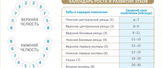
Normally, a person grows 52 teeth over a lifetime: 20 milk teeth and 32 permanent teeth. Supernumerary teeth are “extra” dental units that appear in addition to some incisors, canines, premolars or molars. In various medical sources, the phenomenon is called hyperdontia, supradontia or polyodontia. A superset can include 1–2 teeth or reach several hundred dystopic structures, be true or false.
Supernumerary teeth: causes of anomaly
The main cause of polyodontia is a violation of the formation or development of tooth germs. Doctors are considering several theories for this phenomenon:
- Heredity – hyperdontia develops as a result of genetic abnormalities that can be transmitted from parents to children. In this case, the pathology should manifest itself in the patient’s closest blood relatives.
- Splitting of the root germ of the tooth in the embryonic period of development. Usually observed after the mother becomes ill during pregnancy. Characteristic of single supernumerary teeth (1–2 pcs.).
- Failure during the embryonic period of tooth formation with the formation of multiple dental buds (up to several hundred). It is observed when the mother lives in unfavorable environmental conditions, during treatment with dangerous drugs, taking drugs, or alcohol abuse.
In more rare cases, polyodontia develops as a result of odontoma, a benign tumor in the area of the jaw arches. The disease is typical for children and young people during the period of active growth of tooth germs. Such a tumor structure may contain several tens or even hundreds of highly deformed dentin-enamel formations.
Provoking factors in the prenatal period:
- malfunction of genes - primarily the Msx1 gene (responsible for the formation of tooth germs);
- viral infections;
- taking teratogenic and potentially dangerous drugs;
- smoking, drug use, alcohol use;
- poor environmental conditions;
- exposure to radiation - not only direct exposure, but also increased general background radiation in the place of residence/work.
Reference! Some scientists consider the presence of supernumerary teeth as an atavism. Thus, it has been scientifically proven that various species of the genus Homo could boast a set of teeth of 36–44 pieces. However, this theory does not explain in any way the appearance of multiple rudiments covering all spaces of the sky (a typical example is an Indian boy who had to remove 230 supernumerary units).
Polyodontia is an abnormal number of teeth.
In medicine, this disease is often called hyperdontia, and “extra” dental elements are called supernumerary teeth. Research is still being conducted into why this pathology occurs. Most scientists associate it with disturbances in the formation of tooth germs.
Nature provides that a person grows no more than 20 milk teeth and 32 permanent teeth in a lifetime, but exceptions occur, and in our time quite often. According to statistics, on average, dental anomalies occur in 2% of the world's population, most often in men.
In 2014 alone, two operations were performed, in one of which 80 teeth were removed, and in the other, a record 232 teeth. Until this time, the maximum figure was 37 teeth.
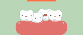
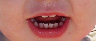
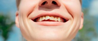
The most common hyperdontia (anomaly in the number of teeth) is an anomaly of the upper incisors. Supernumerary teeth are less common among the lower incisors and in other parts of the jaw. They can come in a wide variety of shapes and sizes. These are usually small, cone-shaped teeth.
Extra teeth lead to deformation of the dentition, so it is recommended to remove supernumerary elements. Another reason for removal is that most patients with this pathology have a lisp.
The formation of extra teeth is quite common today. According to statistics, 70% of patients have only one extra incisor, in 25% of cases – 2 supernumerary elements, and only 5% of all patients have 3 or more teeth during examination.
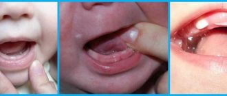
Symptoms of pathology
The main symptom is the presence of teeth beyond the natural set. They can have different positions, shapes and numbers. The process of teething of abnormal teeth is often accompanied by pain, fever, and inflammation of areas of the oral cavity. If measures are not taken in a timely manner, complications may develop:
- redness and swelling at the site of the impacted tooth;
- problems with chewing food and digestion;
- injuries to the mucous tissues of the oral cavity;
- violation of the position of the main teeth (dystopia) and the formation of malocclusion;
- various speech defects - mainly problems with the pronunciation of hissing sounds;
- loosening of adjacent normal teeth;
- deformation of the jaw bones.
In young children and adults, the pathology has its own nuances. They are associated with age-related characteristics of the body and affect the subsequent development of maxillofacial structures. Statistics on the spread of pathology in children and adults:
- 60% of cases are children and adolescents during the period of replacement of milk teeth with permanent ones;
- 35% of cases are adults;
- 5% of cases are small children during the period of growth of baby teeth.
Reference! Supernumerary teeth, as a rule, have a standard structure, sometimes with a smaller crown size. Less common are deformed formations of a teardrop-shaped, lumpy, chisel-shaped form.
Prevention of dystopia
Dentists are confident that if you carefully monitor your child’s dental health from early childhood, you can avoid most bite problems. Dystopic teeth are no exception, because their appearance is not always associated with hereditary factors. To prevent dystopia, doctors recommend the following:
- proper nutrition, adherence to the daily routine of a pregnant mother;
- prevention of jaw injuries from an early age;
- regular visits to the dentist, wearing devices to correct malocclusion;
- giving up bad habits (including weaning off the pacifier after a year and from thumb sucking in sleep), etc.
If you and your child regularly visit a dentist, he will give recommendations on what exactly to do in your case.
Features of polyodontia in children
Possible symptoms of hyperdontia in childhood:
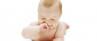
- intrauterine formation of teeth (a child is born with several teeth);
- the appearance of teeth in the first months, weeks and even days of life;
- delayed eruption of baby teeth.
This has unpleasant consequences:
- problems with feeding the baby - he does not latch onto the nipple well and sucks weakly;
- swelling of the mucous tissues can spread to the nasal area and cause breathing problems;
- poor closure of the dental arches causes severe salivation;
- a prolonged increase in temperature to 38C is possible.
Hyperdontia is especially harmful during the period of speech formation. Teeth located outside the general row prevent the tongue from taking the correct position when pronouncing most consonant sounds. Without eliminating the pathology, any speech therapist will be powerless.
In addition to true hyperdontia, children (less often adults) can develop the so-called false form of the disease. It is characteristic of the period of tooth change and develops during the eruption of molars next to the milk teeth that have not yet fallen out. This is a fairly common phenomenon that usually goes away on its own as the dentition finally forms. In some cases, orthodontic treatment may be required.
Dystopic wisdom tooth: what is it?
A dystopic wisdom tooth is a third molar, also known as a “figure eight”, which is positioned incorrectly relative to the rest of the dentition. Such a tooth almost always needs to be removed.
A special case of a dystopic wisdom tooth is an unerupted, but fully formed figure eight, which turns out to be turned parallel to the gum. In this case, the tooth is there, although it is not visible, and it is in an incorrect position, so we can talk about dystopia.
Two examples of dental dystopia
Features of polyodontia in adults and adolescents
In adults, after complete replacement of complete teeth, the presence of additional units can lead to additional pathologies:
- chronic rhinitis, sinusitis when the wall of the maxillary sinuses is perforated by the roots of supernumerary impacted structures;
- interdental caries - due to teeth fitting too closely to each other.
Retention and dystopia
Adults are characterized by 2 main types of supernumerary teeth:
- Dystopic teeth are the name given to teeth with deviations in the direction of growth. The peculiarities of the formation of supernumerary units very often lead to dystopia. This is due to the fact that the space on the dental arch line is limited, and the roots of normal teeth simply push the “intruder” towards the cheek or palate.
- Impacted teeth – impaction occurs when a tooth loses its growth impulse and remains embedded in the jawbone. Impacted teeth can cause the adjacent normal teeth to become loose and cause them to shift and change the bite. Often cause pain.
On a note! According to statistics, hyperdontia accounts for up to 2% of cases of dental problems. Of these, 70% are associated with the appearance of single supernumerary teeth, 25% with a couple of such formations, and only in 5% complex multiple complexes of 3–4 or more teeth can be found.
What is polyodontia?
Supernumerary teeth are extra units in the dentition. There can be 33,34,36 or even more than 100. Normally, the number of teeth in a child’s primary occlusion should not exceed 20, and in an adult – 32 units.
Most often, additional incisors or canines appear. It is much less common for a person to grow extra wisdom teeth, and they usually remain impacted.
Supernumerary teeth in an adult differ from the rest in size, development of the crown and root. They can also have a different shape, resembling, for example, a thorn or a drop.
If the extra units have not erupted, then polyodontia usually does not cause any unpleasant sensations. Therefore, we often learn about the existence of supernumerary teeth only during radiography.
Diagnostic methods
The main indication for diagnosis is the presence of an “extra” tooth in the patient’s oral cavity or obvious signs of its eruption (painful tubercle or swelling in the gum or palate). All of these signs can be identified through a routine visual examination, during which the dentist evaluates the condition of the oral cavity. To confirm the diagnosis and carry out differential diagnosis, an X-ray examination is performed - an orthopantomogram. If it is necessary to examine the dentition in several planes, computed tomography (CT) is additionally prescribed.
On a note! The patient may not be aware of the presence of impacted supernumerary teeth. In this case, hyperdontia is detected only by the results of an X-ray or CT scan of the maxillofacial region.
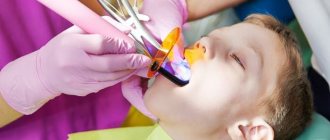
Removal of impacted teeth
In order for the operation to be successful and polyodontia to be cured without any complications, the doctor must fully examine the patient and plan his further actions.
- To begin with, X-rays and/or computed tomography are performed to determine the exact topography of the anomaly.
- Removal is performed under local anesthesia, but there are cases when general anesthesia can be used on the patient.
- First, the mucous membrane is peeled off, then the bone tissue is opened and the root and crown parts of the tooth are removed.
- If necessary, bone defects are covered with osteoplastic material, and the mucous membrane is sutured.
After tooth extraction, the patient continues treatment at home: takes antibiotics (if prescribed by the attending physician), rinses the oral cavity with antiseptic solutions.
Until the wound heals after surgery, it is not recommended to eat too hot, hard or spicy food. You should also brush your teeth carefully, especially on the operated side.
Do supernumerary teeth need to be removed?
There is no clear answer to the question of what to do with “extra” teeth. Much depends on the shape, position and quantity. The following are subject to mandatory removal:
- baby teeth that interfere with normal growth and formation of permanent teeth;
- strongly dystopic structures - located on the palate or at a large angle to the lateral side of the gums;
- impacted formations that put pressure on the adjacent roots of normal teeth and provoke periodic inflammatory processes of soft tissues.
If the additional tooth does not cause discomfort and does not disrupt the development of the dentition, removal may not be necessary. Moreover, such a superset can play the role of a strategic reserve in case of damage to nearby normal teeth.
How does the removal work?
The removal procedure can be simple or complex. In the first case, the doctor is dealing with a fully erupted tooth, the root of which is not intertwined with the surrounding structures. Simple removal steps:
- The oral cavity is prepared - treated with antiseptics, anesthesia is given.
- Grasp the crown with forceps (in some cases, an additional incision of the mucous tissue is required to facilitate access).
- The elevator destroys the retaining ligaments.
- The root is freed and the tooth is removed.
- Clean the hole from tooth fragments and bones.
- Apply stitches (if necessary) and a healing bandage.
Complex extraction is prescribed in the case of impacted, semi-impacted or dystopic teeth with complex root shapes. The procedure is carried out with a deep incision of soft tissues, in especially severe cases - with opening of the jaw bone to gain access to the roots. The latter option is relevant for deep bone occurrence, position in close proximity to the cranial sinuses and orbits, as well as for irregularly shaped roots and a high risk of damage to complete teeth. A complex removal operation is performed by a dental surgeon.
ICD-10 (Dentistry)
The article presents the international classification of diseases, tenth revision, relating to the dental profile.
K00—K14 Diseases of the oral cavity, salivary glands and jaws (click on the appropriate block to expand subcategories)
K00 Disorders of development and eruption of teeth
Excluding: impacted and impacted teeth (K01)
| K00.0 | Edentia
|
| K00.1 | Supernumerary teeth
|
| K00.2 | Anomalies in the size and shape of teeth
Excluding: Carabelli tubercular anomaly, considered as a normal variant and subject to coding |
| K00.3 | Mottled teeth
Excluding: deposits (growths) on teeth (K03.6) |
| K00.4 | Tooth formation disorders
Excludes: Hutchinson's incisors and mulberry-shaped molars in congenital syphilis (A50.5), mottled teeth (K00.3) |
| K00.5 | Hereditary disorders of dental structure, not classified elsewhere
|
| K00.6 | Teething disorders
|
| K00.7 | Teething syndrome |
| K00.8 | Other dental development disorders
|
| K00.9 | Dental development disorder, unspecified
|
K01 Impacted and impacted teeth
Excluding: impacted and impacted teeth with malposition of them or adjacent teeth (K07.3)
| K01.0 | Impacted teeth An impacted tooth is a tooth that has changed its position during eruption without obstruction from an adjacent tooth. |
| K01.1 | Impact teeth An impact tooth is a tooth that has changed its position during eruption due to an obstacle from an adjacent tooth. |
K02 Dental caries
| K02.0 | Enamel caries
|
| K02.1 | Dentin caries |
| K02.2 | Cement caries |
| K02.3 | Suspended dental caries |
| K02.4 | Odontoclasia
|
| K02.8 | Other dental caries |
| K02.9 | Dental caries, unspecified |
K03 Other diseases of hard dental tissues
Excluding: bruxism, teeth grinding NOS (not otherwise specified) (F45.8), dental caries (K02)
| K03.0 | Increased tooth wear
|
| K03.1 | Grinding of teeth
|
| K03.2 | Tooth erosion
|
| K03.3 | Pathological tooth resorption
|
| K03.4 | Hypercementosis
|
| K03.5 | Ankylosis of teeth |
| K03.6 | Deposits (growths) on teeth
|
| K03.7 | Change in color of hard tissues of teeth after eruption Excluding: deposits (growths) on teeth (K03.6) |
| K03.8 | Other specified diseases of dental hard tissues
If it is necessary to identify the radiation that caused the injury, use an additional code of external causes (class XX). |
| K03.9 | Disease of hard dental tissues, unspecified |
K04 Diseases of the pulp and periapical tissues
| K04.0 | Pulpitis
|
| K04.1 | Pulp necrosis
|
| K04.2 | Pulp degeneration
|
| K04.3 | Improper formation of hard tissue in the pulp
|
| K04.4 | Acute apical periodontitis of pulpal origin
|
| K04.5 | Chronic apical periodontitis
|
| K04.6 | Periapical abscess with cavity
|
| K04.7 | Periapical abscess without cavity
|
| K04.8 | Root cyst
Excludes: periodontal lateral cyst (K09.0) |
| K04.9 | Other and unspecified diseases of the pulp and periapical tissues |
K05 Gingivitis and periodontal diseases
| K05.0 | Acute gingivitis Excludes: acute necrotizing ulcerative gingivitis (A69.1), gingivostomatitis caused by herpes simplex virus (B00.2) |
| K05.1 | Chronic gingivitis
|
| K05.2 | Acute periodontitis
Excluding:
|
| K05.3 | Chronic periodontitis
|
| K05.4 | Periodontal disease
|
| K05.5 | Other periodontal diseases |
| K05.6 | Periodontal disease, unspecified |
K06 Other changes in the gingiva and edentulous alveolar margin
Excluding: atrophy of the edentulous alveolar margin (K08.2), gingivitis: acute (K05.0), chronic, NOS (not otherwise specified) (K05.1)
| K06.0 | Gum recession
|
| K06.1 | Gingival hypertrophy
|
| K06.2 | Lesions of the gums and edentulous alveolar margin caused by trauma. If necessary, identify the cause, use an additional code for external causes (class XX) |
| K06.8 | Other specified changes in the gingiva and edentulous alveolar margin
|
| K06.9 | Changes in the gingiva and edentulous alveolar margin, unspecified |
K07 Maxillofacial anomalies (including malocclusions)
Excludes: atrophy and hypertrophy of the half of the face (Q67.4), unilateral condylar hyperplasia or hypoplasia (K10.8)
| K07.0 | Main anomalies in jaw size
Excludes: acromegaly (E22.0), Robin's syndrome (Q87.0) |
| K07.1 | Anomalies of maxillo-cranial relationships
|
| K07.2 | Anomalies of dental arch relationships
|
| K07.3 | Anomalies of teeth position
Excludes: impacted and impacted teeth with normal position (K01) |
| K07.4 | Malocclusion, unspecified |
| K07.5 | Maxillofacial anomalies of functional origin
Excludes: bruxism, teeth grinding NOS (not otherwise specified) (F45.8) |
| K07.6 | Temporomandibular joint diseases
Excludes: current case of jaw dislocation (S03.0), sprain and strain of jaw joint(s) (S03.4) |
| K07.8 | Other maxillofacial anomalies |
| K07.9 | Maxillofacial anomaly, unspecified |
K08 Other changes in teeth and their supporting apparatus
| K08.0 | Exfoliation of teeth due to systemic disorders |
| K08.1 | Loss of teeth due to accident, extraction or localized periodontal disease |
| K08.2 | Atrophy of the edentulous alveolar margin |
| K08.3 | Delayed tooth root (retentive root) |
| K08.8 | Other specified changes in teeth and their supporting apparatus
|
| K08.9 | Changes in teeth and their supporting apparatus, unspecified |
K09 Cysts of the oral region, not elsewhere classified
Including: lesions with histological features of an aneurysmal cyst and other fibro-osseous lesion Excluding: radicular cyst (K04.8)
| K09.0 | Cysts formed during the formation of teeth
|
| K09.1 | Growth (non-odontogenic) cysts of the mouth area
|
| K09.2 | Other jaw cysts
Excludes: occult bone cyst of the jaw, Stafne cyst (K10.0) |
| K09.8 | Other specified cysts of the oral area, not classified elsewhere
|
| K09.9 | Oral cyst, unspecified |
K10 Other jaw diseases
| K10.0 | Jaw development disorders
|
| K10.1 | Giant cell granuloma, central
Excludes: peripheral giant cell granuloma (K06.8) |
| K10.2 | Inflammatory diseases of the jaws
If necessary, identify the radiation that caused the injury, use an additional code of external causes (class XX) |
| K10.3 | Alveolitis of the jaws
|
| K10.8 | Other specified diseases of the jaws
|
| K10.9 | Disease of the jaw, unspecified |
K11 Diseases of the salivary glands
| K11.0 | Salivary gland atrophy |
| K11.1 | Salivary gland hypertrophy |
| K11.2 | Sialadenitis Excludes: mumps (B26), Hereford uveoparotid fever (D86.8) |
| K11.3 | Salivary gland abscess |
| K11.4 | Salivary gland fistula Ex: congenital salivary gland fistula (Q38.4) |
| K11.5 | Sialolithiasis
|
| K11.6 | Salivary gland mucocele
|
| K11.7 | Disorders of the secretion of the salivary glands
Excludes: dry mouth NOS (R68.2) |
| K11.8 | Other diseases of the salivary glands
Excludes: sicca syndrome (Sjögren's disease) (M35.0) |
| K11.9 | Salivary gland disease, unspecified
|
K12 Stomatitis and related lesions
Excluding:
- decaying mouth ulcer, gangrenous stomatitis, noma (A69.0)
- cheilitis (K13.0)
- gingivostomatitis caused by herpes simplex virus (B00.2)
| K12.0 | Recurrent oral aphthae
|
| K12.1 | Other forms of stomatitis
|
| K12.2 | Cellulitis and oral abscess
Excluding:
|
K13 Other diseases of the lips and oral mucosa
Including: changes in the epithelium of the tongue Excluding:
- some changes in the gingiva and edentulous alveolar margin (K05-K06)
- cysts of the mouth area (K09)
- tongue diseases (K14)
- stomatitis and related lesions (K12)
| K13.0 | Lip diseases
Excluding: |
| K13.1 | Biting cheeks and lips |
| K13.2 | Leukoplakia and other changes in the oral epithelium, including the tongue
Excludes: hairy leukoplakia (K13.3) |
| K13.3 | Hairy leukoplakia |
| K13.4 | Granuloma and granuloma-like lesions of the oral mucosa
|
| K13.5 | Submucosal fibrosis of the oral cavity
|
| K13.6 | Hyperplasia of the oral mucosa due to irritation Excludes: hyperplasia of the edentulous alveolar margin due to irritation (denture hyperplasia) (K06.2) |
| K13.7 | Other and unspecified lesions of the oral mucosa
|
K14 Diseases of the tongue
Excluding:
- erythroplakia, focal epithelial hyperplasia, leukedema, leukoplakia of the tongue (K13.2)
- hairy leukoplakia (K13.3)
- congenital macroglossia (Q38.2)
- submucosal fibrosis of the tongue (K13.5)
| K14.0 | Glossitis
Excludes: atrophic glossitis (K14.4) |
| K14.1 | "Geographical" language
|
| K14.2 | Median rhomboid glossitis |
| K14.3 | Hypertrophy of the tongue papillae
|
| K14.4 | Atrophy of the tongue papillae
|
| K14.5 | Folded tongue
Excludes: congenital cleft tongue (Q38.3) |
| K14.6 | Glossodynia
|
| K14.8 | Other tongue diseases
|
| K14.9 | Tongue disease, unspecified
|
Dental diseases from other sections.
| A69.0 | Necrotizing ulcerative stomatitis
|
| A69.1 | Other Vincent infections - Fusospirochetous pharyngitis - Necrotizing ulcerative (acute): • gingivitis • gingivostomatitis - Spirochetal stomatitis - Vincent's ulcerative film sore throat: • tonsillitis • gingivitis |
| B00.2 | Herpetic gingivostomatitis and pharyngotonsillitis |
| B26 | Parotitis:
|
| B37.8 | Candidiasis of other localizations Candidiasis: • cheilitis • enteritis |
| D86.8 | Sarcoidosis of other specified and combined localizations:
|
| E22.0 | Acromegaly and pituitary gigantism Excluded:
|
| E53.0 | Riboflavin deficiency
|
| F45.8 | Any other disturbances in sensation, function, and behavior that are not associated with physical disorders and that are not mediated through the autonomic nervous system are limited to specific systems or parts of the body and are closely related in time to stressful events or problems. Psychogenic:
|
| J36 | Peritonsillar abscess
Excluded:
|
| L55—L59 | Diseases of the skin and subcutaneous tissue associated with exposure to radiation
|
| M35.0 | Sicca Sjögren's syndrome Sjögren's syndrome with:
|
| Q38.2 | Macroglossia |
| Q38.3 | Other congenital abnormalities of the tongue
|
| Q38.4 | Congenital anomalies of the salivary glands and ducts:
|
| Q67.4 | Other congenital deformities of the skull, face and jaw |
| Q87.0 | Syndromes of congenital anomalies affecting primarily the appearance of the face
|
| R68.2 | Dry mouth, unspecified Excluded: decreased secretion of salivary glands (K11.7) dry mouth caused by:
|
| S03.0 | Jaw dislocation:
|
| S03.4 | Sprain and strain of the joint (ligaments) of the jaw Temporomandibular joint (ligament) |
| class XX | External causes of morbidity and mortality:
|
Orthodontic treatment
Depending on the situation, it is used after tooth extraction or instead of it. In children, it is used to stimulate the loss of baby teeth and eliminate false hyperdontia, as well as to facilitate and accelerate the eruption of a normal set. In addition, orthodontic treatment with the installation of braces, mouth guards, dental plates or other auxiliary structures can straighten crooked teeth and return them to their proper place.
Before prescribing treatment, the dentist carefully evaluates the condition of the complete and supernumerary structures and, depending on the overall picture and prognosis for the future, can use both formations or leave the strongest and most durable tooth (not necessarily complete).
Normal deletion
If the dentist decides that in a particular case, polyodontia can only be treated by removing an extra tooth, the patient should count on the following procedures:
- First of all, the patient should be sent for radiography. This is necessary in order to determine the size and number of roots, as well as the ratio of supernumerary and normal teeth.
- After collecting research, the doctor gives the patient anesthesia and removes excess teeth.
- In some cases, soft tissue sutures may be necessary after surgery.
Classification of injuries to children's teeth
The nature and severity of the damage is determined based on a comprehensive examination of the patient using radiological, clinical and other methods. To make a diagnosis, dentists today use different systems for diagnosing trauma to children’s teeth.
Standard ELLIS classification
This system of dividing acute injuries into separate types, developed by Ellis, is the very first and most used in practice. It includes 9 categories of dental damage:
- I – slight fracture of the crown within the tooth enamel;
- II – moderate crown fracture extending into enamel and dentin;
- III – severe fracture of the crown with exposure of the pulp;
- IV – loss of pulp viability in the presence/absence of a crown fracture;
- V – complete dislocation of the injured tooth;
- VI – damage with root fracture;
- VII – slight subluxation of an injured tooth;
- VIII – fracture of the crown in the cervical area;
- IX – trauma to baby teeth.
Types of Traumatic Injuries WHO
Actively used in modern dentistry along with ELLIS. This classification was developed based on the Andresen system and includes, in addition to dental injuries, damage to other tissues (oral mucosa, gums, periodontal tissue, bones).
I. Damage to pulp and hard tissue:
- cracks in tooth enamel (A);
- fracture of the crown without opening the pulp (enamel affected - B, enamel and dentin affected - C);
- fracture of the dental crown exposing the pulp (D);
- combined root and crown fracture with pulp exposure (E);
- trauma with root fracture (F).
II. Periodontal damage:
- shock of the tooth with a weak response of periodontal tissue to percussion without increasing its mobility and displacement (A);
- subluxation with change in mobility without displacement (B);
- penetration of the injured tooth deep into the tissues (C);
- exit of the tooth from the anatomical socket (D);
- displacement in a direction other than axial (E);
- complete dislocation of the injured tooth (F).
III. Bone injury:
- tight connection of the tooth with the socket with penetration deep into the tissue (A);
- fracture of the wall of the anatomical recess (B);
- fracture of the alveolar bone tissue (C, D);
- fracture of the jaw bone (upper – E, lower – F).
IV. Injury to the gums and oral mucosa:
- acute soft tissue injury (A);
- bruise of the mucous membrane or gums (B);
- soft tissue detachment (C).
Classification of injuries to children's teeth according to ICD-C
This is an international system for defining acute injuries, based on ICD-10 (1997). Used in pediatric dentistry to classify various injuries, including fractures of primary and permanent teeth (S02.5):
- fracture (chip) within the thickness of the enamel (S02.50);
- fracture of the dental crown without injury to the pulp (S02.51);
- crown fracture with injury extending to the pulp (S02.52);
- fracture of the root part of a permanent tooth (S02.53);
- double fracture in the area of the dental crown and root (S02.54);
- multiple fractures of different locations (S02.57);
- unspecified fracture of primary or permanent tooth (S02.59).
Procedure for providing assistance to children with dental damage
Immediately after receiving an injury, the patient is provided with primary medical care, which includes a general assessment of the child’s condition, pain relief, prescription of antibiotics, anesthetics and other medications, as well as a preliminary diagnosis. After this, the patient’s parents are given a recommendation to make an appointment with a pediatric dentist (therapist), who will carry out full dental treatment. The doctor providing specialized care carries out:
- registration of a medical history (including legal and social aspects of the case);
- collecting anamnesis (origin of injury, presence of concussion, local symptoms);
- clinical examination of the injury (visual examination, percussion, palpation, temperature tests of the pulp, instrumental methods);
- additional diagnostics (electroodontodiagnostics (EDD), transillumination, targeted radiography, occlusal radiography, computed tomography);
- making a diagnosis based on examination, clinical and other studies;
- choice of treatment tactics taking into account the nature of the damage, possible risks, benefits and costs.
The period of rehabilitation of a child after an injury, starting from the moment of emergency assistance, can take 1-3 or more days. Given the complexity of treatment, the period of complete restoration of the integrity and functions of damaged teeth often takes a longer period - several months or even years.