Dental X-ray is a method of examination that helps the dentist make a correct diagnosis and build a treatment plan. The photographs show the dentist what is hidden from view; with their help, he can easily see whether there is caries, gum disease, or the general condition of the teeth. This diagnosis is very valuable for both the patient and the doctor. It allows you to quickly determine the scope of work and carry out high-quality therapy, relieve pain and unpleasant symptoms. You can familiarize yourself with the features of this survey in a few minutes.
Why do you need a dental x-ray?
Radiation diagnostics is needed in most cases during dental treatment. Without accurate photographs of the oral cavity, the doctor cannot determine the entire affected area and the spread of the disease. Many problems cannot be assessed visually alone, such as nerve damage. X-rays reflect bone tissue well, so this is a very effective technique for examining teeth. Soft tissues also cast a shadow on the photo; using them, the dentist can detect an abscess, cyst or tumor.
Content:
- Why do you need a dental x-ray?
- How it's made
- In what cases is an x-ray needed?
- Where can I do it?
- Examination of children
- X-ray for pregnant and lactating women
- Frequently asked questions from patients
During a visual examination, the dentist can only guess the cause of pain, bleeding or other problems. For an accurate diagnosis, he needs an image of the internal structure of the oral cavity: gums, teeth, roots, canals. The procedure is always carried out before a diagnosis and treatment regimen are made. X-ray examination is necessary not only at the first stage; it is used to determine the effectiveness of therapy.
Be sure to take a series of photographs during cleaning and filling of the canals. They are used to determine the depth of each canal and the tactics for filling it. If the dental treatment is carried out incorrectly, for example, the canal is not completely sealed, this will lead to re-inflammation and various complications are possible, in the form of new dental problems. You cannot do without such technology after prosthetics, complex removal, or treatment of periodontal disease. That is, radiography of the jaw is needed to make a correct diagnosis and to monitor the progress of therapy.
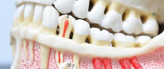
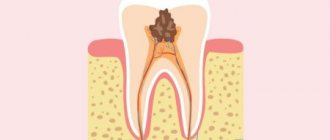
Symptoms of periodontitis
A clear clinical picture is characteristic of the acute stage and periods of exacerbation in the chronic form:
- The pain in the initial stages is aching and intensifies when chewing food or pressing on the crown. As inflammation progresses, the intensity of the pain attack increases sharply. The sensations become unbearable, pulsating.
- A feeling of a tooth protruding from the gums. The causal segment becomes higher than other units in the series.
- Presence of deep caries.
- Previous treatment of pulpitis on the causative tooth.
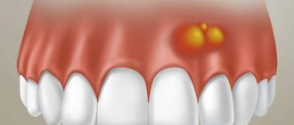
- Swelling of the gums.
- The appearance of an inflammatory tubercle in the projection of the root apex on the gum.
- Formation of a fistula near the diseased segment, discharge of serous or purulent exudate from it.
- Loosening of the unit.
Chronic periodontitis during remission is asymptomatic or has mild symptoms.
How it's made
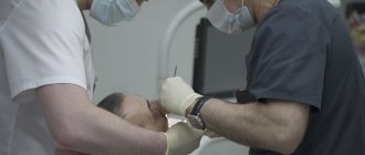
The process takes place in a separate room; most dental clinics have such X-ray areas. The walls and floor in this room are covered with lead material, which protects other rooms from the rays. The office is equipped with equipment, sometimes several types of equipment, as there are different types of dental x-rays.
No preparation is required before the procedure. The patient removes jewelry so that it does not distort the image. To protect other parts of the body, the patient is asked to put on a special cover from the neck to the waist; in medicine this is called a lead apron. Special fabric does not transmit radiation rays, so the radiation dose is significantly reduced. Then a series of pictures are taken or just one, depending on the doctor’s goals and the patient’s complaints. The procedure for obtaining images varies, depending on the type of equipment used in the X-ray room, as well as on the scope of tasks.
Bitewing radiography
This is the easiest and fastest way to obtain images of the internal structure of the jaw. This requires the simplest technique, which is why this technique is very popular in many dental centers. The patient sits on a chair, applies a small film to the gum in the desired area of the mouth, on which the image is then displayed. The film is placed on the side of the tongue and held there with the help of a bite plate. The laboratory technician presses a remote button and an image appears on the film. One such picture shows several teeth, the bone between the teeth, a canal, and periodontium. For complete information about the problem area, 4 photographs are taken. The results usually need to wait no more than 5-10 minutes; immediately after the procedure, the patient receives a series of images and gives them to his dentist.
A variation of this technique is a procedure called the “full set”. The picture is taken in exactly the same way, but in a larger quantity. To examine the entire jaw, the doctor needs 14 to 20 separate image films. From this “mosaic” a clear picture is formed. “The Complete Set” is recommended during your first visits to the dentist to examine all possible problems and prevent them.
Periapical radiography
This type of diagnosis is carried out to examine a separate part of the jaw; the image shows the entire tooth with the root and the gums near it, and the canals are clearly visible. The process is no different from the previous type of examination: a photo is taken on film, which is applied to the diseased tooth. In fact, this is another type of bitewing x-ray. This method is also used to control the treatment of a specific tooth, determine the cause of pain and bleeding. The advantage of the method is that the patient receives a lower dose of radiation.
Occlusal X-ray
This picture is taken when an image of an entire row of teeth is needed. In the office, the patient does everything the same, but the film is clamped with his teeth from a different angle. The plate is placed between the top and bottom rows and bitten. The photo shows all the teeth of the upper jaw or lower jaw, or both at once. This technique is indispensable for the treatment and diagnosis of malocclusion.
Orthopantomogram (OPTG)
A more informative method is a panoramic photo. For this we need a new generation device. The patient is still required to remove jewelry and wear a protective lead membrane.
Stages of the procedure:
- The patient is taken to an orthopantomograph and told how to position himself correctly.
- You need to hold on to special handrails with your hands; some orthopantomograph models also have a head holder. It is very important that the subject does not move during the examination, as the results will be distorted.
- There is a plastic tube in the chamber of the device; the laboratory assistant puts a disposable cover on it. The patient clamps the tube with his teeth and assumes a fixed position.
- The camera will begin to rotate around the patient's head and take pictures.
- Sometimes the radiologist may ask you to reposition your head or body for a different projection.
As a result, the dentist receives a panoramic image of the dentition, roots, and jaw bone. One such session replaces 14-20 bitewing radiography images.
Cone beam computed tomography
New technologies are increasingly replacing film X-ray machines. In dentistry, the most advanced diagnostic method is CBCT or 3D dental x-ray. The procedure is exactly the same as a panoramic shot. The difference is only in the results. CBCT provides comprehensive information about the patient's jaw. While the camera of the device rotates around the patient’s head, data from it is transmitted to a computer monitor. In this case, the image is obtained not in the form of photographs, but in the form of a three-dimensional model. The dentist can examine in detail the soft and bone tissue in any part of the oral cavity. Moreover, the result is transferred to a disk or flash card; when viewing, each individual section is enlarged many times over. Therefore, this method allows you to find even the smallest pathologies and prevent most problems. This method is not used often, since it is more expensive than all other types of examination.
In what cases is an x-ray needed?
During a visual examination, the dentist can see approximately 40% of the oral cavity. However, most problems lie inside: in the canals, roots, interdental space. Therefore, most patient complaints can be resolved only after X-ray diagnosis.
Indications for examination:
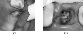
- caries in any part of the jaw, including under the crown;
- root destruction: fractures, cracks, bone depletion;
- periodontitis - destruction of bone, while the gums become inflamed and bleed, teeth begin to loosen and hurt;
- broken bite;
- pathology of the dental joint;
- abscesses and tumors, cysts;
- periodontitis is an inflammation of the root, accompanied by the appearance of a cyst that enlarges.
Painful sensations do not always appear, so patients often delay treatment. Advanced periodontitis leads to gumboil and tooth loss.
In addition, canal cleaning, prosthetics and implantation, bone grafting, and orthodontic treatment are not performed without a detailed examination.
Features of prevention
The main condition for the prevention of dental granulomas is timely assistance from a dentist when caries occurs. You should not allow severe tooth decay or the development of pulpitis. The peri-root tissues are healthy until the pulp becomes inflamed. Therefore, if symptoms of caries or pulpitis appear, it is important to immediately consult a doctor.
Endodontic treatment also increases the likelihood of developing periodontitis. Therefore, it is better to eliminate caries in the early stages and avoid the need for root canal filling. If this is unavoidable, it is important to carefully choose a dental clinic - the professionalism of a specialist will help eliminate possible mistakes and prevent complications.
Where can I do it?
Most dental clinics have an X-ray room where the patient can undergo this procedure without leaving the building and return to his dentist with the images. State clinics also have such installations, but they are exclusively of the old type. This means that you will have to go through the procedure several times and pay less. Old equipment also has high radiation doses. Unfortunately, many private dentistry are also equipped with such equipment. Sometimes a private doctor may refer you to another diagnostic center to obtain data. Cone beam computed tomography is only available in progressive private clinics, but the accuracy of the results fully pays for itself.
The price of the service will depend on the type of examination. Thus, bitewing radiography costs from 3 to 10 dollars, depending on the number of images. At the same time, prices are almost the same in both public and private institutions. A panoramic image will cost approximately $20-25. It can only be done in private institutions, but some clinics can provide this service for free if the patient is being treated by them. The most expensive diagnostic is CBCT, which is done in single diagnostic and dental centers. Its cost will be 50-60 dollars.
Examination of children
Dental x-rays are often prescribed for young children. You should not be afraid of this, since children sometimes need such examination more than adults. Baby teeth are often subject to carious problems, and caries appears in inaccessible places. As with an adult, the dentist cannot fully assess the condition and depth of the problem in a child. To accurately assess the situation, treat and prevent diseases, X-rays are needed. Also, a photo of baby teeth helps determine the process of eruption of molars, the quality of bone tissue, and the formation of a bite. In childhood, it is easiest to prevent improper formation of the dentition. If such problems are not resolved in time, in later life this will happen longer and more severely.
For small patients, only minimal doses of radiation are used. The doctor builds treatment and diagnostic tactics so as not to expose the child to radiation again. The procedure is the same as for adults: the rest of the body is covered with a protective apron, the session lasts a couple of minutes, no pain or discomfort occurs. In addition, some clinics use a collimator - a tube that is attached to the device. This device shrinks the X-ray beam and changes its contour, so children receive even less radiation.
What does an orthopantomogram show?
The overview photograph is a mirror image of the dental system: its right side is shown on the left, and the left side on the right. To facilitate orientation, the image is often marked: L and R (left, right).
To number the teeth, the image is divided into 4 segments. The 2 top ones - left and right, are “tens” and “twenties”, and the 2 bottom ones, the same left and right, are “thirties” and “forties”. The teeth are numbered starting from the center of the dentition, so in the center at the top there are incisors 11 and 21, and at the bottom - 41 and 31, and the row ends, as a rule, with wisdom teeth with numbers 18 and 28 at the top and 48 and 38 at the bottom.
In addition to the dentition, the following images are clearly visible in the panoramic image:
- mandibular canal with neurovascular bundle;
- maxillary sinuses and nasal passages;
- TMJ - temporomandibular joints;
- fillings and sealed canals (for this purpose, radiopaque components are introduced into the filling materials);
- wisdom teeth, supernumerary teeth, and in a child’s photograph - the rudiments of permanent teeth;
- hard palate with zygomatic bones, hyoid bone, nasal septum, conchae and other structures.
X-ray for pregnant and lactating women
Women during pregnancy should see a dentist as part of a routine checkup. The fact is that destructive and inflammatory processes in the gums and roots of teeth can lead to negative effects on the entire body. This situation is extremely undesirable when carrying a child, so a pregnant woman is sometimes prescribed x-rays. To examine the expectant mother, only minimal doses of radiation are used; they try not to take panoramic photographs, replacing them with targeted ones. However, radiation is still present, which raises the question among patients: is it possible to do an X-ray on a pregnant woman?
During pregnancy, of course, it is better to avoid such diagnostics. Manufacturers of new technology claim that it is absolutely safe for women even during this period. But still, the opinions of doctors on this matter remain skeptical. Such an examination can be prescribed only when the disease threatens life more than the possible negative impact of radiography on the fetus. In the third trimester, such diagnostics can be carried out without harm to the fetus. In the process of obtaining images, the patient’s chest, neck, abdomen, and pelvic area are tightly covered.
After childbirth, women often experience dental problems, mainly due to the loss of beneficial microelements. Nursing mothers should not be afraid of x-rays. This does not affect the structure of milk at all (this does not apply to studies of the abdominal and thoracic cavity). During lactation, women undergo X-rays under a protective apron. You can feed your baby after the x-ray on the same day.
Types of targeted shots
Modern innovative equipment allows you to take 2 types of pictures:
- Intraoral. It is done directly in the mouth. They are used when it is necessary to check the quality of the installed filling and the depth of caries damage.
- Interproximal. It is used to monitor the process of prosthetics and the condition of dental crowns.
In general, two devices are used to take pictures:
- X-ray machine (outdated model). The photographs are taken on film. May lose image after some time.
- Digital radiovisiograph (improved analogue). Electronic photographs allow you to magnify and examine every detail of a dental element. They are stored for a long time. The radiation emitted is much less than that of an X-ray machine.
Frequently asked questions from patients
Are x-rays harmful?
Radiography is based on radiation exposure. This sounds quite threatening to the average person. Meanwhile, every person receives a dose of radiation every day, even our bodies are radioactive. The background radiation level per day is 10 μSv (microsieverts). To obtain 4 photos with a bitewing X-ray, the patient receives 20-51 μSv. A panoramic image gives 5-25 μSv. CBCT is accompanied by a higher radiation dose; in one session a person receives from 20 μSv to 700 μSv. The level of radiation will depend on the settings and type of device, and the width of the area being studied.
Thus, there is no direct threat in the procedure. However, radionuclides can accumulate, so diagnostics are prescribed if necessary. After the session, the radiologist must write down how many sieverts the patient received, this will make it possible to calculate the next dosage with minimal harm to the person. After the examination, it is advisable to eat more carrots, apples, radishes, beans and citrus fruits. These products will help remove radionuclides from the body.
How often can it be done
Best materials of the month
- Coronaviruses: SARS-CoV-2 (COVID-19)
- Antibiotics for the prevention and treatment of COVID-19: how effective are they?
- The most common "office" diseases
- Does vodka kill coronavirus?
- How to stay alive on our roads?
The type of x-ray and its frequency depends on the condition of the oral cavity and the complexity of the treatment. It is better to do CBCT no more than 3 times a year. Bitewing film photographs are prescribed no more than 7 times a year; the new technology uses lower radiation doses, so there may be more digital diagnostics. The acceptable norm is 7 diagnostic studies per year. When changing dentists, it is not necessary to take new photographs; it is enough to take the ones you already have with you to the appointment. If digital data has been lost, it can be requested from the clinic that conducted the examination. CBCT results are stored for up to a year in the archives of medical centers.