Competition “Bio/Mol/Text”-2020/2021
This work was published in the “Free Topic” category of the “Bio/Mol/Text” competition 2020/2021.
The general partner of the competition is the annual biotechnology conference BiotechClub, organized by the international innovative biotechnology company BIOCAD.
The sponsor of the competition is SkyGen: a leading distributor of life science products on the Russian market.
Competition sponsor: the largest supplier of equipment, reagents and consumables for biological research and production.
"Book" sponsor of the competition - "Alpina Non-Fiction"
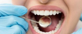
We would all like to have brilliant, healthy white teeth so that we can proudly show them off to others, and those around us would immediately fall into delight from such a demonstration. But, alas and ah, the problem of caries lies in wait for our teeth literally around every corner. However, if you are “armed and dangerous”, namely, you know where the roots of caries come from, that’s already half the victory! Therefore, let's understand the biological subtleties of its occurrence.
Where do the roots come from?
Caries (caries - “rotting”) is one of the most common human diseases, which is increasingly gaining momentum. About 42% of the world's population has dental caries [1]. However, this problem is far from exclusively human (Fig. 1).
Figure 1. Caries occurs quite widely in the animal kingdom, especially in ungulates. Above is a photo of a horse's teeth, below is a photo of a human.
Idade dos cavalos pelos dentes, “Complications in the treatment of dental caries in children”
It is now reliably known that the vital activity of oral bacteria leads to the occurrence of caries. Bacteria, like all living organisms, must eat, grow and reproduce, and also maintain their metabolism (metabolism) at the proper level. But not all bacteria in our mouths lead to tooth decay. Cariogenic bacteria include streptococci (Streptococcus mutans, Str. sanguis, Str. mitis, Str. salivarius) and some lactobacilli (Lactobacillus acidophilus) [2–4].
You can learn about who lives in the oral cavity and how individual representatives of its microflora affect our health from the article “Who lives in our mouth?” [5] special project “Biology, medicine and cosmetology of the oral cavity”. - Ed.
Like many other diseases, caries develops in several stages.
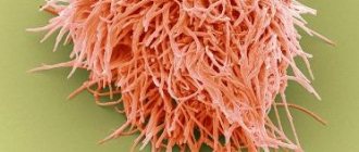
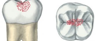
Anaerobic bacteria live in the form of a thin biofilm on the enamel of teeth - plaque. Fixing on the surface of the tooth, they produce extracellular heteropolysaccharides necessary to create comfortable living conditions for themselves, namely to reduce contact with oxygen. Often, the synthesis of such extracellular polysaccharides requires exclusively sucrose, as occurs in Streptococcus mutans (Fig. 2).
Figure 2. Streptococcus mutans is a species of gram-positive facultative anaerobic bacteria whose activity is directly associated with the development of caries. Streptococci, due to their structure (form chains) and the presence of a special receptor that allows them to attach to the smooth surface of the enamel, are most often found in dental plaque. Using sucrose, they synthesize sticky polysaccharides based on dextran, forming a biofilm on the tooth, thereby providing themselves with anaerobic living conditions.
"Wikipedia"
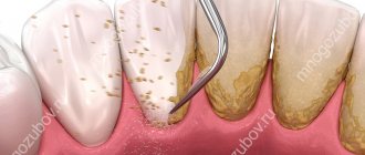
A person eats carbohydrates, and streptococci “eat” them along with him. Hence the direct relationship: the more sugar you ate, the more plaque formed. And the sweeter a person’s life, the better for streptococci: during the fermentation of carbohydrates (glycolysis), they receive energy for their vital functions, and from sucrose they build their protective coating - a “shell” from oxygen. The average time for visible and palpable plaque to form is 3–6 hours, and it takes about 24 hours for a dense bacterial biofilm to form.
For people, the activity of plaque bacteria has its disadvantages. They produce a large number of by-product organic acids, including pyruvic, formic, acetic and propionic acids [3]. And lactobacilli also produce lactic acid. The combination of all the released acids inevitably reduces the pH (increases the acidity) of that part of the oral cavity where the most active fermentation of carbohydrates occurs - in the folds of the enamel. At first, excessive acidity is neutralized by saliva, but the longer bacteria live on the tooth (you must understand that time flows differently for bacteria), the more heteropolysaccharides they secrete, and the more difficult it is for saliva to break through the growing shell of plaque. The critical pH value for tooth enamel is considered to be 5.5. It is at this value that caries begins to develop and demineralization of tooth enamel occurs (Fig. 3).
Figure 3. pH is very important for teeth. The normal pH of the oral cavity in the range of 6.8–7.4 is ensured by the buffering properties of saliva. A shift in the indicator to the acidic side to 5.5 is critical and indicates an active process of caries formation.
drawing by Anastasia Prokhorova
Why is caries dangerous if left untreated?
In this section we will talk about the complications of untreated dental caries in a timely manner, which without exception lead to the need to remove the nerve from the tooth and fill the root canals. And in case of purulent complications, in addition, contact a surgeon to make an incision on the swollen gum.
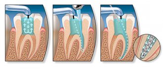
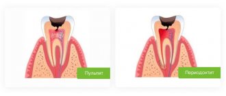
- Development of pulpitis - as the carious process spreads to the deep layers of dentin surrounding the tooth pulp, cariogenic microorganisms penetrate into the tooth pulp. The pulp is the neurovascular bundle of the tooth. As a result of infection, purulent inflammation of the pulp occurs - pulpitis. Pain due to pulpitis is usually acute and paroxysmal in nature. You can see schematically how pulpitis develops in the following pictures (Fig. 7-9) –
- Development of periodontitis – if pulpitis is also not treated in a timely manner, then pulp necrosis occurs, i.e. her death. This stage of inflammation is called periodontitis. In Fig. 11 you can see how the infection, spreading from the carious cavity through the root canals, penetrates beyond the tooth - into the periodontium and bone tissue. And as a result of this, a focus of purulent inflammation appears in the area of the apex of the tooth root (it can be designated by the term “periodontal abscess”).
On an x-ray, such an inflammatory focus looks like intense darkening around the apex of the tooth root. In addition, in Fig. 10 you can schematically see the differences between pulpitis and periodontitis. - Development of a cyst at the apex of the tooth root – as a result of infection of the near-apical tissues, their gradual destruction occurs: the bone tissue is resorbed, and in its place inflammatory granulation tissue is formed, and jaw cysts can also form. Such cysts are called radicular - from the Latin word Radix, which means “root” (24stoma.ru).
A radicular cyst is a cavity in the area of the apex of the tooth, the inner surface of which is lined with a membrane, and the cavity itself is filled with pus. Sometimes they can reach very large sizes. For example, I had to remove such cysts up to 5 cm in diameter. You can see what a radicular cyst looks like in the diagram of a section of the upper jaw (Fig. 12), as well as in the x-ray (Fig. 13), in which the cyst looks like an intense darkening at the root of the tooth.
- The development of gumboil is also a complication of pulpitis and periodontitis and includes a disease called “periostitis”, which is popularly called gumboil. Periostitis is always accompanied by severe swelling of the gums and/or cheeks. Inflammation and swelling in this case are a consequence of the spread of infection from the apex of the tooth root to the area of the periosteum, which covers the jaw from the outside. As a result, a purulent abscess occurs between the periosteum and the jaw (when the periosteum melts, pus breaks through under the mucous membrane of the oral cavity). Treatment of periostitis involves the need to make an incision in the projection of the swelling (to evacuate pus). After this, antibiotic therapy is prescribed. Before starting treatment, the issue of the need to remove or preserve the causative tooth that caused the inflammation is also decided. If it is decided to save the tooth, then after the acute symptoms have subsided, the dentist will need to treat this tooth appropriately so that it does not cause purulent inflammation next time.
Our teeth are like stones
Tooth enamel is the outer protective shell of the tooth, which is 97% composed of hydroxyapatite crystals (Fig. 4) - Ca10(PO4)6(OH)2 - and foundation proteins: amelogenins and emellines, on which during the development of enamel (amelogenesis ) and hydroxyapatite crystals are strung [2], [3], [6], [7].
Figure 4. This is what the “closest relative” of our enamel looks like - hydroxyapatite. It’s good for him, because he doesn’t know anything about caries!
"Wikipedia"
Hydroxyapatite, although the hardest part of man, is still very sensitive to acids. Acids “stuck” in dental plaque, especially in the area of fissures (Fig. 5) - folds of enamel - begin to destroy the mineral, leading to demineralization of dental tissue, in other words, to softening. The mineral decomposes, forming calcium salts with other acids [3].
Ca10(PO4)6(OH)2 + acid → 10Ca2++ 6HPO42– + 2H2O
Notably, hydroxyapatite can remain dissolved for up to two hours after ingestion. In addition to carbohydrates, the main role here is played by fruit acids. That is why after eating fruit you feel a feeling of cleanliness of the enamel. Over time, saliva neutralizes the effects of acids due to its buffering properties [2]. The more often high-carbohydrate foods are consumed, the more constant the acidification process in the oral cavity becomes. Thus, tooth decay does not depend on the amount of sugar you take, but on the frequency of its consumption: do not eat a bag of sweets during the day, but eat a large dessert once a day.
Figure 5. Enamel folds - fissures - are the favorite place for the “seeds” of caries. It is here that demineralization processes occur especially actively.
drawing by Anastasia Prokhorova
Complications of caries in children -
As we said above, in children under 3 years of age, newly appeared caries can turn into pulpitis (inflammation of the nerve in the tooth) in just 2-3 months, which is also due to the too small thickness of enamel and dentin.
The resulting pulpitis can have either an acute or chronic course. The acute course of pulpitis in primary teeth is characterized by the occurrence of spontaneous paroxysmal pain, especially at night and in the evening. The pain is usually provoked by hot or cold food, and after eliminating the irritant it can last from 15 minutes to several hours. In the chronic course of pulpitis of primary teeth, pain symptoms may either be completely absent, or pain may occur during meals (due to irritation of the receptors in the area of the bottom of the carious cavity by the food lump). This form of pulpitis can be very difficult to distinguish from average caries, which often leads to medical errors. Mistaking pulpitis for average caries, the doctor traditionally treats it with silvering or filling, which inevitably leads to the development of periodontitis (24stoma.ru).
But in the absence of treatment, pulpitis in any case sooner or later transforms into periodontitis (purulent inflammation at the apex of the root). The following symptoms are characteristic of periodontitis in primary teeth: 1) an abscess periodically appears on the gum in the form of a lump filled with pus, 2) fistulas with purulent discharge periodically open in the projection of the root of the diseased tooth on the gum. What purulent inflammation looks like during periodontitis (Fig. 15-16).
The occurrence of fistulas and purulent lumps on the gums is an absolute indication for tooth extraction (regardless of how much time is left before its physiological change). The fact is that purulent inflammation at the apex of the root of a baby tooth can most likely cause inflammation of the germ of a permanent tooth, which is located only 1-2 mm from the tops of the roots of a baby tooth. In addition, bacteria and toxins will constantly enter the blood, leading to deterioration of immunity and the development of a number of chronic diseases (for example, diseases of the ENT organs, bronchial asthma, allergies, etc.).
You can read about what needs to be done if your child has a fistula or purulent abscess on his gum in the article at the link above.
White caries - black caries, time plays against teeth
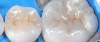
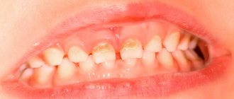
The enamel is translucent and normally ranges in color from light yellow to grayish-white. The color of dentin and any material underneath the enamel greatly affects the appearance of the tooth. Surprisingly, caries begins in the form of the formation of a barely noticeable white matte spot on the enamel (Fig. 6) [3].
Figure 6. “Dental problems.” Over time, the white spots on the enamel caused by caries darken.
drawing by Anastasia Prokhorova
Further, such a stain darkens from frequent meals: the area of the tooth softens more and more, and in the lucky ones a painless hole is formed - a carious cavity [7]. For some unfortunate people, caries is accompanied by inflammation of the tissues around the tooth, infection, and sometimes even an abscess, which can lead not to the small aesthetic problem of “black spots”, but to the loss of the tooth itself.
What do the main bacteria that cause tooth decay look like?
We cannot see what the bacteria that provoke the development of dental caries look like. But visible changes on the teeth provide an understanding of what stage the pathological process is at.
First, a barely noticeable light spot forms on the enamel. At the second stage, the spot becomes darker, its size increases, and the formation of a carious cavity begins. At the third stage, the carious cavity covers not only the enamel, but also the dentin. At the fourth stage, the disease spreads even deeper and goes beyond the dentin.
Sources:
- bsmu.by
- kraszdrav.ru
- en.wikipedia.org
Does genetics have an effect?
Some researchers attribute individual susceptibility to dental caries to genetic factors. By studying the development of caries in animal models, Japanese scientists suggested that loci on chromosomes 1, 2, 7, 8 and 17 contribute to susceptibility to caries. Genetics does not directly influence the onset of the disease, but determines its severity and duration. This is explained by the peculiarities of the structure of the oral cavity: genes involved in the development of enamel, in the formation and composition of saliva, and also those involved in the formation of the immune response are closely studied [4], [8], [9].
What dental diseases are caused by bacteria?
The most common problem that a person faces when pathogenic microflora develops in the mouth is caries. According to experts, caries is the most common chronic disease in humanity. Judging by the photographs taken of this disease, the treatment of advanced caries is too expensive for a person, since his teeth can be almost completely destroyed.
If you do not prevent caries and do not remove pathogenic microorganisms, they can destroy teeth even from childhood.
Another problem associated with pathogenic microorganisms on teeth, clearly visible in photos and videos under a microscope, is bad breath. Moreover, sometimes it can be so strong that it significantly limits communication. This is how bacteria can interfere with a person's social activity and even negatively affect his career.
Numerous photographs taken under a high-quality microscope confirm that just one toothbrush, especially if hygienic rules are not followed during its storage, can contain up to hundreds of millions of microbes.
Is it possible to reverse tooth decay?
If you have already noticed the first stages of enamel destruction, then caries cannot be reversed, but it can be slowed down, stretching the process over decades. Ion exchange reactions always occur on the surface of the enamel, allowing one to maintain a delicate balance between demineralization (destruction) and remineralization (restoration) of the enamel. Calcium and magnesium ions, phosphate ions, carbonate ions, strontium and fluorine ions can penetrate into the hydroxyapatite crystal. Of all the listed ions, we are especially interested in the fluorine ion F⁻ . Fluorine can replace the hydroxyl group (OH⁻) of enamel hydroxyapatite, forming fluorapatite Ca₅(PO₄)₃F . Fluorapatite is more resistant to acids, so the vital activity of bacteria has little effect on it [6].
In the article “Tooth Strength: How Do Pastes Make Our Teeth Better?” [10] of the special project “Biology, Medicine and Cosmetology of the Oral Cavity” talks in detail about fluoride and other useful components of toothpastes. - Ed.
Fluoride therapy is used to prevent tooth decay. The most pronounced effect is observed with optimal intake of fluoride ions into the body during the development of primary and molar teeth, that is, in childhood, but also in the first stages of caries - in the white spot stage - such therapy can slow down its course [6], [8] . Eating a balanced diet will also help keep tooth decay at bay. Cereals (bran, whole grain flour, sprouted grains), vegetables and fruits, spices (parsley, cumin, spinach), as well as animal products (fish, meat, especially bone broth, liver, seafood) contain large amounts of fluoride.
Well, if caries is already noticeable with might and main, then radical measures will be required to eliminate it in the dentist’s chair. By the way, the filling material also contains fluorapatite.
Take care of your teeth!
This short review article has already been published by the author here: https://vk.com/@biovk-temnye-delishki-nashih-zubov-karies.
Two-faced fluoride
If the first way to combat caries - giving up sweets - is not suitable for everyone, then making tooth enamel more resistant to acids is much easier. Today, the only internationally recognized and most effective way to strengthen enamel is still fluoridation.
For the first time, fluoride was added en masse to milk to prevent caries in schools and kindergartens in Switzerland in 1953. After 60 years, 95% of the world's toothpastes contain fluoride. If you read the ingredients in your toothpaste, you will most likely find sodium fluoride, monofluorophosphate, or aminofluoride in it. Or maybe there will be several fluorides. The mechanism by which all these substances help protect teeth from caries is very simple. Fluoride ions are introduced into the crystal lattice of mineral prisms of enamel, after which its solubility in acids decreases.