Symptoms of this disease will be:
- hyperemia, swelling, burning and hemorrhages from the gums;
- hypersensitivity to cold, hot, sour foods;
- aesthetic defect of the gums.
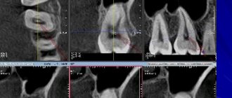
Often, hypertrophied gums act as a mechanical obstacle when chewing food, which contributes to injury, penetration of pathogenic microorganisms into damaged areas and aggravation of the inflammatory process. Diagnosis of hypertrophic gingivitis is carried out through examination, palpation examination of the patient’s gums, determination of dental indices, as well as based on the results of additional studies (radiography and CT of the dental system). Therapeutic tactics include the use of local anti-inflammatory drugs, physiotherapeutic procedures, sclerotherapy, diathermacoagulation of the gum papillae and surgical excision of hypertrophied tissue (gingivectomy).
According to statistical data, the children's category of the population suffers from this disease most often, especially children and adolescents in the pre-pubertal and puberty period (from the beginning to full puberty), since the phenomenon of hypertrophic gingivitis is often associated with hormonal development.
Hypertrophic gingivitis is one of the forms of gingivitis (inflammation of gum tissue) and is less common in comparison with other forms (catarrhal, atrophic, ulcerative necrotic).
Classification of gingivitis according to ICD-10
- K05.0 Acute gingivitis
- K05.1 Chronic gingivitis
- K05.11 Hyperplastic
- K05.18 Other specified chronic gingivitis
- K05.19 Chronic gingivitis, unspecified
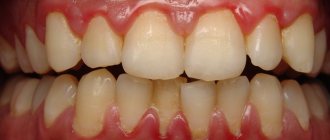
Normally, the gum reaches the crown of the tooth without covering it. It is pale pink, does not hurt or bleed. There is no noticeable distance between the tooth and the gum, however, with hypertrophic gingivitis, due to the enlargement of the gum, a “false pocket” appears between the tooth and the gum, i.e. The gum is attached to the tooth, and its enlarged part forms a pocket.
Principles of treatment
Treatment of gingivitis begins with hygiene: professional teeth cleaning. It is important to remove soft and hard dental plaque. For this, hand tools, an ultrasonic scaler, and the powder blasting method can be used. Subgingival dental plaque can be removed using the Vector device.
It is important to eliminate foci of infection - to fill teeth affected by caries, to undergo endodontic treatment in the presence of pulpitis, to remove the roots of teeth that cannot be restored and are not involved in the prosthetic process.
It is necessary not only to pay attention to the causes of the disease, but also to reduce the influence of harmful factors:
- stop smoking;
- consume food and drinks only at a comfortable temperature;
- remove spicy and smoked foods, marinades, especially those with vinegar, from the diet;
- During the treatment period, try to eat less solid food so as not to injure the loose gum tissue.
If the source of injury to the mucous membrane is the sharp edges of teeth or dentures, the doctor will immediately take measures to prevent further damage or recommend contacting a dentist of another profile - an orthopedist, an orthodontist.
Causes of hypertrophic gingivitis
Hyperplastic gingivitis can be caused by both internal disorders and local factors.
Local factors
- copious amounts of tartar and plaque
- trauma to the gums due to the edge of a crown, filling, or denture that puts pressure on the gum
- improper use of dental brushes and toothpicks also leads to injury
- malocclusion
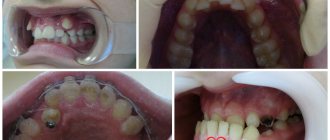
- orthodontic devices (braces, plates): they irritate the gums and complicate oral hygiene
- old and low-quality fillings, crowns or veneers put pressure on the gums and cause them to enlarge
General factors
- taking medications to treat epilepsy, diabetes, reproductive system diseases and heart disease
- taking contraceptives
- physiological hormonal changes in adolescents, pregnant women, and women after menopause
- vitamin deficiency, especially vitamin C
- blood diseases
- endocrine diseases such as diabetes
Diagnostic features
Gingivitis can be recognized visually—sometimes one examination by a doctor is enough. But you should make sure that there are no more serious pathologies, so diagnosis may include not only a visual assessment of the condition of the oral cavity, but also other measures:
- collecting anamnesis, assessing the condition of structures in the oral cavity;
- probing of periodontal pockets if present;
- determination of tooth mobility;
- electroodontodiagnosis to determine the condition of the dental pulp;
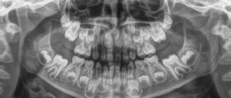
- panoramic x-ray or targeted x-ray - to exclude periodontitis, periostitis and other pathologies of deep structures, jaw bone tissue, etc.
It is important to take into account the presence of chronic diseases and medications. Only with complete information can a doctor make an accurate diagnosis and develop an effective treatment regimen.
Classification of hypertrophic gingivitis by severity2
| DEGREE OF SEVERITY | A COMMENT | ||
| severity: | Lightweight | a comment: | The gum covers less than half the height of the tooth crown |
| severity: | Average | a comment: | The edge of the gum covers the crown up to half |
| severity: | Heavy | a comment: | The gum covers more than half of the crown |
Hypertrophic gingivitis
The essence of the disease is reflected in the name - the gums and gingival papillae grow and become oversized. This occurs either through tissue swelling or due to internal proliferation of connective tissue with the formation of scars - fibrosis.
Accordingly, hypertrophic gingivitis can be edematous and fibrous.
The incentives are:
- Hormonal changes in the body during pregnancy;
- Injury to the gums by the edges of dentures and crowns;
- Progression of catarrhal gingivitis;
- Changes in hormonal levels during adolescence.
Edema form of gingivitis
This type of disease is often associated with internal changes in metabolism in women and growing children. The interdental papillae look loose and engorged. At the beginning of treatment, the oral cavity is sanitized, plaque and stones are removed, then anti-inflammatory procedures begin.
Sclerotization is most often used. After applying anesthesia, a special solution containing calcium chloride, magnesium sulfate and glucose is injected into the patient’s gums. The course takes 6-8 days with one or two day breaks.
Advanced swelling of the gums can change; the patient’s mucosa becomes scarred with the formation of fibrous cords.
Fibrous form of gingivitis
The source of troubles are internal changes in living tissues. Surgery is indicated here; unfortunately, fibrosis is not amenable to conservative therapy. First, external irritants are corrected - tartar is removed, the shape of the crown is corrected. Then the hypertrophied tissues are excised. Subsequently, healing bandages are applied.
Classification of hypertrophic gingivitis by location2
| DEGREE OF SEVERITY | A COMMENT | ||
| severity: | Localized form | a comment: | The gums are enlarged only in a certain group of teeth; it occurs due to trauma, crowded arrangement of a separate group of teeth |
| severity: | Generalized form | a comment: | The entire gum is affected and occurs when taking medications or diseases. |
About tooth loss and how to avoid it
Last year, we, at Star Smile, released a cartoon video in which the heroine was thinking about what to do - either start bite treatment when she was young, or wait until all the teeth begin to decay. Here he is, by the way
The video received quite a lot of views and responses, many patients asked us to continue the topic of consequences - to show readers what inattention and carelessness when caring for teeth and gums can lead to. Therefore, we will continue to educate you and hope that our articles will help you contact doctors in a timely manner and avoid most of the problems that we write about.
Symptoms depend on the form of the disease:
Edema form
The gums between the teeth are red, swollen, glossy, and a false pocket forms between the enlarged gum and the tooth. There is a change in the appearance of the gums, it looks swollen, bleeds, hurts when brushing teeth, and there is a feeling of burning and swelling. The edematous form develops as a complication of catarrhal gingivitis under the influence of an irritating factor (crown edge, fillings, braces, etc.). During pregnancy, hypertrophic gingivitis develops if the woman already had catarrhal gingivitis before pregnancy. The disease may resolve spontaneously after childbirth.
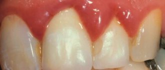
Fibrous form
The gum, as normal, is pale pink, there is no pain or bleeding, but its growth is observed, there are false pockets between the tooth and the gum. Abundant dental deposits are found above and below the gum. The fibrous form often develops when taking medications or in the presence of general diseases, i.e. this is the reaction of the gums to internal changes.
Types of gingivitis
Gingivitis can be acute or chronic. In the first case, noticeable symptoms occur. Chronic forms are characterized by mild symptoms, pain is absent or minimal. Periodic slight bleeding of the gums during brushing and halitosis may occur. It is important to understand that sluggish gingivitis is characterized by periodic exacerbations.
Inflammation of the gums is classified not only by its form, but also by the nature of its course.
Catarrhal gingivitis
Catarrhal gingivitis is characterized by redness of the gum area. Itching and bleeding due to mechanical action and mild pain may occur. This is the most common and easiest to treat type of inflammation. Quite often it acts as the initial stage of development of other forms.
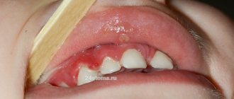
Ulcerative gingivitis
Necrotizing ulcerative gingivitis is characterized by the formation of open ulcers, death of areas of gum tissue, and strong bad breath. One of the typical symptoms is the appearance of a grayish coating. The disease is more difficult to treat; in the absence of timely assistance, purulent foci and severe necrosis may appear.
Hypertrophic and atrophic gingivitis
Ulcerative-necrotic gingivitis is followed by hypertrophic gingivitis - excessive growth of tissues occurs that cover the crowns of the teeth. There is keratinization of areas of the gums.
The atrophic form of the disease, on the contrary, is characterized by a decrease in the level of the gums and exposure of the necks of the teeth. This condition is dangerous due to the loss of healthy teeth.
Desquamative gingivitis
This type of inflammation is characterized by abundant desquamation of the gum epithelium. Distinctive features are pronounced redness and noticeable peeling areas of the surface of the mucous membranes.
Colored plaque
The brightest color is painted on the denser plaque; it is in these areas that teeth are most often not cleaned, which leads to gum inflammation.
After the final diagnosis, the dentist selects treatment tactics depending on the identified form of gingivitis.
Drugs for the treatment of gingivitis
Treatment for gingivitis may involve the use of medications in different forms. Mouth rinses based on antiseptics allow you to solve two problems at once: mechanically remove food debris and bacterial plaque, and also deliver active ingredients to inflamed tissues. Dentists recommend using ready-made pharmaceutical products; in each specific case, a specialist will prescribe a medicinal solution to quickly alleviate the condition. Ready-made formulations are more convenient to use, and the concentration of active components in them is known, this distinguishes them from traditional methods.
The most popular are rinses based on the following antiseptics:
- chlorhexidine;
- furacillin;
- chlorophyllipt;
- Metronidazole.
Calendula, chamomile, yarrow, oak bark, and St. John's wort have antiseptic properties. Doctors do not recommend preparing alcohol infusions; it is better to choose decoctions. The recipes call for using one teaspoon of dry raw materials per glass of boiling water. It is important to cool the broth to a comfortable temperature. Take into account possible allergic reactions, give preference to those herbs to which there was no previous intolerance. It is necessary to discuss with your doctor the possibility of using traditional methods.
Effective drugs for the treatment of gingivitis are ointments and gels. Multicomponent local products for applications not only help relieve inflammation, but also have an analgesic, decongestant, and antipruritic effect. The active components of such drugs can be lidocaine, antibacterial, antifungal substances, and antiseptics. The doctor will select a gel or ointment taking into account the shape and type of gingivitis. Thus, for ulcerative inflammation, it may be advisable to use regenerating agents, and acute catarrhal gingivitis will require the use of a powerful anesthetic gel.
For hypertrophic gingivitis, surgery may be required - a simple gingivectomy. This operation involves excision of excess tissue and application of a bandage, and is performed under local anesthesia.
Answers to popular questions
How to stop bleeding gums at home?
Herbal rinses (for example, oak bark) will help stop the bleeding, but they only give a temporary effect and mask the problem.
Can a hard brush cause gingivitis?
No, the brush itself does not cause gingivitis, but too hard bristles can damage the enamel. For most people, medium-hard brushes are suitable.
I don’t have problems with my gums, but my gums often hurt and bleed in only one place, what should I do?
It's worth seeing a doctor. Most likely, there is an old filling in this place, in which food gets stuck and inflammation begins.
Is gingivitis curable or is it permanent?
Fortunately, yes, if you follow all the recommendations, you will not only cure gingivitis, but also not see it again.
Prevention of gingivitis
It's always easier to prevent something than to treat it. Therefore, it is very important to follow the recommendations below:10
- You need to brush your teeth 2 times a day: morning and evening
- After each appointment, it is not necessary to brush your teeth, but you should rinse your mouth with water
- It is very important to use all personal hygiene products and do it correctly, you need dental brushes and dental floss
- Visit your dentist once or twice a year for a checkup and professional cleaning. It will remove plaque and be able to recognize gingivitis at an early stage
- Treat all concomitant diseases of the body.
- If installed structures in the mouth (fillings, dentures, braces) injure the mucous membrane, consult a doctor.
- Lead a healthy lifestyle: exercise, exercise, eat right
These measures will help you avoid gum disease and keep your mouth healthy.
Diagnosis of gingivitis
It is always important to collect a complete history of the disease, because gingivitis is often a chronic disease that occurs in childhood.6
It is also worth paying attention to the general condition of the body; the dentist may refer you to other specialists.
Consultation with an endocrinologist – in diseases of the endocrine system (diabetes mellitus, hyperfunction of the thyroid gland and adrenal glands), gum inflammation is more active.
Consultation with a hematologist – in case of blood diseases, gum inflammation is only a symptom of the underlying disease. Consultation with a hematologist is required for differential diagnosis and comprehensive treatment.
Consultation with a gastroenterologist – gingivitis is usually accompanied by chronic diseases of the digestive system.
Related doctors, in turn, prescribe a number of other tests to treat pathology, for example, a general blood test, biochemical blood test, gastroscopy, ultrasound, etc.
The dental office conducts diagnostic tests such as:
- Probing is a diagnostic method in which the characteristics of the attachment of the gums to the teeth are determined.
- Staining the gums with iodine-containing solutions: this shows whether the gums are inflamed. With gingivitis, the gums turn shades of brown.
- Determining the level of hygiene, that is, how well a person brushes his teeth.
- Panoramic X-ray of the teeth: This is necessary to see whether the inflammation has affected the bone or not, and also shows the condition of all teeth, fillings and crowns.
Symptoms of gingivitis
- Bleeding gums are the main symptom of gingivitis. Bleeding can occur both while brushing your teeth and when eating. Spontaneous bleeding also occurs
- Swelling and swelling of the gums, which can make teeth appear smaller.
- Pain of varying severity
- Change in gum color: it becomes bright red or blue.
- Deterioration of general condition, fever, weakness, headache
The presence of symptoms and their severity depends on the form and severity of the disease.
Catarrhal gingivitis: symptoms
Among all patients with gingivitis, this form accounts for more than 97% of cases. Those. This is the most common form of this disease. The term “catarrhal” means that the inflammation affects only the mucous membrane of the gums, i.e. flows superficially. Thus, with gingivitis, there is no destruction of the bone tissue around the teeth, as well as destruction of the periodontal attachment. In Fig. 3-5 you can see what catarrhal gingivitis looks like in the oral cavity.
The cause is exceptionally insufficient oral hygiene, as a result of which soft microbial plaque accumulates in the area of the necks of teeth, and the formation of tartar occurs. Plaque bacteria produce toxins and pathogens, which trigger inflammation in the gum mucosa. At the same time, various chronic diseases or vitamin C deficiency are not the direct causes of the development of gingivitis, but may be a predisposing factor that increases the impact of microbial plaque.
Catarrhal gingivitis: photo
Symptoms –
- swelling of the gingival margin and interdental papillae,
- redness or bluishness of the gums,
- bleeding gums when brushing teeth,
- pain when brushing teeth,
- itching in the gums,
- Usually, accumulations of microbial plaque are visible at the necks of teeth.
The appearance of bleeding is associated with an increase in the permeability of capillary walls, capillary fragility, as well as a decrease in the thickness of the epithelium of the gum mucosa. All this arises as a result of the influence of toxins and microbial plaque pathogens on the mucous membrane of the gums. Most often, bleeding occurs when the gums are exposed to mechanical factors (for example, when brushing teeth or chewing rough, hard food).
Pain when brushing teeth is also associated with thinning of the epithelium of the gum mucosa. Thinning of the epithelium against the background of gum inflammation is a natural process, and develops due to an increase in the rate of desquamation of epithelial cells (24stoma.ru).
Forms of catarrhal gingivitis –
There are 2 variants of the course of the catarrhal form of gingivitis. Firstly, there is acute catarrhal gingivitis (Fig. 3-4), which is characterized by bright red gum color, acute development, and sometimes significant bleeding and pain when brushing teeth. With this form of the disease (due to pain when brushing teeth), patients sometimes abandon oral hygiene altogether, which leads to an even greater increase in the amount of microbial plaque, and as a result, to an increase in gum inflammation.
Secondly, the chronic form of the disease (Fig. 5-6), which is characterized by sluggish symptoms over a long period of time. Chronic catarrhal gingivitis symptoms of bleeding will be quite minor, and pain during brushing may be completely absent. The marginal gum and periodontal papillae will have a bluish color. However, exacerbation of symptoms may periodically occur, which usually occurs against the background of a decrease in immunity during the period of colds.
Chronic catarrhal gingivitis –
Causes
Chronic hypertrophic gingivitis is accompanied by active growth of basal cells of the gingival epithelium. The occurrence of the disease can be caused by congenital malocclusions or low attachment of the frenulum, as well as the presence of low-quality dentures, fillings, and dental plaque.
Some common factors also contribute to the occurrence of the disease:
- the presence of chronic diseases of the nervous and endocrine systems;
- long-term use of medications, including antibiotics and hormonal drugs;
- lack of vitamin C in the patient’s body, which is responsible for gum health;
- the presence of hormonal imbalance, including during pregnancy.
Mechanical trauma to the gums from a tooth fragment, poorly fitting filling or prosthesis can also cause the disease.
Most often, the disease is localized on the upper jaw, in the frontal area.
Gingivitis: treatment at home
Gingivitis can result not only from the accumulation of plaque on the edge of the gum due to the use of inappropriate hygiene products, but also develops due to internal problems in the body. At home, it is quite possible to combat the results of the initial stages of catarrhal gingivitis, but you will not be able to get rid of stones with rinses and prepared solvents. You still have to sit in the doctor's chair.
In addition, self-medication of ulcerative or hypertrophic gingivitis is unacceptable. There is an extremely high risk of harming a person and worsening the patient’s condition.
Doctors also advise critically perceiving information about miracle cures presented in advertising. The phrase “Specialist consultation required,” which is now demonstrated in every video, took a lot of persistence from doctors. The fact is that any product is advertised as effective, but it is not specified that we are talking about symptomatic effects. The patient will be able to get rid of the symptoms, but the advertising product is not able to cure the cause that causes them - periodontal plaque, mineralized deposits. Therefore, consultation with a professional is absolutely required.
Treatment with folk remedies
All “grandmother’s” advice for gingivitis is of an auxiliary nature. You cannot refuse traditional treatment, relying on “time-tested recipes.” This is not stated anywhere, but in ancient times, people, using exclusively natural raw materials, lost their teeth extremely early - by modern standards. It is worth taking advantage of the achievements of science to preserve a beautiful and healthy smile for many years.
As maintenance therapy, it is allowed to use rinses with tinctures and decoctions of herbs (excluding alcohol). Chamomile, St. John's wort, and eucalyptus are beneficial. A side effect of this treatment will be rapid darkening of tooth enamel. Therefore, special herbal remedies produced on an industrial scale will be a worthy alternative to home preparations.