Diagnostics plays an important role in any field of medicine, and dentistry is no exception. It is very important for the doctor to see the full picture of the patient’s condition in order to make an accurate diagnosis and select the appropriate treatment. In dentistry, diagnosing something is not so easy, which is explained by the difficulty of accessing some parts of the oral cavity. In some cases, a 3D image of the teeth helps clarify the picture. What is it and how is it made?
Methods for diagnosing dental condition
Most methods for diagnosing the condition of the teeth and oral cavity fall into the category of visualization, that is, they can clearly show everything that happens in this part of the body. At a minimum, it is now completely impossible to imagine dentistry without radiography. Now this type of examination is included in the standard set of procedures that a patient undergoes when visiting the clinic with any complaint.
On a note! An examination is necessary not only when diagnosing the condition of a specific tooth, but also if the patient is faced with problems with the functioning of the entire maxillofacial apparatus.
Table. Main types of diagnostics in dentistry.
| View | Description |
| This is the most common diagnostic option, which can be carried out in any, even the most modest clinic. It provides the opportunity to correctly and accurately assess the condition of the canals in the roots of the tooth and identify developing pathologies in the area of hard tissues. Often used for endotonia. An X-ray image is a small black and white image on a special film or on a CD. | |
| This is otherwise called a panoramic image of the dental system. Using it, the doctor will be able to identify pathologies in the development of the jaw in general and teeth in particular, detect all carious cavities, understand how much hard tissue is destroyed during periodontal disease, clarify whether endotonic treatment helped the patient, etc. The image will also allow you to see whether there are any or deviations or changes in the condition of the lower sections of the maxillary sinuses. An orthopantomogram is also done in relation to the temporomandibular joints. It is often prescribed before prosthetics. | |
| This is a picture of not just the jaw, but the entire skull in one projection or another. It is necessary for measuring parts of the facial part (cephalometry). Based on this image, you can plan treatment with an orthodontist. | |
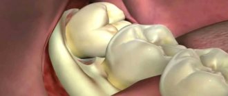
| It is this picture that is called 3D, which is discussed in the article. We'll talk about it separately below. |
Prescribing a CT scan of the upper and lower jaw before installing implants
Diagnosis of teeth and jaw structures
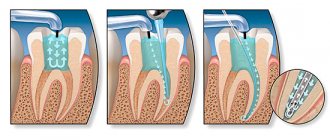
3D X-rays make it possible to examine areas of the maxillofacial system - from one tooth to a complete set of cross-sectional sections of the jaw and maxillary joint. Tomography before implantation allows:
- identify hidden carious cavities of neighboring teeth, curvature, length of canals, number of broken tooth roots to draw up a treatment plan before implantation;
- detect impacted, supernumerary teeth that may interfere with the installation of implants;
- evaluate bone septa, areas of pathological reorganization of bone tissue;
- visualize inflammation, cysts, granulomas, dental abscesses near the planned implantation area, which must be treated before surgery;
- identify inflammation in the maxillary sinuses and lacrimal ducts, which can become a temporary obstacle to implantation;
- evaluate bone tissue parameters - volume, density, degree of resorption, alveolar process inclination, thickness of cortical plates in order to correctly select the size and shape of the implant;
- clarify the anatomical structure of the maxillary sinuses, mandibular canal and other bone structures in order to plan the angle of inclination when installing an artificial rod;
- identify anomalies of the dentofacial system, pathologies of the temporomandibular joint for the correct modeling of the orthopedic structure for the implant;
- control the quality of implant and crown installation;
- assess the bone density around the installed implant;
- clarify the severity of the injuries received if the dentition is being restored after an injury
The information received in digital format is recorded on a medium (disk, flash drive) and sent by e-mail.
3D modeling of the operation CT results allow you to simulate the installation of implants of the required size, determine the angles of implementation bypassing the anatomical structures and simulate the final result of implantation.
The tomograph data is loaded into a computer program, and a 3D model of the jaw is created. The implantologist creates a virtual operation plan - selects the shape and size of artificial roots, their number, determines the installation location and angle of inclination. This allows you to take into account the nuances in advance.
A customized surgical template is printed using a 3D printer. This is an overlay with guides for future implants, which accurately determine the location and angle of installation of artificial roots. During the operation, the template is tightly applied to the gums, and the implants are installed with maximum precision.
What is a 3D photo?
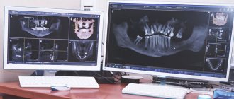
A 3D image or computed tomography of teeth is an x-ray that is performed using a special device - a tomograph (CBCT). The picture is a three-dimensional image that demonstrates the entire dental system in detail. The resolution of the image is high, which means that even the smallest details can be seen in it. The doctor, using a computer program, will be able to thoroughly examine absolutely any part of the patient’s jaw, moreover, at any depth and at any angle. The snapshot is provided on disk, where it is stored. If you need to view the disc, you can insert it into your computer and see everything you need on the monitor. Moreover, sometimes this diagnostic option saves the patient from a number of unpleasant manipulations on the part of the doctor.
On a note! The scanning volumes of such an image vary from 5x5 to 13x15 cm.
An orthodontist, implantologist, maxillofacial surgeon and other doctors can give a referral for such a plan. The technique is very accurate, and often without it, doctors simply will not do most of the manipulations. And the effectiveness of treatment thanks to this image increases significantly.
The advantages of taking a 3D photo are as follows:
- high image accuracy and detail;
- the ability to view all parts of the mouth - the image is three-dimensional;
- errors are excluded when identifying the location of pathology development, since there are no distortions in the image;
- the ability to assess the condition of the jaw from any side;
- the picture is taken quickly - a few seconds are enough to get a detailed image;
- taking a picture is safe for health, since the radiation exposure experienced by the patient’s body is extremely small. Especially if you compare its level with the level with the same x-ray.
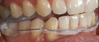
What is an intraoral scanner used for?
Using an intraoral scanner, you can immediately scan the patient’s teeth directly and immediately receive a digital 3D model of the teeth online. That is, teeth are scanned instead of taking classical impressions.
What are the advantages of intraoral scanners? First: convenience and comfort for the patient. There are a number of patients who cannot tolerate conventional impression materials. They develop a gag reflex - for example, it is very common in children.
And for such patients, intraoral scanning is probably the only possible way to make a digital impression of their teeth. Second, intraoral scanning allows for significantly higher accuracy than traditional impressions.
For example. When we make a high-precision A-silicone impression of teeth, then cast a plaster model for 3D scanning in a laboratory scanner, at the stage of taking the impression and casting the plaster model, an error of about 100 microns is obtained. Modern intraoral scanners provide 5-7 times higher accuracy – up to 12 microns.
Accordingly, the digital model obtained using intraoral scanning is significantly more accurate. This is very important for orthopedics, in the production of crowns, and it is also important in the production of aligners. Why? Because the aligner works by fitting the teeth very well. And if the impression is not accurate enough, this means that in some places the aligner will not fit the teeth enough, will not press as expected, and, accordingly, some of the movement of the teeth may not happen because of this.
This rule is confirmed by the statistics of our company. We see that clients who use intraoral scanners in their practice, in their clinics, have a significantly lower revision rate than those clients who work with traditional impressions, even if they use high-precision A-silicone.
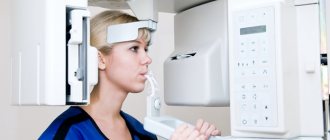
How is a 3D photo taken?
The image is taken using a special tomograph. The procedure is carried out as follows:
- a person takes a sitting or standing static position;
- then a special protective apron is put on his chest - the same as used during radiography;
- there is a special small plate in the mouth that you need to bite with your teeth;
- then you need to rest your forehead against a special support, and grab the handrails with your hands to avoid unnecessary movements;
- then you need to freeze for about 15 seconds at the doctor’s command to get an accurate image;
On a note! The tomograph will make only one full revolution, but will have time to take about 200 mini-images.
- After this, the pictures are processed and compiled into one overall picture - this is how the desired three-dimensional image is obtained. The treatment only takes a few minutes.
At the exit, the patient receives a disk with an image recorded on it, which he brings to the doctor to assess the condition of the jaw system.
Advantages
Indeed, doctors used to do without computed tomography. However, the possibilities of prosthetics or orthodontics were then limited: it’s easy to recall the terrifying false jaws, in which it was impossible to eat or speak, or dental plates, in which only a few decided to walk due to the colossal inconvenience and terrible external kind.
Modern advances in dentistry also require cutting-edge diagnostic methods. Dental CT is just such a method. Many experts call modern tomographic examination the term “3D computed tomography.”
The main advantages of this diagnostic method are:
- identification of infection localizations;
- diagnosis of maxillodental anomalies or defects (for example, identifying bone pockets, joint diseases, determining the degree of gum inflammation);
- diagnostics for complex injuries or congenital pathologies;
- the need to calculate down to the millimeter the movement of teeth when using braces;
- control over the development of jaw pathologies;
- clarification of the location of the lesion before surgery.
Dental CT is often prescribed for the following disorders:
- previously undiagnosed jaw diseases (developmental defects, difficult or spontaneous movements of the jaws, the appearance of clicks when moving, pain during chewing, etc.);
- injuries;
- prosthetics of bone organs;
- complex tooth extraction;
- spasms of the jaw muscles when moving the mouth;
- complications after treatment or tooth extraction;
- identification of hidden diseases of the pulp, teeth, gums;
- detection of cysts or tumors;
- hidden cavities in the mouth;
- before implantation in combination with reconstructive operations;
- malocclusion;
- unerupted teeth;
- examination of any soft tissues of the mouth.
Indications for this type of examination
3D tomography has a lot of possibilities, for which it is valued by doctors. Thus, it will allow you to assess the condition of not only the jaw, but also the gums, see the quality of previously installed fillings, and reflect all pathologies and anomalies. Indications for this may include:
- abnormalities in the structure of the jaw and the shape of the teeth;
- suspicions of pathological changes in dental tissues;
- identification of the rudiments of permanent teeth in children, assessment of the condition of milk teeth;
- the presence of cysts, inflammation, cracks in the roots of the tooth;
- suspicion of traumatic injuries to the jaw;
- preparation for implant installation;
- the presence of tumors in the jaw area;
- preparation for plastic surgery;
- preparation for operations in the jaw area.
Thus, a detailed three-dimensional image is necessary in all complex cases that require detail and a thorough assessment of the patient's current condition. It is especially important in assessing abnormally growing teeth, as well as in case of complex fractures and dental pathologies.
On a note! A CT scan is also often performed during preparation for implant placement. Moreover, its efficiency and effectiveness are much higher than that of orthopantomography. It is often necessary when performing a so-called sinus lift.
Table. Areas of application of CT.
| Sphere | Explanation |
| Orthodontics | The ability to assess how distorted the bite is. |
| Surgery | Assessment of the possibility of certain manipulations by the surgeon, as well as the possibility of installing teeth. |
| Maxillofacial Surgery | Assessment of the condition of the jaw and surrounding areas of the skull. |
| Therapy | The ability to detect deep-lying caries and choose the right treatment option. |
Painlessness of the computed tomography procedure
Modern medicine offers many options for oral examinations to obtain detailed information about the condition of periodontal tissues, all of which are safe and painless. While standing near the device, the patient does not experience any discomfort or pain, there is no dizziness or nausea. It only takes one minute to take pictures, so no need to worry.
Many patients are afraid of having a CT scan because they mistakenly compare it to an MRI. However, these are two completely different procedures! During a CT scan, a person is in an open space with only a small device in front of his eyes, while during a magnetic resonance imaging scan he is placed in a closed device. That is why even claustrophobia is not a contraindication to computer examination.
You can obtain the necessary data without psychological or physical inconvenience, pain and fear. In addition, the state of health does not change in any way even some time after diagnosis. The patient can drive a personal vehicle without fear, his consciousness is not confused, there is no loss of coordination of movements.
Are there any contraindications?
This procedure also has contraindications. Whatever one may say, there is an effect on the body from x-rays. In general, the load is small, especially if you remember the SanPiN standards (1 mSv/year), and is only 0.045-0.06 mSv. However, there is still a load with CT. So the indications for a CT scan must be justified - it is not advisable to just take a picture.
A 3D photo cannot be taken or is permitted with caution under the following conditions:
- pregnancy, especially the first months when the fetus is actively developing;
- breastfeeding period;
- problems related to the thyroid gland;
- diabetes;
- certain forms of allergies;
- chronic renal failure.
Attention! In some cases, a doctor, despite contraindications, may prescribe a 3D image, but only if the risk of the study is completely justified, and doctors have no other choice.
There is also a so-called tomography with contrast agent. It makes it possible to obtain a more detailed picture of soft tissues, so it is used extremely rarely to take photographs of the jaw. But it has many more contraindications than conventional CT. Substances that may cause undesirable effects should not be used as contrast. It is important to inform your doctor or specialist before the examination if you have any allergic reactions.
How to prepare for a 3D photo?
Step 1. The first thing you need to do is visit a dentist to understand whether it really makes sense to spend money on such an expensive examination. In some cases, a dental CT scan may not be strictly necessary.
Step 2. Next, if the doctor has confirmed the need for an examination, it is important to evaluate the market for this service in your city and choose a clinic based on the price-quality ratio.
Step 3. Then it is important to sign up for a convenient day and arrive at the clinic at the appointed time.
Step 4. Before taking the photo, it is recommended to clean your mouth by chewing gum or brushing your teeth.
Step 5. After the picture has been taken, you need to receive the results and take them to an appointment with your doctor.