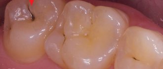
Early detection of caries is the main and priority task of the dentist. Using a caries detector and a high-tech intraoral camera VistaCam IX, detection of the disease is possible at an early stage, in the form of a spot. If detected and treated in a timely manner, such a case will not require grinding off the hard tissues of the tooth and opening the cavity.
The appearance of a caries detector in the dental field has improved the quality of early diagnosis and treatment of caries. When using this technique, the affected areas of the enamel are stained. The boundaries of the carious spot are clearly identified, which contributes to the early and most effective treatment of caries.
Patients of the innovative dental clinic "Implantmaster" in Moscow have the opportunity to use the latest digital caries detector - the VistaCam IX intraoral camera. The camera has a wide range of capabilities - from detecting the initial stage of caries on visible surfaces to determining the process in the interdental space, invisible to the eye. Our specialists regularly improve their skills in the best clinics in Europe, participate in scientific conferences and symposiums. We are accustomed to solving complex problems in the shortest possible time, setting loyal prices for the services provided.
Next, we will tell you in more detail about what caries detectors are and what types there are. You will also learn about the VistaCam IX intraoral camera and its benefits. Let's take a closer look at the stages of diagnosing teeth with a caries detector.
Get a consultation
We will answer all your questions before visiting the clinic!
+7
Online registration
Content
1 What is a caries detector?
2 Types of caries detectors
3 Intraoral camera VistaCam IX 3.1 Possible applications of the intraoral camera
3.2 Benefits of the VistaCam IX intraoral camera
3.3 Stages of diagnosing teeth with a caries detector
4 Caries marker
5 Cost of diagnosing teeth with a caries detector
Prices
Chemical indicators are an inexpensive diagnostic method, common in economy-class dentistry. The use of a caries marker does not increase the cost of treatment, but it greatly facilitates the dentist’s work.
Cost of known drugs:
- “Voco” (Germany) – about 450 rubles per 1 gram;
- “Kuraray” from a Japanese manufacturer – 2500 rubles. for 6 ml;
- “Color test No. 2” of domestic production – 100 rubles/20 ml;
- “Omega” (Russian company) – 200 rubles/8 ml.
Caries marker Omega
A caries marker is a universal tool, with the exception of diagnosing secondary caries under a filling. In areas close to the pulp, it is dangerous to use a marker because of the risk of confusing the carious areas stained with the reagent with the pulp (also pinkish in color). For such a case, they resort to alternative diagnostic methods - targeted X-ray or computer visiography.
What is a caries detector?
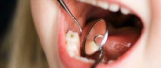
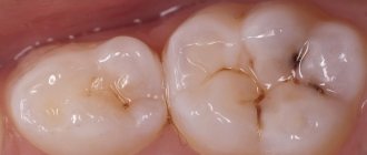
Initially, dentists examined the structure of tooth tissues during a visual examination using probing with a sharp instrument. It is not always possible to detect damage in the initial stages in fissures or between teeth. The method is not accurate, especially in difficult situations. Very sharp tools and excessive pressure can cause harm. Probing may even be painful. To make the diagnosis accurate and painless, caries detectors .
Types of caries detectors
There are 3 types of caries detectors: chemical, optical and laser.
Chemical
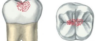
These are caries markers. When the active substance (acid fuchsin or methylene blue) is applied to tooth tissue, a chemical reaction occurs that stains areas susceptible to caries.
Optical
Diagnostics are carried out using a computer. Light waves with a length of 405 nanometers are produced. Depending on the condition of the tooth enamel, the reflection of light is different. A specialized program on a computer analyzes the data and produces a color picture, where the boundaries are clearly defined and at what stage the development of the carious process is.
Laser
The mechanism of action is the same as that of optical caries detectors, but instead of light a laser beam is used. Laser indicators are expensive to use.
Advantages and disadvantages
Advantages of using special colored markers:
- accurate indicators of healthy teeth and teeth affected by carious diseases;
- the natural composition of the markers makes the procedure safe;
- the opportunity to demonstrate to the patient the condition of his teeth;
- using a marker not only for the treatment of caries, but also for examining dental canals;
Disadvantages of using special colored markers:
- staining occurs even in areas with a minimal level of infection, which can be treated with remineralizing drugs;
- When grinding down stained areas, a smeared color may appear, making it difficult to examine the tissues being cleaned and distinguish them from completely healthy teeth.
Our team of doctors
Maxillofacial surgeon, Implantologist
Bocharov Maxim Viktorovich
Experience: 11 years
Dental surgeon, Implantologist
Chernov Dmitry Anatolievich
Experience: 29 years
Orthopedist, Neuromuscular dentist
Stepanov Andrey Vasilievich
Experience: 22 years
Endodontist, Therapist
Skalet Yana Alexandrovna
Experience: 22 years
Orthopedic dentist
Tsoi Sergey Konstantinovich
Experience: 19 years
Dentist-orthodontist
Enikeeva Anna Stanislavovna
Experience: 3 years
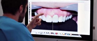
Intraoral camera VistaCam IX
The VistaCam IX intraoral camera is a digital device for obtaining dental images using various physical and optical methods, which make it possible to make an accurate diagnosis, as well as determine the location and extent of the lesion. The modular system of interchangeable nozzles helps to analyze the clinical situation from different angles. High image resolution and color rendering quality, high magnification, the use of advanced methods of UV fluorescence and infrared illumination, absolute safety for the patient and staff, have made the VistaCam IX intraoral video camera the most popular diagnostic device for the modern dentist.
Patients of the Implantmaster dental clinic have a unique opportunity to attend an appointment with our highly qualified specialists who use the latest technologies and equipment, including the VistaCam IX intraoral camera.
Possibilities of using an intraoral camera
The VistaCam IX dental intraoral camera has:
- Replaceable nozzles, which are quickly replaced and each has its own specific area of application;
- convenient location of on/off buttons on both sides;
- the ability to rearrange a convenient work surface;
- energy saving mode;
- digital data transmission.
1. Cam attachment for intraoral images.
- High-quality image transmission;
- uniform illumination of the working area;
- photo/video modes.
2. Macro attachment for detailing under high magnification, while maintaining image quality.
- Magnification 120 times.
3. Poly nozzle for polymerization of all modern composites.
- The presence of a soft start (gradual increase in light) for deep curing.
4. Proof attachment – diagnostic.
- Detection of plaque and analysis of the degree of carious lesions;
- fluorescence of cariogenic bacteria in the UV spectrum highlights places in the tissues and on the surface of the tooth in red, and healthy enamel is highlighted in green.
- The diagnostic result is assessed on a color scale with numerical values.
- 0 – 1.0 healthy enamel (green)
- 1.0 – 1.5 (blue)
- 1.5 – 2.0 (red) superficial caries (initial stage of enamel caries)
- 2.0 – 2.5 dentin caries (orange)
- 2.5 – 3.0 and deeper dentin caries (yellow)
5. Proxi head for detecting proximal caries.
- No harmful radiation;
- detection of caries between teeth, on contacts in the stain stage;
- Safe infrared radiation is reflected from caries-affected areas and is identified as lighter, opaque areas.
Benefits of the VistaCam IX intraoral camera
- At the same time, the consultation involves visualization of processes on the screen, creation of computer documentation and diagnostics. All this encourages the patient to trust the doctor.
- The proprietary camera system is a valuable assistant to the doctor during the appointment. The VistaCam IX intraoral video camera helps explain the need for treatment and professional cleaning.
- The intraoral camera in dentistry fully meets the requirements for clarity and quality of photographs when transferring data from the device to a computer.
- The presence of specialized replaceable nozzles is unique for a specific clinical case.
- An optical caries detector is a significant aid to the doctor during an appointment when determining the carious process.
- Caries and plaque are indicated by a color scale, supplemented by a digital value of 0-3, in a special computer program.
- Indispensable in detecting initial caries, as well as controlling the spread of plaque.
- The camera, ergonomic in shape, is functional when used in the workflow.
- It can be installed on almost any surface, and the standalone version can be used even without installing a PC.
- With the ability to be removed from the tip, the camera can be used without being tied to a specific point in the room.
- The motion sensor activates the device when picked up, and turns off when placed in place. This promotes efficient use of the device and saves battery power.
- Due to the absence of ionizing radiation, it is used at children's appointments, for pregnant women, and other categories of patients for whom the use of x-rays is undesirable.
Possible complications and ways to eliminate them
In some cases, patients after prosthetics complain of severe pain. The reason may be incomplete sealing of the dental canals, due to which an inflammatory process develops. Pain under the prosthesis can also be caused by the formation of a cyst or the presence of a piece of instrument in the canal. To fix the problem you need:
- remove the prosthesis
- open the channels,
- clean them,
- reseal,
- install a crown.
If there is structural damage to the tooth, cracks or holes at the junction of the crown with living tissue, prosthetic stomatitis may develop, which occurs due to strong pressure on the gums. This leads to poor circulation and bedsores. In this case, re-installation of the crown is also required.
Some patients experience allergic reactions to the material from which the prosthesis is made. Galvanic syndrome is observed if crowns made of different materials are installed in the mouth. Such a proximity is fraught with the appearance of a galvanic current, which stimulates the oxidation process. The patient feels a metallic taste in the mouth and weakness. He has a change in the color of the crowns and teeth next to them.
Removing dental crowns is quite difficult, especially if you need to save the structure for later reinstallation. Help to preserve the denture: special tools, which are hooks, with the help of which dentures are cleaned. In addition, modern dentistry allows crowns to be removed using ultrasound. After treatment, the crown can be easily returned to its place.
Our clinic
Dentistry "Implantmaster"
5.0 – 79 reviews
- Maly Sukharevsky Lane, 9, building 1, Moscow 115280
- Tsvetnoy Boulevard
- Pipe
- Sukharevskaya
- Mon-Sat: from 09:00 to 21:00; Sun: Closed
More details
Sign up for a consultation
Stages of dental diagnostics using a caries detector
The VistaCam IX intraoral camera is a multifunctional imaging system from Durr Dental that provides the doctor with valuable diagnostic support during the treatment process. The functionality of the camera, tip and interchangeable attachments simultaneously uses several methods for identifying carious lesions, increasing the accuracy of diagnosis and treatment plan. Disposable covers reliably protect the camera and patients without interfering with the operation of the optics.
1. An area that worries the patient or one that was noticed by the doctor during a visual examination, or a prepared cavity for further examination with a caries detector is determined.
2. A nozzle suitable for the stage of treatment is selected.
- For taking photos or recording videos, use the Cam attachment, which can shoot from any angle without glare or shadows.
- For deep detail, use the Macro attachment, which magnifies the image 120 times. The accuracy and quality of work increases.
- The Proof tip helps identify the location of early carious lesions that are not visible to the eye. Photo-stimulation of cariogenic bacteria is carried out in the UV spectrum. The brightness and color of fluorescence depend on their quantity, determining the extent and depth of the lesion. The attachment is used for the initial diagnosis of caries and for control during preparation. Plaque that turns pink motivates patients to visit a hygienist for cleaning.
- The Proxi attachment illuminates tooth tissue in the infrared spectrum, producing a b/w image. Healthy enamel appears transparent, while caries appears matte with white areas.
- The Poly attachment is used for polymerization of composites at the final stage of treatment.
3. The device is inserted into the oral cavity. The procedure is painless and harmless.
4. The camera is activated. There is a multifunction button on the handle of the device. When you press it, you can switch between photo and video modes. The accuracy of the work is ensured by the system's feedback to the operator through vibration of the tip.
5. A clear demonstration of the entire oral situation during the consultation makes the treatment plan sound, accurate and consistent. Promotes the formation of a trusting relationship between doctor and patient.
Caries marker
A caries marker is a chemical that is used to instantly color plaque and carious areas.
The coloring pigment is applied by the doctor to the dried surface of the teeth. Penetrates into damaged areas of enamel and dentin, where mineral loss has occurred and the carious process has begun. In this case, healthy tissues and non-carious lesions are not affected.
Advantages:
- availability;
- visibility - the doctor shows the patient the full extent of the damage and the level of oral hygiene;
- residual caries is detected, which was not visible during visual inspection;
- economical - no additional equipment required.
Flaws:
- When interacting with the oral mucosa, local irritation may develop;
- There are natural low-mineralized zones in the teeth, they also become stained, this misleads inexperienced doctors;
- If accidentally contacted with clothing or skin, dyes leave stains that will be difficult to remove;
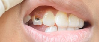
- The use of a caries marker for the frontal group of teeth is recommended with caution.
- Large amounts of dye may change the color of subsequent restorations if they are needed.
This type of caries detector is not used in the Implantmaster clinic. We use the VistaCam IX intraoral camera, which is the most advanced optical caries detector.
A familiar diagnosis
We are taught how to brush our teeth properly since kindergarten. However, more than 90% of people have faced and are still facing this problem.
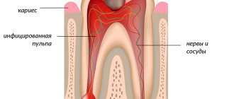
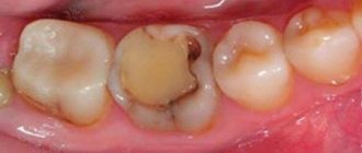
The disease develops asymptomatically at the initial stage, affecting only the superficial layers of the enamel, and gradually penetrates deeper, reaching the nerve endings and blood vessels. As the process progresses, severe dental caries appears, which causes toothache and dentin damage.
Important! This is the insidiousness of the disease - the patient comes to the clinic when the pain becomes unbearable - which means complications, pulpitis and removal of the nerve.
The cost of diagnosing teeth with a caries detector
When treating dental caries at the Implantmaster clinic, diagnosis with a caries detector is included in the cost of treatment.
Also, during comprehensive professional hygienic teeth cleaning, diagnosis with a caries detector is carried out free of charge .
Professional hygienic teeth cleaning under a microscope
9,600 ₽ -50% 4,800 ₽ + Dental diagnostics with caries detector
Until 01/31/2022 Submit a request
Treatment of caries by filling tooth surfaces
- Anesthesia
- Cavity preparation (preparation, medicinal treatment, application of a gasket)
- Filling with light-curing composite
- Grinding and polishing the filling
- X-ray images
Price : from 3,000 ₽ to 8,050 ₽, depends on the number of tooth surfaces affected.
Treatment of caries by placing a ceramic inlay “E-MAX” or “CEREC” or “EMPPRESS”
| from 20,000 to 28,000 ₽ |
Unsealing all tooth canals
| from 4,000 to 8,500 ₽ |
Filling all tooth canals
| from 6,500 to 11,500 ₽ |
| Removing a foreign body from a tooth canal (pins, fragments of instruments, etc.) | 3 000 ₽ |
| Application of medicinal paste into the tooth canals | 2 000 ₽ |
| Sealing tooth fissures | 2 000 ₽ |
| Treatment of caries without drilling the tooth using the “Icon” method | 3 000 ₽ |
| Complete restoration of a tooth with a light-curing composite (Tooth restoration) | 9 500 ₽ |
Author:
Caring for your teeth after crowns are installed
Dentures are immune to tooth decay, but plaque can accumulate on them. With insufficient oral hygiene, it causes an unpleasant odor and the formation of tartar. As a result, healthy teeth may begin to decay.
Standard care for crowns boils down to preventative brushing of teeth twice a day and visiting the dentist once every six months. It is recommended to use pastes with a low abrasiveness coefficient, and remove food particles between the teeth using an irrigator or dental floss. It is also advisable to rinse your mouth after every meal.
Sudden temperature changes lead to the formation of microcracks in solid materials. To ensure that your crowns last as long as possible, you should avoid eating foods and drinks that are too hot or cold.