April 5, 2020
Neoplasms (neoplasia) is the medical name for tumors, i.e., excessive growth of any tissue in the body. Tumors are the result of uncontrolled proliferation of cells that have not yet reached maturity and therefore have lost their ability to fully perform their functions.
Tumors can occur in internal organs and on the surface of the skin. Many people, not knowing what types of skin tumors there are, when any skin tumor appears, mistakenly believe that it is cancer. In fact, this is not always the case.
According to the main classification, skin tumors are divided into benign and malignant. There are also precancerous formations - borderline between the two main types. Each type has its own subtypes and characteristics, and correct diagnosis is needed to make an accurate diagnosis.
Benign skin tumors
In benign neoplasms, the ability to differentiate cells is usually not impaired. That is, they retain their original functions and are similar in structure to normal cells. Such cells also grow slowly and can put pressure on neighboring tissues, but never penetrate them.
Types of benign skin tumors:
- Atheroma is a tumor of the sebaceous gland that is formed due to its blockage. It most often occurs where there are most sebaceous glands: on the neck, back, head, and groin area.
- Hemangioma is a vascular tumor that is formed from cells of blood vessels. It has a color from red to bluish-black.
- Papilloma and warts. Formation in the form of a small nodule or papilla. The cause is human papillomavirus (HPV). They usually occur against a background of stress, decreased immunity, and autonomic disorders. Often appear in the armpits and groin area. Papillomas are also the most common neoplasms of the skin of the eyelids.
- Lymphangioma is a tumor from the walls of lymphatic vessels that forms inside the womb. Externally, these are small formations with a lumpy surface of a bluish or red-brown color.
- Lipoma is a tumor of the fatty layer (“wen”). It is located in the subcutaneous layer, most often in the upper back, shoulder girdle, and outer thighs. The tumor is painless, soft and mobile.
- Fibroma is a formation of connective tissue. More common in young and middle-aged women. They look like a new growth on the skin in the form of a ball protruding above its surface.
- Neurofibroma is a tumor of nerve sheath cells. Outwardly it looks like a dense tubercle measuring 0.1-2.3 cm.
A separate group of neoplasms includes nevi (moles). These are skin growths of different colors: brown, red, black, purple, etc. In most cases, nevus is a congenital malformation of the skin. But moles can appear throughout life, most often under the influence of sunlight. Nevi do not have a tendency to malignant degeneration, but in some cases this can occur due to damage or trauma to the skin on the mole.
Despite the absence of direct danger, all benign types of neoplasms on the skin of the face, arms, legs and other parts of the body require constant monitoring. It is necessary to ensure that tumors do not grow, increase in size, or change color. Otherwise, you need to consult a doctor.
Reasons for education
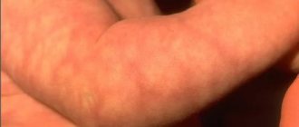
After conducting numerous studies, scientists have not been able to determine an exhaustive list of factors that provoke the development of pathology. However, some conditions and phenomena still occur most often when diagnosing this disease. For example, this is possible with a hereditary predisposition to fibroids on the gums (shown in the photo below), tongue, inside the cheeks and other areas of the mucous membranes. In this case, the disease develops in childhood in young patients from 1 to 10 years.
Often, in the presence of this diagnosis, a history of regular traumatic exposure is established. There may be constant biting of a certain area of soft tissue. They can also be injured by sharp edges of crowns, orthodontic structures, dentures, especially poorly fixed ones, and hard food.
Taking certain medications can also be a provoking factor. Some medications can cause the appearance of benign lumps in representatives of different age categories, not only in children and adolescents. Drugs that may cause fibromatosis after use:
- Cyclosporine. It is indicated to prevent organ rejection during transplantation.
- Valproate, used by epileptics.
- Verapamil and other calcium channel blockers.
- Estrogens of synthetic origin in oral contraceptives or other hormonal pills.
The culprits in the development of the disease may be inflammatory processes in the oral cavity. These include stomatitis, glossitis, periodontal disease, gingivitis, etc.
It is impossible to exclude a hereditary factor, but the risk of formations can be significantly reduced if you follow simple preventive rules:
- do not forget about hygiene;
- Carry out regular professional cleaning to remove thick plaque and tartar;
- protect soft tissues from traumatic effects (solid food, biting), and also avoid sudden temperature changes;
- treat dental diseases in a timely manner;
- consult with doctors before starting to take medications and avoid self-medication;
- visit the dentist for preventive maintenance at least once every six months.
Precancerous neoplasms (precancrosis)
Precancerous neoplasms are those that, under the influence of congenital or current causes, have acquired a tendency to malignant degeneration. As a rule, these are chronic conditions that are observed in a person for a long time.
Thus, precancerous tumors are dangerous neoplasms on the skin that can lead to the development of oncological processes. These include:
- Actinic keratoma is a keratosis in which dry crusts and scales appear on the skin of older people. When they peel off, slight bleeding may occur.
- Xeroderma pigmentosum is a hereditary tumor that develops due to increased sensitivity of the skin to ultraviolet radiation. Rarely encountered, these are pigmented spots that become warty growths.
- The cutaneous horn is a cone-shaped tumor that looks like a horn. Has a yellow or brown color. occurs in exposed areas of the body that are regularly subjected to friction or pressure. Typical for older people
- Bowen's disease is an intraepidermal cancer. Without treatment, it can transform into invasive skin cancer. In the early stage, Bowen's disease appears as a small reddish-brown spot measuring 2-50 mm. has a flaky surface and raised, uneven edges. After removing the scales, a weeping but not bleeding surface remains.
Methods for removing skin tags
There are several known ways to remove skin growths:
- Simple cutting with scissors or a scalpel;
- Removal with radio knife;
- Removal with a coagulator;
- Removal with liquid nitrogen;
- Laser removal;
- Plasma removal.
Rice. 5A. Stages of removing skin growths with a laser. Anesthesia
Rice. 5 B. Stages of removing skin growths with a laser. Laser vaporization
Rice. 5V. Stages of removing skin growths with a laser. Laser vaporization.
Rice. 5G. Stages of removing skin growths with a laser. Laser vaporization
Rice. 5D. Stages of removing skin growths with a laser. Final result
Each of them has the right to exist. However, since skin growths in most cases are removed solely for cosmetic reasons, removal methods should be chosen that will not subsequently leave a visible mark on the skin. These methods include: laser and plasma vaporization.
Barley
Styes are more common than chalazions. This type of lump on the lower or upper eyelid is caused by inflammation of the follicle (bulb) of the eyelash. This also clogs the sebaceous gland duct. Styes develop over several days or even hours and can occur in both adults and children. More often, the systematic appearance of barley is observed in people with weakened immune systems or who have changed their place of residence to an area with a more severe climate, as well as in people exposed to constant stress factors.
Based on their origin, there are two types of barley. Inflammation can be external (when the sebaceous gland suppurates) and internal (when the source of inflammation is located in the membolic gland).
The development of external styes is characterized by subjective sensations similar to a foreign body entering the eye. The initial stage may also be accompanied by stabbing pain. External stye visually manifests itself as redness and swelling of the eyelid. The internal one is usually not so noticeable, but it causes even more discomfort and pain.
Without treatment, barley develops within a few days into an abscess, which opens with the release of purulent contents. This brings relief, but an open wound is dangerous due to the possibility of re-infection.
It is better to start treating barley without waiting for the abscess to spontaneously break through. This allows you to get rid of the painful lump faster and with less risk of complications. If you still don’t have the courage or time to visit an ophthalmologist, you should remember that prolonged suppuration of the eyelid is very dangerous. If the stye does not open for more than two weeks, surgical treatment is necessary. An ophthalmic surgeon will remove the abscess under local anesthesia and give recommendations for further treatment of the eyelid. Most often, therapy for developing or already opened barley includes drops and ointments that contain antibiotics (albucid, gentamicin, erythromycin, tetracycline ointment).
Chalazion
Cones of this type are quite common. They develop from a sebaceous gland whose duct is blocked. This formation is also called a “grading lump” or “cold barley.” The continued production of sebaceous gland secretion leads to the accumulation of a viscous mass in the capsule, which stretches and thickens, taking the form of a dense lump. On palpation, the contents under the skin feel like a moving ball.
Cold barley develops at a slow pace, so it does not cause pain. Only a formed hard capsule can cause pain when squeezed. If a chalazion is not treated, it can develop into a cyst. As the lump develops, the risk of complications increases: inflammation, formation of a purulent fistula, granulation.
Video from our specialist about the disease and its treatment
There are cases where chalazion spontaneously resolved without medical intervention. However, most often this formation does not develop back and requires prompt and conservative help. Treatment for such a lump on the eyelid is prescribed by an ophthalmologist. If the chalazion is small and not old, you can limit yourself to UHF therapy, ointments and eye drops. More severe cases are treated by injecting corticosteroids into the capsule cavity. Local drugs (ofloxacin, dexamethasone, sodium sulfacyl, hadrocortisone, levofloxacin, tetracycline ointment) can also be used as an addition to the injection.
If drug therapy is ineffective, the doctor decides on surgical treatment. The operation to remove a chalazion is performed under local anesthesia and lasts no longer than 15 minutes.
What is the best way to treat?
A large number of papillomas certainly causes inconvenience and does not look aesthetically pleasing. However, this is not a reason to use old-fashioned methods for removal with thread, pliers or a blowtorch.
For single small (1x1 mm) formations, celandine juice
- a proven folk remedy.
When there are many papillomas or they are large, this method is not suitable
, as it can last for months and lead to severe inflammation.
Removal using radio wave surgery is much more convenient and effective
The operation does not require special preparation, is practically painless, and after it you can drive a car.
Once I had the opportunity to remove about 70 papillomas from one patient at once. It is difficult to convey all the joy of a person who got rid of them, whom they tormented for several years.
Treatment of oral fibroids
Surgery remains the most effective and most common therapeutic method. The seal is excised using a laser or radio waves. This procedure lasts about half an hour. If the tumor is very large, after its removal the wound is covered with a flap, which is formed by the doctor from the surrounding tissue.
When pathology is caused by taking certain medications, they should be completely eliminated and replaced with alternatives with similar properties. After discontinuation of drugs in such cases, the appearance of the mucous membranes is often restored without outside help, and the likelihood of relapse approaches zero. However, this does not apply to situations where the disease is advanced.
Surgery can also be avoided in case of traumatic effects of orthopedic structures. For example, when a crown, filling, or prosthesis puts pressure on the tissue. Elimination of the provoking factor often leads to a decrease or complete disappearance of a benign formation. Most likely, it will be necessary to dismantle the old structures and replace them with new ones.
On the Internet you can find stories of healing using home remedies. It is worth remembering that the disease cannot be treated with the help of folk recipes. Herbal decoctions and infusions, and other compositions are used only as an auxiliary element of complex therapy.