Changing the color of tooth enamel is an alarming signal that cannot be ignored. Dark spots can be associated not only with the consumption of coloring foods and drinks, but also with the development of various pathological processes. If the tooth has turned black at the root, you should contact your dentist as soon as possible. Do not wait for pain and other symptoms to appear, as they indicate extensive damage to hard tissues or inflammation of the dental nerve. It is easier to treat any disease in the early stages, so do not delay visiting a doctor.
Chromogenic components (food, drinks, other products)
The food we eat and other substances we use contain coloring agents (coffee, tea, cola, wine, tobacco, etc.) that can cause yellow, brown or bright orange spots to appear on the skin. teeth In most cases, the discoloration of teeth is generalized, which means that stains tend to form with equal success both on the entire surface of the tooth and in individual areas. If stains appear, it is better to make an appointment with an inexpensive dentist near your place of residence.
What is dental granuloma, the reasons for the formation
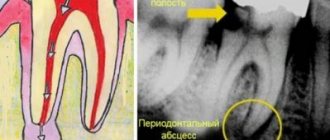
Dental granuloma is a pathological neoplasm of periodontal tissues affected by inflammation, appearing in the upper part of the tooth root. Externally, the granuloma looks like a dense small nodule (up to 0.5 cm in diameter). If you do not urgently treat a dental granuloma, it will continue to develop and deform into a radicular cyst - a capsule that is filled not only with dead tissue cells, but also with pus and bacteria.
Some people believe that a cyst and a dental granuloma are the same thing. But these are different diseases. The tooth cyst is larger in size and does not show any symptoms longer. As granuloma develops, the tooth will become very painful.
What causes dental granuloma? There are a number of reasons that contribute to the occurrence of pathological formation at the apex of the tooth root:
1. Granuloma under the tooth can occur as a complication of caries, pulpitis, periodontitis, the treatment of which was not carried out on time. It is important to understand that no dental disease goes away on its own! If you don’t treat your teeth, they will continue to decay, and the inflammatory process will spread to the internal tissues of the tooth and to its root!
2. Poor quality of dental treatment can lead to the appearance of a granuloma on the root of a tooth. If mistakes were made during the treatment of caries or other dental diseases, for example, the tooth canals were poorly cleaned and treated, then in the future there is a high risk of various complications, including the formation of granulomas on the root of the tooth.
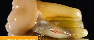
3. Severe tooth trauma, leading to infection penetration to the root.
These are the main reasons that can provoke the growth and development of granulomas on the roots of the tooth. If you do not want to face such an unpleasant disease, never delay dental treatment, which you can undergo without pain and stress in our dental clinic in Moscow - Firadent.
It is worth learning and remembering that granuloma can appear after tooth extraction. Most often this happens when the tooth was removed incorrectly or after the operation the patient did not follow the doctor’s recommendations on hygiene. Bacteria easily enter the open socket that remains after the tooth is extracted from the gums, and as a result of their active reproduction, an inflammatory process begins and a granuloma is formed.
Tooth decay
In the early stages of tooth decay, white spots or spots appear on the tooth enamel. The affected areas lose their natural shine and become dull. A thorough examination reveals the presence of damage to the enamel. If the process of tooth decay progresses, the affected area becomes brown or black, it is necessary to diagnose dental treatment. The damage is initially noticeable as a small dark spot or spots that gradually increase in size (over several months to several years) and often lead to actual tooth decay.
Why isn't every spot a black tooth decay?
There is no need to think that dentists are being cunning, wanting to deliberately turn an initial carious cavity into mid- or deep-stage caries.
After all, the more complex the problem, the more expensive its treatment. No doctor does this. It’s just that the identified violation has a completely different origin. In reality, dark spots can represent:
- Consequence of hypoplasia. This is a dental disorder in which, during the formation of a tooth, its crown part is underdeveloped. The disease is a consequence of metabolic disorders in the buds of teeth. It can also develop due to improper metabolism of proteins and minerals in the fetal body during its intrauterine development. Rickets, somatic pathologies, diseases of the gastrointestinal tract, severe infectious diseases - all these are also factors that provoke hypoplasia.
- The result of fluorosis. This disease occurs due to the constant and prolonged ingestion of large amounts of fluoride into the body. First, whitish spots form on the crowns, and then depressions resembling black caries. During examination, the doctor records destructive and erosive changes in the enamel.
- Erosion of teeth. Another type of non-carious lesion of tooth enamel. Defects occur on the outer layer of the crown and can even affect the dentin. Erosive defects are most often localized symmetrically. They provide an unsightly cosmetic effect and therefore require urgent correction.
In many non-carious pathologies, it is possible to improve the aesthetics of a smile through remineralization. The patient is prescribed vitamin and mineral complexes, calcium and phosphorus supplements, and local applications. Electrophoresis is used in many clinical situations.
Only with voluminous cavities reminiscent of black caries do they resort to filling material. If the damage is very severe (which happens when a person puts off visiting the dentist for a long time), even prosthetics may be required.
It is important to understand that caries is always a cavity of microbial origin. That is why it has to be drilled out to remove all damaged tissue and prevent further spread of pathogens. From this position, not every even point on the tooth requires the use of a drill.
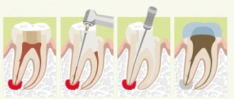
Conservative (medicinal) treatment of dental granuloma
This technique is used only for those cases in which the development of granuloma has not yet progressed far. The essence of conservative therapy for tooth root granuloma will be the intake and use of various drugs - antibiotics, antiseptics, drugs that have an anti-inflammatory effect. All medications are prescribed by a dentist and the treatment process takes place under the strict supervision of a specialist. This treatment option for granuloma is effective with early diagnosis of the disease and, if you strictly follow the doctor’s instructions, the granuloma will resolve within 1-2 weeks.
In some cases, conservative treatment of granuloma is complemented by root canal therapy. Working with tooth canals for root granuloma is indicated:
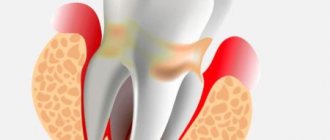
- For constant toothaches, the intensity of which does not subside from taking painkillers;
- Swelling of the gums, spreading to the cheek and jaw;
- Treatment of tooth canals is clearly indicated when large granulomas are detected - from five millimeters.
What to do if large or small spots appear on your teeth?
Any changes in the color or structure of tooth enamel are a reason to be wary and seek advice from a dentist. Only a doctor, after diagnosis, will be able to accurately answer the question: is a blackhead a symptom of caries? Or the darkening occurred for another reason.
If a stain on the front teeth occurs as a result of a bruise, the dentist will remove the affected tissue, process and fill the canals, and, if necessary, recommend installing veneers or endo-whitening (lightening the tooth from the inside).
If the cause of dark spots is poor hygiene or poor plaque removal, professional cleaning using ultrasound or laser will help solve the problem. These methods are good at removing plaque and tartar from the enamel surface and returning the teeth to their natural shade.
If food coloring is the cause of teeth staining, professional hygiene combined with a subsequent whitening procedure can help.
Unfortunately, dark spots on teeth are caries in the vast majority of cases, and the carious process at any stage requires immediate treatment.
Modern understanding of pathogenesis (mechanism of disease development)
In addition to Miller's theory, there are many other scientific explanations for the causes and mechanisms of caries development. The modern approach to this topic is based on three conditions, the coincidence of which leads to the development of carious tooth damage. This:
- Consumption of easily digestible (fast) carbohydrates. These include sugar, honey, starch, some cereals, sweet drinks, and confectionery. Products high in sucrose, glucose and lactose are especially harmful. They form a plaque on the enamel that interferes with the neutralizing effect of saliva.
- Growth of cariogenic microflora. Now there is no doubt that without pathogenic microorganisms, caries does not develop. In addition to the main “culprit” Str. mutans, cariogenic bacteria also include Str. Sanguis, Str. Salivarius.
- Low enamel resistance. The resistance of a tooth to damaging factors depends on the quantity and quality of the organic and inorganic components of the enamel.
Treatment methods for initial caries
In most cases, treatment of caries in the spot stage is carried out without drilling. For this purpose, in particular, remineralizing therapy or deep fluoridation of enamel can be used.
When the chewing surface of a molar or premolar is damaged, the doctor often suggests invasive fissure sealing, that is, sealing the enlarged natural grooves with a sealant.
If the stain has acquired a dark shade, it is sanded or prepared, then the defect is filled.
One of the modern technologies for treating caries in the spot stage is the Icon technique. When using it, the affected tooth surface is treated with a flowing material that penetrates the enamel, fills its pores and neutralizes bacteria.
Additional symptoms
Typically, the appearance of secondary caries is accompanied not only by darkening, but also by a number of other symptoms that are difficult to miss. What should you pay attention to if your teeth turn black? Let's consider additional symptoms:
- change in enamel color, appearance of a black dot on the surface, darkening and other visual signs of destruction;
- bad breath, especially in the morning after waking up, is the result of the development of pathogens and tissue destruction;
- mobility of the filling, which may eventually partially crumble or fall out completely;
- pain, especially if the nerves are not removed;
- redness of the gums caused by the presence of an inflammatory process.
Even if the blackening of the filling is not accompanied by the additional symptoms described above, it is recommended to undergo a preventive examination so that a specialist can identify the causes of the problem and determine the tactics for further action.
Diagnosis of dental discoloration in CELT
Before making a diagnosis, our dentists conduct comprehensive diagnostic tests, which include:
- Taking anamnesis and performing a dental examination;
- X-ray (allows you to identify violations of the internal structure);
- Rheodentography (determines the condition of the pulp and the depth of the lesion);
- Computed tomography (providing images of teeth in three-dimensional format);
- Microscopy (multiple magnification makes it possible to assess the condition of the enamel);
- Electroodontodiagnostics (determines the condition of the tissues located around the tooth root).
Pigmentation must be differentiated from necrosis and erosion of tooth enamel, as well as from Capdepont disease.
Types and classification
Depending on the location of the defect formation, caries in the spot stage is divided into types:
- Fissure. The discolored area is located on the chewing surface of a molar or premolar.
- Contact. Changes are detected on the lateral parts of the tooth, which are in contact with each other.
- Cervical. The stain is found in the area bordering the gum.
The form of the disease can also be different. Progressive caries develops quickly and after a short time passes into the superficial stage. The intermittent form is characterized by periodic exacerbations and attenuation of the pathological process. Suspended caries spots can persist for years without moving to the next stage.