General characteristics of the procedure
Relatively recently, it became possible to accurately diagnose pathologies of the dental system.
This can be done using a new orthopantomograph with cephalstat. This device has two examination programs: pediatric and adult. It is distinguished by low radiation exposure and high accuracy of the resulting images. An X-ray examination carried out using such a device is called “teleradiography” (what it is and how important such an examination is in orthodontics will be described below), and such a procedure today is an integral part of orthodontic treatment.
Types of installations for teleradiography
The procedure is carried out using devices with high-power X-ray tubes that work as quickly and accurately as possible, with minimal radiation exposure:
- digital cephalostat is the best option;
- computed tomograph;
- an orthopantomograph equipped with a craniostat, that is, a device for fixing the head in a stationary state.
What can be determined?
Using a teleroentgenogram (TRG), you can obtain an X-ray image of the skull
in a lateral projection (as well as in other projections, which are described below), as close as possible to the real dimensions. Such scale and realism of the image allow us to make the most accurate calculations of the facial skull parameters and create a detailed picture of the pathology.
Determine the size of the cranial bones, their structure, location, connection
Using TRG, the exact dimensions of the jaws are determined and their location in relation to each other is assessed. This is very important when diagnosing bite pathologies. Also, a teleroentgenogram allows you to determine the size of the cranial bones as a whole, find asymmetry, disruption of their connections or other deviations from the norm. Such information may be required not only as part of dental treatment, but also, for example, when examining the skull of a patient who was injured in an accident, fell from a height and received a traumatic brain injury.
Detect a lesion with pathological tissue growth, its location
TRG allows you to identify foci of pathological tissue growth. In other words, this procedure allows you to diagnose various tumors, cysts and other disorders in the structure of bone tissue and gums. Based on the TRG image, an analysis is made of the size of the neoplasms, their localization, the cause of their occurrence is determined, and so on.
Determine the direction of tooth displacement
Using TRG, the angle of inclination of the front upper and lower teeth is determined, which helps to identify anomalies in their location.
As a rule, such a pathology is initially visible during the initial examination, but x-rays help to find out exactly how much the current position of the teeth deviates from the norm. Without such an image, the orthodontist will not be able to provide high-quality treatment. For example, when there is a strong protrusion of the front teeth, it is important to understand how much they need to be moved back. To do this, you may need to remove a couple of teeth from the front row and then install braces. After about a year of wearing braces, the teeth will move back, taking the places of the removed ones. The result is an aesthetic smile, as well as a fully functioning dental system.
Another option for correcting this pathology is to correct the type of tooth with extensions.
There are other solutions to this problem, and the TWG will help determine the most optimal one.
Determine the location of tissues in relation to the skull
A teleradiogram makes it possible to identify and study anomalies in the location of bone tissue, correlating it with the size and anatomical features of the patient’s skull.
Find the factors that affect malocclusion
TRG allows us to identify specific factors that influenced the formation of malocclusion in a patient:
- direct genetic predisposition;
- for children: long-term attachment to the pacifier, habit of thumb sucking;
- mouth or mixed breathing (for example, due to adenoids, chronic rhinitis, etc.).
Identify abnormalities that are difficult to treat
When orthodontic treatment aimed at correcting the bite does not produce results, TRG is prescribed in order to identify abnormalities that contribute to this. Perhaps these are the consequences of a birth injury or a broken jaw, etc.
When is TRG performed?
Most often, a TRG image is needed at the planning stage of orthodontic treatment. Based on special calculations made, the doctor makes a decision on which treatment method is suitable in each specific case. In the image you can see many obvious and hidden pathologies, and based on the calculations, draw conclusions about the state of the dental system, determine the diagnosis and find out which treatment methods may be contraindicated in this case. TRG is often a mandatory and sometimes priority examination method for an orthodontic patient.
For a surgeon and traumatologist, such an examination can help detect trauma to the cranial bones and see the displacement of fragments due to the fact that the bones are visible in different projections. Timely identification of injury and its location can radically change treatment tactics and prevent complications.
Indications for use
Teleradiography in orthodontics, without exaggeration, is considered the most effective and informative diagnostic procedure and is prescribed for a number of indications.
When correcting the bite
Today, malocclusion can be completely eliminated if high-quality and effective orthodontic treatment is carried out. It, in turn, cannot be carried out without an x-ray, which will show how complex the pathology is, associated problems, and so on.
Making the correct diagnosis or clarifying it
It is sometimes difficult to understand the causes of toothache, incorrect tooth growth, soreness and bleeding gums during an initial examination of the oral cavity. TRH may be prescribed to clarify the diagnosis.
Planning and prognosis of treatment for various dental anomalies
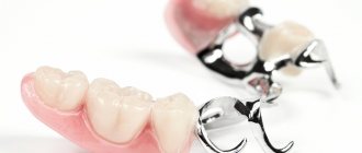
Before installing dentures, braces, implants, sinus lifting, tooth extraction and other dental operations, it is necessary to do a TRG to accurately determine the treatment plan.
Monitoring the ongoing processes of changes in periodontal tissues
Treatment of periodontal disease, pulpitis, periodontitis and other complex diseases of the oral cavity requires monitoring over time. In this case, an x-ray will show how effective the treatment is.
What difficulties does radiophobia create during bite correction?
Every sane person who decides to invest substantial funds in correcting a bite with braces wants to get an answer from the dentist to three main questions during preliminary consultations.
- What's wrong with my teeth?
- How can I improve the situation?
- How to save money on braces treatment?
At the same time, the most demanding clients often refuse an X-ray examination: “I will not be exposed to radiation again. It’s enough that they recently forced me to undergo fluorography.”
What should a potential patient of the orthodontist center understand? Locks glued to tooth enamel alone will not correct the bite. The treatment program is developed, activated and controlled by a person. This is a professionally trained specialist who, even before installing metal or aesthetic ceramic braces, must see the goal and imagine the path leading to it.
Teeth can be moved in different ways. The main task that the doctor solves is to choose:
- the correct direction of their movement;
- a device that can do this in the optimal way - effective and most comfortable for the patient.
For this, in-depth diagnostics are performed, and the provider of the most important diagnostic information is precisely the X-ray examination.
Types of teleroentgenogram
There are several types of teleradiography. The doctor’s task is to choose exactly the option (or several) that will help create the most complete picture of the pathology.
Frontal
Frontal TRG is considered the main diagnostic procedure and is prescribed before the start of orthopedic treatment. An overview image of the head is taken from the back and front and allows you to clearly see the existing inflammatory processes, facial asymmetry and fractures.
TRG in lateral projection
A lateral view of the TRG is performed to diagnose abnormal jaw development and prevent its consequences. In addition, a lateral teleroentgenogram provides the doctor with the opportunity to examine the jaw from the side and calculate the inclination of the lower teeth and those located in the upper row. This x-ray is often taken before a patient is assigned to wear braces.
Axial
Axial TRG (also called “mental”) is an additional diagnostic procedure along with lateral or frontal. Axial TRG is necessary to determine changes in the structure of the cheekbones, maxillary sinus and nasal cavity. Mainly used when implantation of the upper front teeth is necessary.
How is an x-ray performed?
- An x-ray of the knee joint taken in a standing position shows the narrowing of the joint space and the position of the knee joint.
- An x-ray of the knee at a 45-degree angle shows complete loss of the medial joint space, which may not be obvious when the x-ray is taken in a standing position. This type of imaging allows early degenerative changes in functional position to be detected.
- Lateral x-ray of the knee at a 60° angle shows Patellaalta. This position also allows one to see trochleadysplasia.
- X-ray of the knee at a 30° angle
- Stress radiography is sometimes used as a diagnostic tool to assess knee instability in all directions, but because it only provides two-dimensional (2D) and static images, this method of measuring dynamic knee functionality is limited.
- X-ray of passive stress.
Passive stress radiography offers objective, quantifiable, noninvasive, and retrievable data for the diagnosis and evaluation of knee ligament injuries.
- Dynamic stereoradiography and fluoroscopy can provide accurate measurements of knee joint kinematics, but their use is limited by high radiation exposure.
How is diagnosis carried out?
Typically, a TRG image is taken in a standing position (in special cases, when it is difficult for the patient to stand, a lying position is allowed).
The patient stands close to the device, the doctor helps him take the desired head position. To fix the head, special plastic supports are inserted into the ear canals. Before the image, the patient’s face along the midline of the soft tissue (including the soft palate) is lubricated with a barium suspension, this is necessary to create contrast in the image. The photo is taken in a few seconds. The procedure is painless and relatively safe.
Advantages of the technique
A teleroentgenogram (almost every dentist can tell you in more detail what it is) has some advantages over other diagnostic procedures:
- high information content of images (with the help of TRG, especially if it is done in several projections at once, it is possible to identify almost all pathologies of the dental system
); - instant results;
- safety and painlessness (can be done on children and pregnant women in the second trimester).
Contraindications
There are a number of contraindications for performing TRG of the skull in one of the possible projections (lateral or other).
Bleeding and/or pneumothorax
Conditions such as open bleeding and pneumothorax (accumulation of air or gases in the pleural cavity) require urgent medical attention, possibly surgery. Radiography
(in particular, the TRG method), if required, will only be done after solving the problems described above.
General serious condition of the patient
It is not recommended to disturb a patient who is in serious condition once again to perform an x-ray (in particular, for an x-ray of the skull). The condition can worsen even from moving along the hospital corridor, so it is better to either refuse an x-ray or have it done later, when the patient’s well-being improves.
Pregnancy (especially 1st and 3rd trimesters)
The first and third trimesters of pregnancy are contraindications for taking most medications, as well as performing many diagnostic procedures, especially x-rays. There is a possibility (albeit very small) that in the first trimester, when all the organs and systems of the fetus are developing, ionizing radiation can disrupt this process. The third trimester is dangerous because any disturbance in the ongoing development of the fetus (which theoretically could occur due to exposure to X-rays), excessive anxiety of the expectant mother or other negative factors can provoke premature birth.