Implantation today is one of the most popular procedures in dentistry. However, it is not available to everyone. In order for the implant to reliably take root, strengthen and withstand daily loads in the future, it needs a solid base, the role of which is played by its own bone tissue.
Sometimes, for various reasons, there is not enough natural bone tissue for implantation. In this case, osteoplasty helps to cope with the problem.
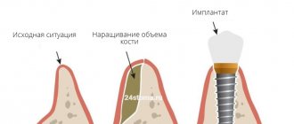
Bone augmentation in dentistry is a surgical procedure in which the missing volume of bone tissue is restored to the patient to install one or more artificial tooth roots.
Indications and contraindications for bone augmentation
The main indication for osteoplasty is the lack of native bone tissue for implantation.
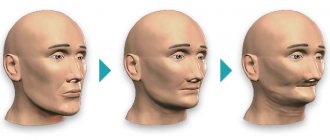
Bone augmentation in dentistry
Bone augmentation is also prescribed in the following cases:
- various types of jaw injuries;
- elimination of complications after tooth extraction, especially if the patient does not take proper care of the oral cavity;
- large areas of damage or atrophy of the alveolar ridge that have arisen against the background of infectious diseases or injuries.
Like any surgical intervention, osteoplasty has a number of contraindications:
- oncological diseases;
- diseases of the immune system;
- some diseases of the circulatory system (for example, bleeding disorders);
- allergy to anesthetics;
- epilepsy or other mental disorders;
- sinusitis or tonsillitis - surgery in this case is postponed until the patient has fully recovered.
Types of bone tissue augmentation
Before deciding how osteoplasty will be performed, it is necessary to undergo a thorough examination.
The choice of method is influenced by several parameters: the height and width of the patient’s bone, its structure, as well as the location of large blood vessels and the mandibular nerve.
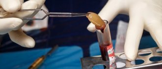
Autogenous transplantation
Autogenous transplantation is one of the most effective methods, since the donor material is the patient’s own bone tissue. The material is taken from the area of the chin or wisdom tooth and used to restore the atrophied part.
The operation is carried out in two stages, since additional surgical intervention is required to extract the donor material. This increases injury and the recovery process (up to one month), but the risk of rejection of structures in this method is minimal.
Xenogeneic and allogeneic transplantation
When using this method, additional surgical intervention is not required. The donor material is bone tissue from other patients or animal bones (mainly cows).
Before transplantation, such materials are carefully sterilized and processed to improve compatibility. During the procedure, only one incision is made, due to which the healing process is significantly reduced.
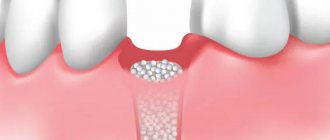
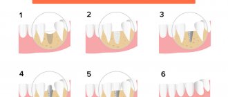
Alloplastic transplantation
This method is also called guided tissue regeneration (GBT). This technique uses two components to build bone: bone material and a barrier membrane made of an ultra-thin elastic film.
The membrane isolates the mucous membrane and prevents it from penetrating into cavities formed due to bone tissue atrophy. The membrane also protects against the effects of bacteria and other adverse factors.
The recovery process during alloplastic transplantation proceeds quickly; membranes made from modern materials dissolve over time or become a framework on which new bone tissue is formed. Since alloplasty does not seriously injure the jaw, it has recently been used much more often.
A significant disadvantage of this method is long-term rehabilitation. It often takes 4 to 6 months for the new bone to form and for the graft to heal.
Sinus lift
Sinus lifting
In modern dentistry, the most popular technique for building bone tissue is sinus lifting. This operation is performed in the lateral parts of the upper jaw, by raising the bottom of the maxillary sinus and increasing the alveolar ridge.
Sinus lifting comes in open and closed types.
The open technique is used in cases where it is necessary to build up a large volume of bone tissue, which entails serious surgical intervention. The healing process is very long and the installation of the implant is delayed for several months.
A closed sinus lift is used when the patient's bone tissue reaches a height of at least 7-8 millimeters. This procedure is not as traumatic as an open sinus lift. The implant is installed immediately after the bone has been grown in the upper jaw.
The advantages of these bone augmentation methods:
- the use of ultra-thin blades and drills makes this method less traumatic;
- relative speed of the operation - depending on the qualifications and experience of the doctor, the duration of the procedure ranges from 30 minutes, if there are no complications, to an hour, if there are complications;
- reliability of the result - in most cases, any sinus lift method gives a high percentage of a favorable prognosis.
One of the most advanced methods of bone augmentation in the upper jaw at the moment is the balloon sinus lift.
This technique allows you to simultaneously create a base and install an implant even on the thinnest bone. All manipulations are performed through a small hole for installing an artificial root. Through this hole, a special balloon is inserted into the maxillary sinus, which is filled with liquid and, increasing in volume, carefully separates the soft tissue from the bone. Then bone material is placed into the resulting cavity and an implant is installed.
One-stage protocol with immediate loading
A one-stage operation is performed if 3 or more teeth are missing. The protocol helps avoid tissue buildup. Single-component (not divided into an abutment and an intraosseous part) implants are installed in the deep layers of bone, which are not subject to resorption. For a larger area of contact between the bone and the titanium root, the product is fixed at an angle, identifying areas of bone tissue that are less susceptible to atrophy.
The implant is implanted using a minimally invasive method: it is screwed through a puncture in the gum. The top of the artificial root (abutment) rises above the gum - the dentures are installed immediately after implantation (on 2-3 days). After a year, the lightweight adaptive prosthesis is replaced with a permanent one.
1
Implantation
Implantation of 8-12 implants into deep bone layers (without bone grafting)
2 - 3 days
2
Prosthetics
Fixation of an adaptive prosthesis with a titanium arch and metal-plastic crowns
in a year
Permanent prosthetics
One-stage dental implantation for abnormal bone deficiency (less than 2.5 mm) is impossible without tissue augmentation. The operation is not performed for large maxillary sinuses, which cannot be avoided when installing implants at an angle.
Among the complexes of the one-step protocol:
- All-on-4.
The technique is used if the patient is missing all the teeth on the jaw. The dentition is restored using 4 implants (2 in the smile area and 2 on the sides). To avoid complications, “all-on-4” implantation for atrophy of the bone tissue of the upper jaw is carried out with caution, taking into account the close location of the maxillary sinuses. - All-on-6.
A modernized version of the All-on-4 method (implantation for severe bone deficiency). The increased number of supports (6 instead of 4) expands the scope of application of the method and allows for more stable fixation of the structure. - Basal implantation.
The method is used for acute bone loss. Suitable for missing three or more teeth in a row. For complete edentia, 8-12 implants are used on one row of teeth. They are fixed into the deep layers of the jaw bone.
All-on-4 and all-on-6 protocols involve the use of surgical templates to determine the location of the implant, without taking into account the condition of the bone. With basal implantation, templates can be dispensed with; this expands the scope of application of the technique. Implantologists at the ROOTT Clinic, using computed tomography, select suitable jaw sites for fixation with sufficient bone volume. If circumstances require it, a couple of additional implants are installed free of charge - if one of the titanium roots does not take root, there are enough implants left for prosthetics.
Possible complications after bone grafting
Even the use of modern technologies does not provide an absolute guarantee against the possible negative consequences of bone grafting.
After the procedure, some patients may experience the following problems:
- The appearance of edema within several days is a natural protective reaction of the body to surgical intervention.
- Bleeding is normal immediately after surgery. If it does not stop within several days, the cause may be loose sutures or medical error. In this case, it is definitely recommended to consult a doctor.
- The appearance of pain in the cervical spine is the result of prolonged anesthesia. It is enough to stretch your neck and avoid heavy physical exertion.
- A feeling of numbness in the jaw area is normal in the first 2-3 days. If numbness persists for a longer period, nerve damage may have occurred. You should definitely consult a doctor.
If the discomfort does not go away for a long time (more than 72 hours), this may indicate the first sign of rejection, it is recommended to immediately go to the hospital, preferably to the doctor who performed the operation.
Dentine
The bone substance of the tooth is its main part. Dentin performs an important function as it protects the pulp from external irritants. Having a loose structure, the bone substance of the tooth ranks second in strength in the body after enamel. The composition of this tissue is 2/3 inorganic substances, 1/5 collagen and 10% water. If you look at dentin under a microscope, you can see that it is a substance that is unevenly covered with calcareous deposits. A huge number of dentinal tubules filled with nerve endings of the pulp pass through it. There are 3 types of bone substance of the tooth: 1. Primary dentin, the formation of which begins even before the first eruption of the tooth. 2. Secondary or physiological, it is formed after the appearance of the tooth. It is characterized by a chaotic arrangement of dentinal tubes and fibers, and their smaller number. 3. Tertiary or, as it is also called, replacement, it is formed as a result of tissue irritation. It is characterized by an uneven appearance with slight mineralization. Dentinal tubules are most often absent in this type. The formation of dentin is individual and depends on numerous factors. This process can be influenced, for example, by tooth wear or other defects, as a result of which dentin replacement occurs at different rates of intensity.
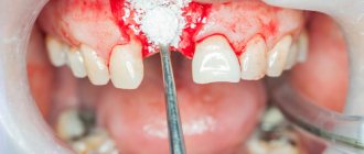
Rehabilitation after osteoplasty
After the bone restoration operation has been performed, the patient begins a rehabilitation period, which can last from 2 to 6 months depending on the chosen method of osteoplasty, as well as on the individual characteristics of the body.
During this period, it is important to strictly follow the doctor’s recommendations:
- Chew food on the non-surgical side. If both sides were operated on, then chew alternately.
- After eating, be sure to rinse your mouth with an antiseptic. Minimum 4-5 days.
- For a week, adhere to a gentle diet - exclude the consumption of any thermal and food irritants (salty, sour, spicy and hot foods).
- Refrain from smoking and drinking alcohol - this can provoke bone rejection.
- In the first week, avoid air travel so as not to provoke surges in blood pressure, as the stitches may come apart.
- Eliminate the possibility of heavy physical activity, avoid visiting saunas and baths.