According to statistics, the 6th tooth implant is installed more often than others, which is due to the characteristics of this first molar. Sixes are the first of all permanent teeth to erupt, they are used more, and therefore deteriorate faster and more often than others. The procedure for implantation above and below differs in the choice of implant and timing of prosthetics. Implantation of a six on the upper jaw takes longer - 4-6 months, on the lower jaw - 2-4 months. Osseointegration occurs with differences due to the structural features of the jaws and different bone densities. The total duration of treatment depends on the volume of bone tissue; if there is a deficiency, bone grafting or sinus lifting is performed first.
Why do sixth teeth deteriorate more often than others?
The sixth teeth (first molars according to dental classification) are molars. The rudiments are laid at the 5th month of fetal development, and at the 9th month their mineralization occurs, which continues throughout the first month after the birth of the child. Their final formation with enamel occurs only by the age of 2-3 years. Therefore, the condition of the root and crown depends on the course of pregnancy and childbirth.
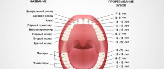
Sixes do not erupt in early childhood, since the function of chewing is not yet needed. When they erupt, they do not have predecessors in the form of milk teeth, so they essentially grow from scratch. Of the permanent molars, they begin to erupt first at 5-6 years, so they last longer than others. Frequent diseases, removals and the need for implants are associated with this.
Crown for a chewing tooth
The main purpose of a crown for the sixth tooth is to restore chewing function. The crown must be made of durable material to withstand heavy loads. Let's look at the types of crowns that are installed on chewing teeth.
Metal-ceramic crowns. Due to the durable metal frame and external ceramic lining, the tooth is firmly held in the oral cavity. But there are also disadvantages: over time, the metal frame begins to show through the gums. The crown itself does not have light transmission properties and can stand out against the background of natural teeth.
Zirconium dioxide crowns have all the properties of natural teeth. The main feature is the ability to transmit light. This makes them indistinguishable from a real tooth. These crowns are considered the best today. They last a long time and do not cause allergic reactions.
Metal crowns. The most budget option, but very unaesthetic. When you smile, others can easily notice your artificial tooth. In addition, metal crowns are gradually becoming a thing of the past, giving way to more modern materials.
Only a doctor can give an exact answer to the question of which crown to put on a chewing tooth after a detailed study of the clinical case.
Is it worth getting an implant?
The absence of the sixth tooth threatens:
- displacement of neighboring units;
- advancement of antagonists on the opposite jaw.
Neighboring and antagonist teeth will slowly shift, which threatens to expose the roots, the appearance of wedge-shaped defects at the gum itself, exposure of the neck, which is sensitive to changes in temperature of water and food, and pain. In addition, there will be a change in the bite, since the teeth will be out of place. Therefore, it is necessary to place an implant to maintain healthy teeth and correct bite.
Haderup system
Some believe that this is the Zsigmondy-Palmer system, which has been improved. It involves the use of numeric symbols from one to eight. The numbering goes from the midline of the jaw to the edge. As additional signs there are plus and minus to indicate the top or bottom row.
Baby teeth are numbered from one to five. Each number must be preceded by a zero. This system has a number of shortcomings. For example, one of them is to indicate the location of the dental element on the jaw.
Zsigmondy-Palmer system
The scheme is not very popular in the world because it has errors. The exception is Great Britain. When created, the idea of Adolf Zsigmondy (a dentist from Hungary) was taken as a basis. It was he who came up with the Zsigmondy cross, that is, dividing the dentition into quadrants.
Each dental element has a specific serial number. The calculation starts from the midline. However, there are no exact indications of the location. Permanent teeth are marked with Arabic numerals - from 1 to 8, and milk teeth - with Roman numerals from 1 to 5. Subsequently, the American dentist Palmer made additions to the system. He changed Roman numerals to Latin letters.
The scheme is mainly used in orthodontics and maxillofacial surgery. For their specific work, it is most suitable.
Operation stages
The six implant is installed in the classical way, as for all other lost teeth. Includes stages:
Preparation
Identification of contraindications, assessment of bone tissue and study of its size and density. Sanitation of the oral cavity, removal of plaque, treatment of gums and adjacent teeth. The stage lasts from several days to a month.
Bone tissue augmentation
If necessary, it is carried out as a preliminary stage in case of lack of bone tissue. The operation lasts an hour. After the planted material has fused with the bone (4-5 months), the artificial roots themselves are installed.
Installation of implants
The gum is peeled off, a bed is formed in the bone, an implant is placed, a plug is fixed, and sutures are applied. On the lower jaw, the implant takes 2-4 months to take root, on the upper jaw - 4-6 months.
Temporary prosthetics
At the patient’s request, lightweight plastic removable Butterfly immediate dentures are installed, which are attached to adjacent teeth.
Permanent prosthetics
The implant is opened, the plug is removed, the gum former is fixed, and after 10-14 days the abutment is installed. Impressions are taken and crowns are installed.
The sixth tooth implant can be placed immediately after removal into the resulting socket, but with a sufficient amount of bone tissue and under the following conditions:
- a pre-planned operation with the possibility of conducting diagnostics and assessing the immediate installation of an implant;
- atraumatic removal without damaging the alveolar process;
- absence of inflammatory and purulent processes at the root.
Features of the structure of teeth, their roots and canals
There are no two identical root dental systems, which is explained by the purely individual structure of a person’s teeth. In addition, the root system of incisors, canines and molars is arranged in accordance with their purpose:
- Ones and twos (incisors) are needed for biting food.
- Fours and fives (premolars) perform the initial chewing function.
- Sixes and sevens completely grind food.
Based on this, it becomes clear that the seventh tooth requires more nutrients than the fifth. It must be strong and hardy, therefore it has a more developed channel system. Despite the fact that the 6th tooth in the lower jaw performs the same functions as the seventh, it usually has fewer canals. This is due to the fact that there is less chewing load on it.
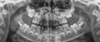
For a detailed study of the structure of the dentofacial apparatus of a particular patient, radiographic examination is used.
Tooth structure
Each dental unit consists of:
- crowns - the area above the gum;
- neck - the area between the crown and the root;
- root - the area under the gum.
Inside the crown is the pulp, which passes into the root canals. At the end of the root there is a small apical opening through which blood vessels and nerve endings pass, starting from the main neurovascular bundle and ending in the pulp.
When a person’s pulp becomes inflamed, not only it, but also all root canals need to be cleaned of infected tissues, since they are “communicating vessels.” If even one canal is left uncleaned, pathogenic microorganisms will continue to develop inside the dental unit, which will lead to its removal. That is why the doctor must know the exact number of canals in the tooth.
How many nerves are there in a human tooth?
Thanks to the nerve, the tooth can respond to external stimuli. After removing the pulp and filling the canals, the dental unit loses sensitivity, as it is deprived of a nerve. But due to the removal of blood vessels, problems begin with its blood supply and mineralization. The crown becomes less durable and more prone to various chips and breaks. The enamel quickly darkens, and it cannot be properly bleached even with strong chemicals.
Before removing the pulp, the patient is sent for an x-ray to find out how many canals are in the operated tooth: a person has only one dental nerve in a tooth, but there may be several canals
. This preparation allows for depulpation to be carried out competently and quickly.
Types of Root Canals
There are several options for the structure of dental canals:
- in the root there is one canal passage, which corresponds to one apical foramen;
- in the root there are several canal branches that connect in the area of a single apical foramen;
- two different branched passages have one mouth and two apical openings;
- canal cavities in one root merge and diverge several times;
- three root canal passages emerge from the same orifice, but have 3 different apical openings.
There can be as many channels as there are roots, but often their number differs. Several types of canals may be present in one molar and premolar.
How many canals in a person’s teeth - table
According to statistics, the number of channels depends on the depth of the tooth.
: The deeper it is located in the jaw, the more canals it has. This is due to the increased load on the molars located at the base of the dentition.
Typically, teeth in the upper jaw have more canals. But this pattern is not observed in all patients.
The table below presents average statistical data on how many canals are in a person’s teeth above and below.
| Dental unit | Number of channel passages | |||
| Fangs | Upper | 1 | ||
| Lower | 2 | |||
| Incisors | Upper | 1 | ||
| Lower | Central | in most cases 1, less often 2 | ||
| Lateral | 1 or 2 (about the same probability) | |||
| Premolars | Upper | First | most often 2, but sometimes first premolars with 1 or 3 canals are found | |
| Second | in most cases 2, sometimes 1 or 3 | |||
| Lower | First | 1 or 2 | ||
| Second | 1 | |||
| Molars | Upper | First | 3 or 4 with equal probability | |
| Second | in most cases 3, sometimes 4 | |||
| Third | around 5 | |||
| Lower | First | most often 3, sometimes 2 or 4 | ||
| Second | usually 3, but there are roots with 4 channels | |||
| Third | no more than 3 | |||
Number of canals in teeth in the lower jaw
The teeth on the lower and upper jaws are significantly different from each other. This is partly due to the uneven load and different functions. Typically, teeth in the lower jaw have fewer canals. But each specific case requires detailed study. Therefore, the dentist first sends the patient for an x-ray, and only then proceeds to open the crown and treat pulpitis.
It is impossible to start treating caries and pulpitis based only on encyclopedic information, because:
- The 6th tooth of the lower jaw can have any number of canals - from 2 to 4;
- in the 5th tooth below there is usually only 1 canal, but in approximately 10% of patients there are quints with 2 canals;
- In the 4th tooth there is usually only 1 canal, but in about a third of cases there are 2.
The eighth tooth on the lower jaw is the most “unpredictable”. Exactly how many canals are in the wisdom tooth located below can only be determined using x-rays. Officially, there are no more than 3 of them, but during the treatment of caries, additional cavities usually open. It is precisely because of its incomprehensible structure and inconvenient location that the figure eight is most often removed.
It is impossible to treat a dental unit without studying the structure of its root and canal system. This can only aggravate the pathology and lead to complications.
Number of canals in teeth in the upper jaw
The root system of the teeth of the upper jaw is more complex and branched. This explains the longer treatment of upper molars and the frequency of repeat visits due to incompletely sanitized dental cavities.
Features in the structure of the canal system of teeth in the upper jaw:
- The 6th tooth of the upper jaw is most often three-channel. But sometimes there are also four-channel first molars.
- The fourth and fifth teeth from above are most often two-canal, but sometimes single-canal and three-canal premolars are found.
- The 4th upper tooth usually has 2 canals, but sometimes there are premolars with 1 or 3 canals.
The “wise” eight on the upper jaw is a four-channel tooth. Third molars with 5 canals are extremely rare. However, in dentistry, even cases of the presence of eight-channel wisdom teeth located at the top have been recorded.
Features of implantation of the 6th lower tooth
The six of the lower jaw bears more loads during chewing than the upper jaw. Therefore, implants are selected taking this fact into account - wide and of sufficient length. The nuances of implantation from below:
- easier to carry out than on the top;
- implants take root faster - in 2-4 months, since the jawbone is denser and anatomically higher;
- but there is a risk of damage to the trigeminal nerve if the protocol is not followed, which is complicated by numbness of the chin and lips.
If the bone is small in height, then a preliminary operation is performed to increase it. The gum is peeled off, bone material is added, and a resorbable membrane is applied to secure it. Recovery takes up to six months.
Without preliminary bone grafting, surgery is possible if the atrophy is moderate. In this case, our Center uses bone growth stimulants. They are fixed to the neck of the artificial root during its installation and trigger the processes of bone tissue regeneration. In this case, implant healing and bone tissue growth occur simultaneously.
What is a human tooth made of?
The visible part or crown is covered with a layer of enamel - the strongest tissue in the human body. Beneath it is dentin, and then the walls of the pulp chamber. The pulp (nerve) is a neurovascular bundle that smoothly passes into the root canals. At the very end of the root there is a small apical opening - nerve endings, vessels and capillaries pass through it.
The photo shows the structure of the tooth
Thus, if for some reason the pulp becomes inflamed, the pathological process spreads very quickly through the canals. Essentially, these are communicating vessels, so if one of the passages is missed during treatment and filling, the inflammatory process will continue to develop and ultimately lead to the need to completely remove the diseased element.
Price
Our Center has a case payment system, which means that the case includes all materials and necessary manipulations.
The cost of implant installation includes:
- implant and superstructures;
- work of an implantologist;
- anesthesia;
- basic bone building complex;
- primary and repeat CT.
The price of implants varies depending on the type of bone. Nobel Biocare PMC (cheaper) is intended for weak tissue, and Nobel Biocare Conical Parallel CC (more expensive) is intended for dense tissue.
The cost of the crown includes:
- production of a prosthesis by a technician;
- taking impressions;
- installation of a crown.
Tooth extraction (for simultaneous implantation), bone grafting or sinus lifting are paid separately. Prices for services can be found here.
How to care for a crown
The service life of crowns varies from 5 to 15 years, depending on the material and compliance with hygiene rules. Despite the fact that the crown is made of artificial material, plaque also accumulates on it. You should take care of it the same way you would take care of your teeth. Particular attention should be paid to the area at the edge of the gum. Dental plaque in these places can cause inflammation and other problems in the body.
Basic recommendations for crown care
- Brushing teeth 2 times a day
- Use of additional hygiene products: brush, dental floss, irrigator.
- Professional teeth cleaning every six months. If the dentist indicates other dates, follow his recommendations.
- Preventative visits to the dentist every six months.
- Monitor the condition of the joints between the tooth and the crown.
- Do not eat cold and hot food at the same time.
If you experience crown mobility, an unpleasant taste, or any other discomfort in the oral cavity, you should make an appointment with your doctor. Otherwise, it can lead to serious complications. You can get to our doctors by calling: 220-86-30
Guarantees
In our Center, implants are installed with a lifetime warranty from the manufacturer - Nobel Biocare. We provide guarantees:
- lifetime for the installation of implants;
- 1 year for crowns.
The warranty program includes a complex for the implant, surgery, bone reconstruction and prosthetics.
The guarantee is valid provided that the patient follows the doctor’s recommendations, care rules and regularly visits the dentist.
Teeth numbering systems, what are they?
Today there is not one, but several schemes at once. Each of them has advantages and disadvantages. Let's take a closer look.
Universal
It was originally adopted by the American Dental Association. Each tooth corresponds to a specific digital designation - from 1 to 32. The countdown starts from the upper jaw (right) strictly clockwise. To number primary dental elements in children, they use not only specific numbers, but also letters of the Latin alphabet.
Two-digit Viola system
It is one of the most popular due to its brevity. Each jaw has a pair of segments. There are four of them in total. For an adult, the numbering is calculated in numbers from one to four, for a child - from five to eight. Each dental element is assigned a two-digit number. The first one means the segment, and the second one means the sequence number. The Viola system is currently in active use, due to its relative simplicity. It allows you to easily transfer information from the dentist to the patient.
Alternatives
An alternative method for restoring sixes is a bridge prosthesis. This is a structure consisting of crowns tightly connected to each other. To restore one tooth, the bridge consists of 3 crowns. The two outer ones are fixed on the teeth on both sides of the defect. The supporting teeth are ground down to form the internal cavity of the outer crowns of the prosthesis. The middle crown is hinged and imitates the lost six.
This option is cheaper than implantation, but has disadvantages - grinding the enamel of living teeth leads to a reduction in their service life. Another important point is that the bone under the hinged crown does not receive stress during chewing, so bone tissue atrophy is inevitable.
Root canal treatment
The treatment plan for dental canals consists of several stages:
- First, access to the problem area is freed: using a special dental instrument, the filling or the area of the crown damaged by caries is removed.
- Then the contents of the pulp are removed, and the canals are cleaned mechanically using antiseptic drugs.
- After this, the root is prepared for filling. At this stage, the dentist can form the correct conical shape of the canal passage.
- Then the canals are carefully sealed. If baby teeth are treated, the dentist uses a special filling paste, which gradually dissolves as the root dissolves.
- After this, a filling is placed on the crown.
This treatment regimen is standard and does not depend on exactly how many canals there are in the diseased tooth. The main thing is that all dental canals are cleaned, treated with an antiseptic and carefully closed. If treated incorrectly, it may be necessary to remove the tooth and visit an oral surgeon.
Teeth can be single-channel, two-channel, three-channel and even eight-channel. If one of the ducts becomes inflamed, it is necessary to clean and seal not only it, but also all other canals, since the infection could penetrate into them.
Teeth numbering for children
In young children, baby teeth differ in location. This is due to the fact that there are fewer of them than in adults (only 20 pieces). Over time, they will be replaced and supplemented by indigenous ones. In the last century, experts developed a numbering scheme specifically for baby teeth. At its core, it is an analogue of the adult Viola system.
Teeth numbering is a very necessary thing in dental practice, which greatly simplifies the work of specialists. They spend much less time and quickly fill out the patient's medical record.
How many canals does a wisdom tooth have?
How many wisdom teeth can there be? This is a difficult question, because this organ has a very unusual structure. If it is located at the top, then it can have four and sometimes even five channels. If this tooth is in the lower row, then it usually has no more than 3 indentations. In most cases, when teething and already at the moment of full growth, the figure eight causes unpleasant sensations and severe discomfort. To clean it, it is recommended to use a special brush, which is designed for hard-to-reach places. Typically, wisdom teeth have narrow sockets that have irregular shapes. This property causes great difficulties when performing medical procedures. Often, if improper eruption or other pathological processes occur, the figure eight is completely removed.
The wisdom tooth is the last one to erupt; it seems to fight for space in the jaw, often shifting the dentition and causing discomfort. The roots of the tooth have a swirling, intertwined shape, so the root canals of the tooth cannot always be treated.
How to remove a rotten tooth root while preserving the crown
A tooth in which only the root remains is not always pulled out entirely. For example, if an inflammatory process develops at the root apex, but the tooth itself can still be saved, resection of the root apex is performed - partial removal.
The procedure is carried out after filling the canals, under local anesthesia. The operation is simple and lasts no more than half an hour. Its main stages:
- Anamnesis collection.
- Preparation of the surgical field.
- Anesthesia.
- Cutting the gum to access the root.
- Delamination of soft tissues.
- Sawing out a “window” in the bone.
- Cutting off the inflamed area of the root with a granuloma or cyst.
- Placing drugs into the cavity that stimulate bone growth.
- Stitching.
Indications and contraindications for molar tooth extraction
Reasons for removing a molar tooth may be:
- purulent periostitis;
- abscess;
- phlegmon;
- large tumor in the tooth area;
- purulent periodontal disease;
- the presence of a cyst in the area of the diseased tooth;
- exposed pulp;
- longitudinal tooth fracture;
- critical tooth decay;
- critical tooth curvature;
- disease of the dental bone tissue;
- sinusitis.
The following reasons are contraindications to the removal of molars:
- acute heart diseases;
- acute viral diseases;
- flu;
- angina;
- renal failure;
- pancreatitis;
- hepatitis;
- acute diseases of the nervous system;
- oncological diseases
- initial and final stages of pregnancy;
- stomatitis;
- gingivitis
- general degeneration of the body;
- alcohol poisoning.


