Author of the article:
Soldatova Lyudmila Nikolaevna
Candidate of Medical Sciences, Professor of the Department of Clinical Dentistry of the St. Petersburg Medical and Social Institute, Chief Physician of the Alfa-Dent Dental Clinic, St. Petersburg
Not only the health of teeth and gums, but also the functioning of the digestive, immune and other systems and organs depends on the state of the oral microflora. Let's figure out what this part of the body is, what affects the conditions of the oral cavity and how to quickly restore the normal functioning of the microflora.
What is the microflora of the oral cavity?
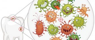
Surprisingly, approximately 160 species of microorganisms live in the mouth of a healthy person. You've probably heard that “the mouth is the dirtiest place in the body.” This statement is partly true: the oral cavity is one of the most populated parts of the human body.
Microorganisms enter the oral cavity with food and water, as well as from the air. It is in the mouth that the most favorable conditions for the development of bacteria are observed. This part of the body always has uniform humidity and temperature (approximately 37 °C). The abundance of nutrients, sufficient oxygen content, the presence of folds in the oral cavity, interdental spaces and gum pockets, and slightly alkaline pH provoke the proliferation of various bacteria.
Microorganisms are unevenly distributed in the oral cavity. Their maximum number is observed on the surface of the teeth and on the back of the tongue. One gram of dental plaque contains approximately 300 billion microbes, and saliva contains approximately 900 million per 1 milliliter.
- 30-60% of the microflora are facultative and obligate anaerobic streptococci;
- part is occupied by Veillonella, coccobacteria, which ferment acetic, pyruvic and lactic acids to water and carbon dioxide. It is Veillonella that neutralizes acidic foods, so many dentists consider them as destroyers of cariogenic bacteria;
- Bacteria of the genera Propionibacterium, Corynebacterium and Eubacterium are also necessarily present in the mouth, which actively produce molecular oxygen, synthesize vitamin K and promote the development of obligate anaerobes. Some types of such bacteria provoke purulent inflammation.
- Lactobacilli are strict anaerobes. There are more than 10 types of similar bacteria in the mouth that form a biofilm in the cavity. The vital activity of these microorganisms creates a favorable environment for the development of normal microflora. Lactobacilli ferment carbohydrates to form lactic acid, lower the pH, and most importantly, prevent the development of pathogenic, putrefactive and gas-forming microflora.
- Rod-shaped lactobacilli in a certain amount are, like streptococci, producers of lactic acid.
- Bifidobacteria are necessary to ferment various carbohydrates, as well as produce B vitamins and antimicrobial substances that inhibit the growth of pathogenic microorganisms. Moreover, bifidobacteria are a bunch of epithelial cell receptors: they form a film that prevents the colonization of pathogenic bacteria.
It would take a very long time to list all the types of bacteria that populate the microflora of the oral cavity. It is important to understand that each person is unique, and to assess the “normality” of the microflora, you need to know the characteristics of a particular organism.
For example, in one case, a large number of lactobacilli in the oral cavity will preserve teeth, and in another, the formation of a large amount of lactic acid during their vital activity will retard the growth of other important microorganisms. The number of staphylococci, dysentery and typhoid bacilli decreases, carious processes become more active, and the microflora of the oral cavity will have to be restored.
References
- Khaitovich, A.B., Voevodkina, A.Yu. Microbiome and its impact on human health. Crimean Journal of Experimental and Clinical Medicine, 2022. - No. 1. - P. 61-70.
- Krasnova, E.I., Khokhlova, N.I., Evstropov, A.N. and others. Streptococcal infection of the oropharynx: approaches to diagnosis and treatment. Infectious diseases, 2015. - No. 4. - P. 30-36.
- Li, N., Ma, W., Pang, M. et al. The commensal microbiota and viral infection: a comprehensive review. Frontiers in immunology, 2019. - Vol. 10.
Disturbance of oral microflora: causes
Oral dysbiosis can be caused by a variety of diseases and problems. Violation of opportunistic microflora of the oral cavity most often causes problems such as:
- Diseases of the gastrointestinal tract. Malfunctions of the digestive organs lead to a slowdown in metabolic processes in the body. The absorption of vitamins and nutrients deteriorates, the balance of the intestinal bacterial environment is disrupted, which provokes problems in other organs and systems.
- Decreased immunity. If the body's resistance deteriorates, the oral cavity automatically becomes more vulnerable to pathogenic microflora.
- Chronic diseases. Often, small caries or stomatitis, if left untreated, can spread from the source of inflammation to the entire oral cavity.
- Bad habits, such as systematic drinking of alcohol and smoking, inevitably affect the quality of the salivary glands. Prolonged drying out or too much moisture in the oral cavity has a detrimental effect on the composition of the microflora.
- Poor nutrition and lack of vitamins worsens the quality of saliva and makes the microflora of the oral cavity more vulnerable.
- Taking antibiotics and certain medications, such as hormones;
- Wearing dentures.
In what cases is research usually prescribed?
Important: the test is prescribed only to women of reproductive age (the test is not suitable for women in menopause, since the interpretation of normal values is based on the ratio of microorganisms characteristic of women of childbearing age.)
- for complaints of vaginal discharge
- in the presence of inflammatory diseases of the pelvic organs;
- for infertility and miscarriage
- when planning invasive manipulations on the pelvic organs;
- when planning pregnancy;
- in case of unprotected sexual contact with a new or non-regular partner;
Stages of dysbiosis
Depending on the degree of development of the disease, dentists distinguish four stages of dysbiosis:
- Latent. The first, latent stage is characterized by subtle changes in the number of microorganisms of one strain. The patient feels well and does not experience any symptoms of inflammation.
- Subcompensated. The number of lactobacilli decreases, the disease has a blurred picture. The patient may feel discomfort in the oral cavity, but does not always understand that this is dysbacteriosis.
- Pathogenic. Lactobacilli are observed in minimal quantities in the oral cavity. The oral cavity begins to be populated by a facultative pathogenic environment.
- Decompensated. In addition to severe inflammation in the mouth, uncontrolled growth of yeast-like fungi occurs. The functioning of the salivary glands is disrupted, and an unpleasant taste and burning sensation occurs in the mouth.
Advanced forms of dysbiosis are characterized by symptoms such as:
- inflammation of the gums and mucous membranes;
- plaque on the teeth and surface of the tongue;
- bleeding gums;
- ulcers and blisters on mucous membranes;
- increased body temperature;
- swelling and soreness of the tongue;
- dry skin, sticking in the corners of the mouth.
Gynecologists
- Levshina Natalya Vasilievna
Experience: 20 yearsObstetrician-gynecologist of the highest qualification category
Rating: 5/5 — 1 votes
Make an appointment
- Mikhailidi Irina Arkhimedovna
Experience: 11 years
Head of the gynecological department/obstetrician-gynecologist, ultrasound diagnostics doctor, Ph.D.
Rating: 5/5 — 1 votes
Make an appointment
- Paneva Svetlana Alexandrovna
Experience: 40 years
Obstetrician-gynecologist of the highest qualification category, Honored Doctor of Russia
Rating: 5/5 — 1 votes
Make an appointment
A reliable and accessible method for diagnosing inflammatory diseases of the urogenital area is to perform a “Gynecological smear for flora” analysis. This is a fairly common test in gynecology, which doctors recommend taking if you suspect inflammatory diseases of the genital organs and during preventive check-ups.
Methods for restoring oral microflora
Treatment of dysbacteriosis depends, first of all, on the nature of the pathogen, which is determined on the basis of dental examination.
Unfortunately, making a diagnosis of dysbacteriosis is often difficult, since at the initial stage the disease does not manifest itself in any way. At the slightest suspicion of a disease, the dentist refers the patient to a smear from the surface of the mucous membranes, blood and urine tests.
Depending on the diagnosed cause of dysbiosis, the dentist may prescribe the following methods of treating the disease:
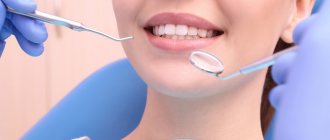
- Sanitation of the oral cavity. The doctor removes tartar and fills all teeth affected by caries, and, if possible, treats the gums and mucous membranes.
- Treating the oral cavity with antiseptics to eliminate pathogenic microorganisms.
- Taking immunostimulants to strengthen the body and activate its defenses.
- A course of probiotics to restore the balance of beneficial bacteria in the oral cavity.
- Taking vitamin complexes for general strengthening of the body with vitamin deficiency. Properly prescribed vitamins promote cell regeneration and strengthen bone tissue.
In rare cases, antifungal agents and antibiotics are prescribed.
As a rule, the duration of treatment for dysbiosis is 2-4 weeks and depends on the patient’s health status, the number of foci of inflammation and existing complications of the disease.
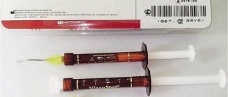
A reliable assistant in the fight against dysbiosis will be the probiotic complex ASEPTA PARODONTAL*, a source of lactobacilli to restore the microflora of the oral cavity. This unique complex with patented strains of lactobacilli and vitamin D has the ability to effectively restore oral microflora. The complex normalizes the bacterial flora in the oral cavity, eliminates bad breath and prevents the formation of biofilms of pathogenic microorganisms.
To improve the effectiveness of the prescribed therapy, dentists recommend giving up bad habits (at least for the duration of treatment), reviewing the diet, paying attention to plant foods, and be sure to take care of the oral cavity after each meal.
Medical and social problems of managing patients with chronic dermatoses
An analysis of the incidence of diseases of the skin and subcutaneous tissue in the Russian Federation over the past decades states their prevalence and consistently high level - 4259 per 100 thousand population [1]. The problem of chronic dermatoses remains one of the most significant due to the prevalence of pathology in all age groups, frequent relapses, lack of stable remission, an increase in the number of patients with severe, treatment-resistant forms of the disease, the frequent development of complications, malignancy, the formation of severe disabling forms, which has a negative impact impact on the quality of life of patients [1–4].
In the Russian Federation, the provision of medical care to this category of patients is carried out in accordance with the Order of the Ministry of Health of Russia dated November 15, 2012 No. 924n “On approval of the procedure for providing medical care to the population in the field of dermatovenerology.” Recommendations for the management of patients with chronic dermatoses are long-term in nature, which requires them to strictly implement treatment and rehabilitation programs. The problem of patient adherence to therapy is relevant; the compliance rate varies widely - from 25% to 75% [5]. Treatment adherence declines over time, especially in chronic diseases, and leads to serious medical consequences [6, 7]. About 10% of all hospitalizations are associated with non-compliance with the attending physician’s prescriptions [8]. Only in hospital settings, where medical and nursing supervision is carried out, is strict compliance by patients with medical recommendations guaranteed.
An important aspect of preventing the frequency of exacerbations and the development of complications of chronic dermatoses is the early detection of diseases and timely taking patients under dispensary observation [9]. Medical examination is a complex of measures, including a medical examination by doctors of several specialties and the application of the necessary examination methods, carried out in relation to certain groups of the population in accordance with the legislation of the Russian Federation [10]. In 2016, in dermatovenerological medical organizations, the proportion of patients with severe skin pathology (atopic dermatitis, psoriasis, discoid lupus erythematosus, localized scleroderma) under dispensary observation averaged 49.5% for this group among adults. adolescents - 48.7%, children 0–14 years old - 40.1% [1]. The joint work of dermatovenereologists and doctors of related specialties to implement dispensary activities for dermatovenereology patients helps to identify comorbid pathologies and makes it possible to develop an algorithm for interdisciplinary interaction aimed at increasing the efficiency of the quality of medical care. A multi-level approach in the form of standardization of a complex of diagnostic, treatment-and-prophylactic and dispensary measures, on the one hand, and high-quality interaction between doctor and patient, on the other, will be the key to a successful outcome of therapy.
Taking into account the above, we present our own clinical observations that reveal the medical and social problems of managing patients with chronic dermatoses.
Clinical case No. 1
Patient G., 30 years old, was admitted to the inpatient department of the State Budgetary Healthcare Institution of the KKVD of the Ministry of Health of the KK on March 26, 2018 with complaints of rashes on the skin of the ears, neck, torso, upper and lower extremities, intense periodic itching, a feeling of dryness and tightness, and an unpleasant odor .
History of the disease: considers himself sick since 2002, when rashes first appeared on the skin of the back and chest after prolonged exposure to the sun. I contacted a dermatologist at my place of residence, where the diagnosis of “Darier’s disease” was first established. The recommended treatment is betamethasone valerate 0.01% cream. After the therapy, he noted an improvement. From 2002 to 2013, he noted exacerbations once a year in the summer, the rashes were limited in nature, the patient was not seen by a dermatologist, he was treated irregularly with “folk remedies,” believing that “it would go away on its own.” For the first time in 2013, due to the spread of a skin pathological process, the patient was hospitalized for inpatient treatment at the State Budgetary Healthcare Institution KKVD of the Ministry of Health of KK. The patient underwent pathogenetic therapy with acitretin with a pronounced clinical effect. Upon discharge, it was recommended: clinical observation with a dermatologist at the place of residence, continued systemic therapy with aromatic retinoids and essential phospholipids, external therapy with topical glucocorticosteroids and emollients, and the use of photoprotective agents. In the period from 2013 to 2022, exacerbations occurred in the spring-summer period, the rashes were widespread, the patient was treated irregularly, only with external agents (betamethasone valerate 0.01% cream, mometasone furoate 0.1% cream, Dexpanthenol ointment 5%) , he visited a dermatologist sporadically, was not under dispensary observation, and did not follow discharge recommendations in 2013, as he believed that full recovery would not occur. The last exacerbation occurred in August 2022, when the patient changed the conditions of his professional activity: he got a permanent job in a smelting shop (heavy metal processing). He was treated independently with antibacterial drugs, without improvement, and only at the end of March 2022, when the skin pathological process became generalized, he went to the clinic of the State Budgetary Healthcare Institution of the KKVD of the Ministry of Health of the Republic of Kazakhstan and was hospitalized.
Allergological history and heredity are not burdened. Not examined somatically. Among bad habits, it indicates moderate alcohol consumption, once a week. Currently not working, until 2022 he was engaged in fishing. Divorced.
Upon admission to the hospital, the consciousness is clear, the position is active. Body weight 80 kg, height 185 centimeters. Peripheral lymph nodes are not enlarged. Temperature 36.8 °C. Breathing is vesicular, no wheezing. The respiratory rate is 17 per minute. Heart sounds are clear and rhythmic. Pulse 70 beats per minute. Blood pressure is 120/80 millimeters of mercury. The tongue is moist, covered with a white coating along the back of the tongue. The abdomen is soft and painless. The liver is at the edge of the costal arch. Physiological functions are normal.
St. specials: the skin pathological process was of a generalized, symmetrical nature. On the skin of the ears, neck, torso (in the chest, abdomen, back, with transition to the skin of the buttocks) there were multiple follicular and perifollicular papules up to 3 mm in diameter, dirty brown in color, covered with a hard, dry horny crust (Fig. 1, 2). On the skin of the upper extremities (in the area of the shoulders, forearms), lower extremities (in the area of the thighs and legs), abundantly located miliary and lenticular follicular papules of a grayish-brown color were determined, some of which were covered with layers of purulent tightly and loosely adjacent crusts, purulent discharge was visualized (Fig. 3, 4). On the skin of the outer surfaces of the forearms and anterior surfaces of the legs, papular elements were formed into verrucous plaques, on the surface of which, after rejection of the purulent crusts, multiple papillomatous growths were clearly visible (Fig. 5). Keratosis punctata were evident on the skin of the palmar surface of the hands.
According to additional research methods, the following results were obtained:
- Complete blood count: red blood cells - 4.7 × 10¹²/l; hemoglobin - 150 g/l, leukocytes - 7.7 × 10⁹/l; band neutrophils - 1%, segmented neutrophils - 68%; eosinophils - 1%, lymphocytes - 29%, monocytes - 2%; ESR - 28 mm/h.
- Biochemical blood test: total protein - 78 g/l; creatinine - 73.93 µmol/l; cholesterol - 5.1 mmol/l; total bilirubin - 33.7 µmol/l; alkaline phosphatase - 120 U/l; amylase - 50 U/l; glucose - 4.4 mmol/l; gamma-glutamyltransferase (GGT) - 35 U/l; alanine aminotransferase (ALT) - 88 U/l; aspartate aminotransferase (AST) - 86 U/l.
- General urine analysis: color - light yellow, transparent, specific gravity - 1015, pH - 5.5, protein - absent; ketone bodies - absent; leukocytes - 3–4 per field of view; bilirubin is absent.
- Enzyme-linked immunosorbent assay (ELISA) of blood for HIV - HIV antigen/antibodies were not detected.
- Blood test for syphilis: microprecipitation reaction (MPR) - negative; ELISA - negative.
- Blood ELISA test for viral hepatitis B and C: HBsAg - not detected, HCV - detected.
- Scraping from the tongue for fungi of the genus Candida: pseudomycelium of a yeast fungus was detected.
- Ultrasound examination of the hepatobiliary region: diffuse changes in the liver and pancreas.
- Consultation with an infectious disease specialist: chronic viral hepatitis C, newly diagnosed.
The final clinical diagnosis was established: “Darier's disease. Gottron's carcinoid papillomatosis. Chronic viral hepatitis C, newly identified. Candidal glossitis."
Analyzing this clinical case, it can be argued that the worsening of the course of dermatosis and the development of its complication in the form of Gottron's carcinoid papillomatosis is natural due to the presence of a combination of socio-psychological factors:
- exposure to unfavorable environmental factors and disturbances caused by lifestyle features (hypothermia, insolation, stress caused by divorce);
- ignoring a healthy lifestyle (systematic alcohol consumption);
- exposure to harmful production factors (skin trauma, working with heavy metals);
- socio-economic factors (lack of a permanent job and permanent income);
- failure to comply with the doctor’s recommendations (for 12 years from the moment of diagnosis, the patient avoided visiting an outpatient dermatologist; did not comply with the prescriptions and requirements for registering with a dermatologist at the doctor’s place of residence in the inpatient department).
Clinical case No. 2
Patient M., 63 years old, was hospitalized at the State Budgetary Healthcare Institution of the KKVD of the Ministry of Health of the Republic of Kazakhstan on October 22, 2018, with complaints of rashes on the skin of the legs and feet, accompanied by intense itching, weeping, an unpleasant odor, and a feeling of heaviness in the legs. The patient is withdrawn, depressed, and has difficulty hearing.
History of the disease: he has been ill for about 20 years, when the diagnosis of “Microbial eczema” was first established. From 1998 to 2014, I regularly noted exacerbations of dermatosis once every 6 months, and visited a dermatologist at the clinic twice. He was treated independently with topical agents: gentamicin + betamethasone + clotrimazole cream, chlorhexidine bigluconate aqueous solution 0.05%, Brilliant Green alcohol solution 1%, with short-term improvement. In 2014, for the first time, he was hospitalized at the State Budgetary Institution of Healthcare of the KKVD of the Ministry of Health of the KK with the diagnosis: “Varicose eczema. Gottron's carcinoid papillomatosis of the skin. Chronic venous insufficiency of the lower extremities." After the course of therapy, he noted an improvement. Recommendations were given on the need to register with a dermatologist at the place of residence; conducting consultations with related specialists: angiosurgeon, dermato-oncologist, therapist; deciding on the initiation of cytostatic therapy. From 2014 to 2022, the patient did not comply with the recommendations, skin manifestations continuously recurred, and the patient was not treated. In October 2022, due to severe deterioration, he went to the clinic of the State Budgetary Healthcare Institution of the KKVD of the Ministry of Health of the Republic of Kazakhstan and was hospitalized.
From the life history it was established that the patient was lonely. He is a visually impaired person of group I. Has a guardian who does not live with him, but visits him periodically.
Upon examination, the skin pathological process is limited and symmetrical. On the skin of the legs, muff-like, with a transition to the dorsal surface and toes, against the background of pronounced lymphostasis, skin infiltration and diffuse erythema of a reddish-bluish color, there are layers of serous-purulent and purulent-hemorrhagic crusts, erosion with a weeping surface (Fig. 6). On the skin of the posterior surface of both legs, single hemispherical formations are visualized, about 3.0 cm in diameter, dense upon palpation (Fig. 7). On the skin in the area of the ankle joints and toes there are verrucous lesions with an uneven, bumpy surface and numerous papillary growths, covered in places with purulent crusts (Fig. 8, 9). When the lesions were squeezed from the sides, a yellowish-white, sticky discharge with an unpleasant odor was released onto their surface.
Based on additional research methods and expert opinions, the following results were obtained:
- Complete blood count: red blood cells - 4.3 × 10¹²/l; hemoglobin - 125 g/l, leukocytes - 8.1 × 10⁹/l; band neutrophils - 7%, segmented neutrophils - 75%; eosinophils - 1%, lymphocytes - 16%, monocytes - 1%; ESR - 18 mm/h, platelets - 433 × 109 g/l.
- Biochemical blood test: total protein - 55 g/l; creatinine - 89.97 µmol/l; cholesterol - 6.4 mmol/l; total bilirubin - 12.8 µmol/l; alkaline phosphatase - 112 U/l; amylase - 26 U/l; glucose - 8.0 mmol/l; GGT - 23 U/l; ALT - 10 U/l; AST - 23 U/l.
- General urine analysis: color - light yellow, transparent, specific gravity - 1015, pH - 5.5, protein - absent; ketone bodies - absent; leukocytes - 1–2 per field of view; bilirubin - absent, mucus +.
- Blood ELISA for HIV - HIV antigen/antibodies were not detected.
- Blood test for syphilis: RMP - negative; ELISA - negative.
- Blood ELISA for viral hepatitis B and C: HBsAg - not detected, HCV - not detected.
- Glycemic profile: 13:00 - 6.5 mmol/l, 17:00 - 13.5 mmol/l, 21:00 - 11.7 mmol/l, 08:00 - 7.6 mmol/l, 13:00 - 7.0 mmol/l.
- Blood hemostasis: activated partial thromboplastin time - 29 sec, prothrombin time 12.1 sec, prothrombin index 94.6%, international normalized ratio - 0.93, fibrinogen 4.4 g/l, thrombin time 19.7 sec, antithrombin determination -III - in plasma 124.80%.
- Biochemical blood test: sodium 144.0 mmol/l, potassium 5.1 mmol/l, chlorine 103.0 mmol/l.
- Consultation with a general practitioner: hypertension, degree 2, stage 2, risk 4. Moderate thrombocytosis. Hypocoagulation syndrome. Chronic venous insufficiency of the lower extremities.
- Consultation with an endocrinologist: newly diagnosed diabetes mellitus.
The final clinical diagnosis was established: “Varicose eczema. Gottron's carcinoid papillomatosis. Hypertension, stage 2, stage 2, risk 4. Moderate thrombocytosis. Hypocoagulation syndrome. Chronic venous insufficiency of the lower extremities. Diabetes mellitus, newly diagnosed.”
The features of the given clinical case are another list of problems that led to the development of complications and chronicity of dermatosis:
- forced socio-psychological maladaptation of the patient (loneliness, visual impairment, hearing loss, depression, communication difficulties, inability to carry out independent activities);
- socio-economic factors (low living standards).
In the clinical cases described, the patients were not registered at the dispensary and therefore did not have:
- proper scope and frequency of examination by a dermatovenerologist;
- regularity of assessment by a medical specialist of complaints, anamnesis, clinical, laboratory and instrumental studies;
- consultations with related specialists (in the described patients, concomitant pathology was identified for the first time only at the stage of inpatient treatment);
- prescription of preventive and rehabilitation measures, including sanatorium and resort treatment.
Summarizing both clinical situations, it should be noted that when providing specialized medical care in the profile of “dermatovenerology” to these patients, such an important stage of work as dispensary observation was missed.
Disclosure of the characteristics and analysis of medical and social problems of patients with chronic dermatoses will allow us to balance and optimize the structure of medical care, determine the nature of priority measures to improve and strengthen the health of patients in the “dermatovenereology” profile.
Literature
- Kubanova A. A., Kubanov A. A., Melekhina L. E., Bogdanova E. V. Analysis of the incidence of diseases of the skin and subcutaneous tissue in the Russian Federation for the period 2003–2016. // Bulletin of dermatology and venereology. 2017; 6:22–33.
- Federal clinical guidelines. Dermatovenereology 2015: Skin diseases. Sexually transmitted infections. 5th ed., revised. and additional M.: Business Express, 2016. 768 p.
- Batkaeva N.V., Batkaev E.A., Gitinova M.M. Comparative assessment of the dermatological index of quality of life in patients with chronic inflammatory dermatoses // Russian Medical Journal. 2018; 82:68–71.
- Tlish M.M., Kuznetsova T.G., Sycheva N.L. Clinical and morphological aspects of Gottron’s carcinoid papillomatosis of the skin // Medical Bulletin of the South of Russia. 2014, no. 2, p. 138–143.
- Cowell W., Fulford-Smith A., Poultney S. Adherence with bisphosphonate treatment for osteoporosis in UK patients. Poster presented at the second joint meeting of the European Calcified Tissue Society and the International Bone Mineral Society. Geneva, 25–29 June 2005.
- Hill MN, Miller NH Compliance Enhancement. A Call for Multidisciplinary Team Approaches // Сirculation. 1996; 93:4–6.
- Horne R. Representation of medication and treatment: advances in theory and measurements. In: Petrie R, Weinlan J, eds. Perceptions of Health and Illness: Current Research and Applications. London: Harwood Academic, 1997, p. 155–188.
- Binhas E. Comment augmenter I acceptance des plans de traitement. Dialgue 1999; 13–15.
- Teplyakova E. D., Shcherbakov S. M. Improving the organization of medical examinations and medical examinations in outpatient settings based on simulation modeling // Kuban Scientific Medical Bulletin. 2015; 4: 124–130.
- Federal Law of November 21, 2011 No. 323-FZ “On the fundamentals of protecting the health of citizens in the Russian Federation” / Collection of legislation of the Russian Federation. November 28, 2011. No. 48. Art. 6724.
M. M. Tlish, Doctor of Medical Sciences T. G. Kuznetsova1, Candidate of Medical Sciences
Federal State Budgetary Educational Institution of Higher Education Kuban State Medical University of the Ministry of Health of the Russian Federation, Krasnodar
1 Contact information
Medical and social problems of managing patients with chronic dermatoses / M. M. Tlish, T. G. Kuznetsova For citation: Attending physician No. 1/2019; Page numbers in the issue: 77-81 Tags: skin, malignancy, disability, quality of life.
Possible complications of dysbiosis
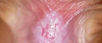
It is important for each patient to closely monitor the state of the oral microflora. Lack of treatment for dysbiosis can lead to such unpleasant diseases as:
- pathological halitosis – bad breath caused by an imbalance of the oral microflora;
- caries – destruction of hard tooth tissues;
- pulpitis - inflammation of the pulp - the internal tissues of the tooth;
- periodontitis – inflammation of the tooth root membrane and adjacent tissues;
- gingivitis – inflammation of the oral mucosa;
- stomatitis – damage to the oral mucosa;
- periodontitis is a deep lesion of the periodontal tissue.
In addition, the close relationship between the state of the oral microflora and the state of the cardiovascular system has been scientifically proven. In 2008, it was proven in the USA that periodontal disease, as a source of chronic inflammation, is an independent risk factor for coronary heart disease (CHD).
So, now you know the role of normal oral microflora in the human body. Treat yourself carefully, and your healthy body will delight you every day.
How to avoid getting sick
Preventive measures include following simple rules:
- balanced diet;
- elimination of traumatic effects on the tissues of the tongue;
- taking an antifungal drug during a course of antibiotics and hormones;
- course use of immunomodulators;
- maintaining oral hygiene;
- timely treatment of emerging dental diseases.
People who take care of their dental health and visit the dentist twice a year are much less likely to experience tongue candidiasis. This is a proven fact. Therefore, do not ignore your doctor’s requests to come for preventive examinations.
Laboratory at home, how to collect material at home?
Cotton swabs for collecting materials.
Various situations happen in life. There are times when it is not possible to take the child to the laboratory or come independently. What to do in this case? Is it possible to collect material for research on your own? Conducting a fence at home is not difficult. To do this you will need:
- Non-sterile gloves – 1 pair
- Cotton wool or cotton swab – 1 pc.
- Sterile container
How to make a fence:
- Prepare container
- Wear gloves
- Apply a cotton swab several times in the anal area
- Place the stick in the container, close the lid tightly and deliver it to the laboratory.
Remember! Biological material is not stored even in the cold. Therefore, you need to take him to the clinic as quickly as possible! While you're getting dressed and getting ready, put a sample in the refrigerator!