Pulp amputation
Unnoticed caries in time risks developing into its more formidable form - pulpitis, and then into periodontitis. When infection reaches the pulp, doctors use different methods of treating it, both biological and surgical, and amputation or extirpation is considered the most effective.
Dental pulp amputation is a gentle treatment method that involves removing only part of the neurovascular tissue and is used primarily in children. It is effective for mild tissue damage or significant curvature of tooth roots. Extirpation is used most often in adult patients and consists of completely cleaning the tooth cavities.
The possibility of amputation arose as a result of understanding the differences in the structure of the root and coronal parts of the pulp. That is why, with minor or superficial damage to the neurovascular bundle, it is possible to remove only its coronal part. This is especially important in childhood, when the roots of the tooth continue to grow and develop, and it is necessary to preserve their nutrition.
According to the method of performing the operation, vital and non-vital pulp amputation are distinguished.
Vital pulp amputation - pulpotomy
The method involves mechanical removal of tissue damaged by caries under local anesthesia, and today its use is widespread. After cutting off all damaged tissue with a bur, the open cavity is treated with medication, the pulp is amputated at the level of the root canals and its surface layers are fixed (in the apical, or root, area it remains alive), then the cavity is closed with a temporary filling and observed for six months. If at this time nothing bothers the patient, and the tooth does not respond with pain to percussion and palpation, the doctor applies a permanent filling.
Vital pulp amputation is especially often used in children, since the developing roots of permanent teeth have complex ribbon-like wriggling tubules with a large number of branches, and if we are talking about baby teeth, it is important to preserve the ability of their roots to resorption and not affect the rudiments of permanent teeth.
The possibility of vital amputation is determined by the viability of the root part of the pulp, the absence of inflammation in the tissues surrounding the tooth and the absence of symptoms of the disease.
Unlike conventional vital pulp amputation, deep amputation is performed not only on the crown part of the teeth, but also on the root part - at different depths of the canals.
Devital pulp amputation
The method of devital amputation is that before removal, the coronal pulp is “killed” with special pastes, after which it becomes more fibrous, making it easier to remove. The root pulp remains in place. Mummification of the root pulp remaining after amputation of its coronal part occurs due to the impregnation of the tissues with a drug, as a result of which it dehydrates, dries out and turns into a sterile cord. Today there are many anti-inflammatory and antiseptic pastes based on a variety of active substances, which have their pros and cons.
Among the indications for the method of devital pulp amputation are acute and chronic pulpitis, as well as the presence of a strong fear of injections and syringes, as well as other phobias regarding dental treatment.
Experts declare that the method of devital amputation of the pulp of permanent teeth is insufficiently effective, since in most cases the periodontal tissue becomes inflamed as a result. This method of treatment is chosen only for the period of root growth, and after their final formation, further cleansing of the root canals is carried out - pulp extirpation.
Vital amputation using ferrous sulfate-based drugs in pediatric dentistry
At the present stage of development of dentistry, treatment methods that involve preserving the vitality of the pulp and restoring its functions are becoming increasingly widespread. This is especially true when treating pulpitis of primary teeth with unformed roots, when it is important to preserve the pulp to complete root growth.
Treatment of pulpitis of primary teeth in children should be timely and adequate. Temporary teeth play an important role in the formation of dentition and jaws, timely eruption and correct placement of permanent teeth, normal development of the functions of the dental system, while early removal leads to a failure of normal formation processes.
The choice of treatment method for pulpitis depends on the diagnosis of the disease, the age of the child and the possibility of establishing psychological contact with him. With the development of new technologies in dentistry, there has been a tendency to increase the frequency of use of vital treatment methods, which have a number of advantages: the number of patient visits to the dental office is reduced, and the use of drugs with a resorptive effect is eliminated. Among the vital methods, the most widely used method is vital amputation (pulpotomy), based on morphological differences in the structure of the root and coronal pulp.
When treating pulpitis of primary teeth using the vital amputation method, preparations based on 35% formocresol and 2% glutaraldehyde are used. However, according to studies, in many cases the use of these drugs was unsuccessful. There is also an opinion about the possible cytotoxic and mutagenic effects of formocresol. In this regard, there was a need to search for new, more effective drugs for the treatment of pulpitis of primary teeth. Today these are preparations based on ferrous sulfate (ViscoStat, Astringedent).
According to the literature (R.E. McDonald, D. Avery, M.S. Duggal), treatment of primary teeth using the vital amputation method using 35% formocresol was unsuccessful in approximately half of the cases: during the study period (12 months) Children in this group complained of pain in the area of the treated tooth, changes were found on the radiograph in 5-6 teeth out of 50 (destruction of bone tissue in the periapical region, in the root furcation zone). Accordingly, the percentage of successful treatment was 89%. When using ferrous sulfate preparations during the entire observation period (1 year), only in one tooth out of 50 changes were detected on the radiograph: destruction of bone tissue in the periapical region and in the root furcation zone. Accordingly, the success of endodontic treatment was 98%. Thus, the percentage of successful treatment when using ferrous sulfate preparations is much higher than when using formocresol-based preparations.
Materials and research methods
Vital pulp amputation is a method of removing inflamed and infected coronal pulp in order to preserve the vital root pulp.
The method of vital amputation consists in removing the coronal pulp, rich in cellular elements, and preserving the root pulp, which ensures the normal physiological course of the growth and development of the temporary tooth and its surrounding structures. The use of pulpotomy is based on differences in the structure of the coronal and root pulp of teeth: the coronal pulp has a looser structure due to the large number of vascular anastomoses and the presence of cellular elements. Consequently, during inflammation, more significant changes in microcirculation occur in the coronal pulp. In the root pulp there are practically no cellular elements, connective tissue fibers predominate, therefore, in the root pulp tissue swelling is less pronounced, there is no compression of blood vessels and the phenomena of congestive hyperemia. This structural feature allows amputation of the coronal pulp with subsequent preservation of the function of the viable root pulp.
Ferrous sulfate preparations used for vital amputation in primary teeth are ViscoStat and Astringedent.
ViscoStat is a viscous hemostatic gel containing 20% ferric sulfate, which has a gentle coagulating effect on soft and hard dental tissues. Hemostasis is achieved mainly due to the formation of coagulation plugs (thrombi) in the lumens of the capillaries. This product is used to stop capillary bleeding and ensures high-quality pulpotomy of primary teeth in one visit.
The effectiveness of the ViscoStat solution (Fig. 1) increases significantly when using the special Dento-Infusor device, since the effect of hemostatic agents depends on the method of their application.
Rice. 1. ViscoStat - viscous, containing 20% ferric sulfate, hemostatic gel, has a gentle coagulating effect.
Using a brush at the end of the Dento-Infusor nozzle, the hemostatic is “rubbed” into the capillaries, which leads to the formation of blood clots. This also removes blood clots outside the lumens of the capillaries. This procedure protects the formed vascular blood clots from being removed when washed off.
As a result, we have a clean, dry surface.
Description of clinical observations
Clinical case: patient A. A. Frolov, 4.5 years old. He complained of pain in the area of the 74th tooth that occurred during meals. When examining 74 teeth, a carious cavity filled with softened dentin was discovered. The tooth cavity was opened at one point, touching which caused pain. The color of the tooth was not changed. Percussion is painless, the mucous membrane and the transitional fold in the projection of the roots of the causative tooth are without pathology. As a result, a diagnosis of chronic fibrous pulpitis of 74 teeth was made and treatment was carried out using the vital amputation method using ViscoStat gel according to the following scheme:
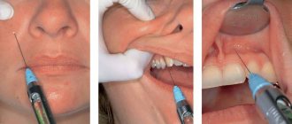
Local anesthesia: Ultracaine D-S 1.7 ml, 1:200,000.
Preparation of a carious cavity and opening of the cavity of 74 teeth. Necrotic dentin is removed from the walls and bottom of the carious cavity. It is important to carefully prepare the carious cavity before opening the pulp chamber. The cavity is then opened wide to create a direct transition into the tooth cavity. Resection of the arch of the dental cavity is carried out with a sterile bur. In temporary molars, after opening the hole with a ball-shaped bur, the overhanging edges are cut off with a cylindrical bur. This manipulation requires the doctor to know the topography of the pulp chamber in order to prevent perforation and provide direct access to the orifices of the root canals. Antiseptic treatment (chlorhexidine bigluconate 0.05%).
Removal of coronal pulp (pulpotomy) using a ball bur at low speed. Next, the mouths of the root canals are processed, forming additional platforms to relieve excess pressure from the root pulp. Then a deep pulpotomy is performed with a sterile carbide bur on an extended stem (Fig. 2).
Rice. 2. Pulpotomy was performed.
Using the Dento-Infusor tip, ViscoStat solution is applied to the orifice pulp. Hemostasis is achieved within 10-30 seconds.
During the process of rubbing the solution, additional water is sprayed so that coagulation clots do not stick to the treated tissue. The work area is thoroughly rinsed and cleaned with a saliva ejector. The amount of hemostatic required for one tooth is 1/3-1/2 of the volume of the tooth cavity. After the bleeding has stopped, the ostial pulp is covered with a brown scab, there is no bleeding (Fig. 3, 4).
Rice. 3. Application of ViscoStat gel to the canal orifices.
Rice. 4. Coagulated ostial pulp.
Applying a thin layer of zinc oxide eugenol cement to the treated tissue and the bottom of the pulp chamber. Next, an insulating gasket made of glass ionomer cement is made (Fig. 5).
Rice. 5. Placement of zinc oxide eugenol cement and a gasket made of glass ionomer cement on the bottom of the tooth cavity.
Carrying out tooth restoration (Fig. 6, 7).
Rice. 6. Tooth restoration.
Rice. 7. The final appearance of the restoration using photopolymer material.
Conclusion
Ferrous sulfate preparations appeared on the dental materials market relatively recently. In the course of research and clinical experiments, the physical and mechanical properties of ferrous sulfate preparations were studied, and their greater effectiveness was proven over formocresol preparations (98% and 89%) in the treatment of pulpitis of primary teeth in children using the vital amputation method.
The percentage of successful treatment with ferrous sulfate preparations is much higher than with formocresol-based preparations. The use of ferrous sulfate preparations (ViscoStat) makes it possible to quickly stop bleeding and ensure high-quality pulpotomy of primary teeth in one visit, avoiding complications and the need for repeat visits for patients.
Literature
- V. K. Leontiev, prof. L.P. Kiselnikov. Pediatric therapeutic dentistry.
National leadership. - M.: GEOTAR-Media, 2010. - Dentistry of children and adolescents /
edited by Ralph E. MacDonald, Daveig R. Avery (translation by T. V. Vinogradova). - M.: MIA, 2003. - M. S. Duggal, M. E. J. Curzon, S. A. Fail, K. J. Toumba, A. J. Robertson. Treatment and restoration of baby teeth
(translation by T. V. Vinogradova). - M.: MEDpress-inform, 2006. - L. S. Persin, V. M. Elizarova, S. V. Dyakova. Pediatric dentistry.
- Medicine, 2005. - R. Beer, M. A. Bauman, Andrei A. Kielbasa. Illustrated guide to endodontology.
- M.: MEDpress-inform, 2008. - T. V. Vinogradova. Guide to pediatric dentistry.
- Medicine, 1987. - M. V. Kuryakina .
Therapeutic dentistry for children . - M., Nizhny Novgorod: NGMA, 2001. - A. A. Kolesov. Pediatric dentistry.
- M., 1991.
A complete list of references is in the editorial office.
Pulpotomy of primary molars
In 1991, Yacobi et al wrote about the then-evolved techniques and materials for performing pulpotomy of primary molars. A year later, Croll and Killian described a technique for performing pulpotomy of primary molars using a mixture of zinc oxide powder and eugenol as a material to fill the pulp cavity. After performing a pulpotomy, the authors recommended covering the teeth with stainless steel metal crowns. This approach was modified from the original technique of Kaare Langeland, who described the toxicity of the use of formocresol in pulpotomy, and stated that the pulpotomy procedure itself was effective without the use of formocresol, and not because of it. If there is a need to achieve hemostasis, the author suggested using ferrous sulfate. Later, the use of zinc oxide eugenol (ZOE) for pulp capping and filling the pulp chamber space was reported to be successful. MTA is another alternative material that can be used for a similar purpose. MTA is a derivative of Portland cement and is currently most often used during pulpotomy.
A systematic review and meta-analysis by Lin et al found MTA to be the first choice material for pulpotomy of primary molars. In this article we will present three cases of pulpotomy of primary molars with their further covering with metal crowns. In one clinical case we performed the pulp cavity with COE, and in the other two with MTA.
Clinical case 1
A 39-month-old female patient and her mother sought advice regarding an acute carious lesion on the occlusal surface of a right lower second primary molar (Figure 1). The patient had already previously obtained an orthopantomogram, which was performed on the recommendation of the pediatric dentist (photo 2).
Photo 1. Occlusal caries of the lower second primary molar in a patient aged 39 months.
Photo 2. Orthopantomogram demonstrating an extensive cavity.
No other pathologies were found in the oral cavity. Their pediatric dentist recommended contacting a specialist to perform general anesthesia, perform a pulpotomy, and then cover the tooth with a metal crown. But at the appointment, the patient behaved absolutely adequately, was somewhat shy, but cooperated with the doctor, and based on this, it was decided to perform a pulpotomy under sedation with nitrous oxide, which was administered through a nasal mask. After the mother signed the informed consent, the patient was scheduled for the procedure the next day after the initial visit. During sedation, nitrous oxide was administered at a ratio of 20% nitric oxide/80% oxygen. The child also listened to music on headphones in order to avoid irritation from the sound of the drill. The area of the lower second molar was anesthetized by infiltrating a 4% solution of articaine hydrochloride with epinephrine in a ratio of 1:200,000. After the rubber dam was installed, access to the pulp was created using a cylindrical diamond bur (photo 3).
Photo 3. Exposure of the pulp chamber area.
During the formation of the access, it was confirmed that caries had penetrated into the area of the pulp chamber. After removal of carious dentin, the pulp tissue was removed to the orifice area using a sterile, low-speed No. 6 bur. Bleeding from the root canal area indicated that the pulp in these areas remained vital. A cotton ball soaked in water was placed over the orifice area and pressed, which allowed good hemostasis to be achieved without the use of ferrous sulfate solution or any other hemostatic agent (Figure 4). After this, the COE powder was mixed until it reached the consistency of a paste, and packed into the pulp chamber area, carefully removing the excess (photo 4).
Photo 4. COE-pulpotomy.
An orthodontic strip was placed around the tooth to stabilize the position of the rubber dam. The area where COE was applied was covered with modified glass ionomer cement, which was then polymerized and prepared for further fixation of the metal crown (photo 5). After 4 years and 8 months, the patient again sought help for the restoration of a permanent maxillary molar, in the area of which there were signs of carious lesions. During the examination, the successful result of the previously performed pulpotomy and crowning of the lower second primary molar was confirmed. However, given the level of root resorption, the degree of eruption of the second permanent premolar and the presence of minor inflammatory changes in the mucosa, it was decided to remove the primary molar under local anesthesia (photo 8).
Photo 5. View after preparation for a crown.
Photo 6. View 4 years and 8 months after the intervention.
Photo 7. Orthopantomogram 4 years and 8 months after the intervention.
Photo 8. View of the tooth after removal.
Clinical case 2
A 9-year-old boy was referred by his family dentist for treatment of an extensive carious lesion in the area of the right mandibular second molar (Figure 9). The area of the adjacent first primary molar was also noted to have caries, as was the area of the maxillary first primary molar. The radiograph showed signs of penetration of carious pathology into the pulp space (photo 10).
Photo 9. Caries of a primary molar in a 9-year-old patient.
Photo 10. X-ray showing an extensive carious cavity.
After obtaining informed consent from the parents, it was planned to perform a pulpotomy in the area of the lower second primary molar of the mandible on the left and perform a composite restoration in the area of the first primary molar. The patient contributed in every possible way to the intervention, and after anesthesia with a 4% solution of articaine hydrochloride with epinephrine 1:200,0000, a rubber dam was installed (photo 11). All further manipulations were performed similarly to those described in clinical case 1. Using a cylindrical bur, the cavity was cleared of caries (photo 12), the space of the pulp chamber was reached and hemostasis was ensured by means of sterile rollers, which were inserted into the pulp space under compression (photo 13-14). The dense MTA mixture (NeoMTA, NuSmile) was then packed using an amalgam condenser over the root canal orifices (Figure 15). Once the material had cured, the excess was removed with a cylindrical diamond bur, and preparation of the first molar began immediately for further restoration (Figure 16).
Photo 11. Insulation using rubber dam.
Photo 12. Removal of carious tissue.
Photo 13. Achieving hemostasis using cotton rolls.
Photo 14. View after achieving hemostasis.
Photo 15. Packaging MTA in the tooth cavity.
Photo 16. View after pulpotomy.
A bioactive composite (ACTIVA BioACTIVE-RESTORATIVE, Pulpdent) was introduced into the cavities of both teeth, after which it was photopolymerized (photo 17-18). After polymerization, a 5% fluoride varnish was applied to the mesial surface of the permanent first molar. When the contour of the restoration on the first primary molar was completely recreated, the rubber dam clamp was reinstalled on the permanent first molar and the preparation of the second primary molar for a metal crown began (photo 19-20). The latter was contoured, cut and polished according to the usual algorithm, after which it was fixed with modified glass ionomer cement. Excess cement was removed using a Hollenback instrument (Figure 21), and in the area of interproximal contacts - using dental floss. The maxillary first primary molar was restored at the next visit. Photographs and radiographs obtained 22 months after treatment are shown in Figures 22 and 23. After 22 months, the patient no longer returned for monitoring.
Photo 17. Filling the cavity with a bioactive composite.
Photo 18. Polymerization of a bioactive composite.
Photo 19. View after contouring the restoration in the area of the first primary molar.
Photo 20. View after preparing the tooth for a crown.
Photo 21. Removing excess cement.
Photo 22. View 22 months after treatment.
Photo 23. X-ray 22 months after treatment.
Clinical case 3
An 8-year-old boy was referred to the dentist due to the need for extraction of the second primary molar of the lower jaw on the left and restoration in the area of the first primary molar on the same side of the jaw (photo 24-25). Taking into account the initial parameters of the clinical situation, the child’s parents were offered a treatment option with a pulpotomy procedure and further covering with a metal crown. Using the same approach as Case 2, the pulp chamber space was completed with MTA in the orifice area, the tooth was then capped with a metal crown, and the first primary molar area was restored with a restoration (Figure 26). Hemostasis was achieved using sterile moistened cotton swabs, and the dental cavity was filled with a modified glass ionomer material over the MTA. 33 months after treatment, clinical examination and radiological monitoring data indicated a successful outcome of the intervention (photos 27-28). 33 months after treatment, both primary teeth of the lower jaw on the left side were also removed according to orthodontic indicators (photo 29).
Figure 24: An 8-year-old patient was referred for extraction of the mandibular left mandibular second primary molar.
Photo 25. X-ray of the patient before treatment.
Photo 26. View after pulpotomy.
Photo 27. View after 33 months.
Photo 28. X-ray after 33 months.
Photo 29. View of the tooth after extraction.
Discussion
The use of a rubber dam when performing pulpotomy of primary molars is one of the key factors to ensure the success of the manipulation. The rubber dam ensures both safety of the intervention and comfort during the procedure. In cases where the pulp is completely damaged, the pulp tissue shows almost no signs of bleeding, therefore, in such cases, it is necessary to perform a pulpectomy or consider the need for tooth extraction. In cases of extraction, it is necessary to remember the need to manufacture a retainer to replace the space, so as not to compromise the process of eruption of permanent teeth.
In the three clinical cases described above, we were able to achieve hemostasis using conventional cotton swabs without the use of any other additional means. In cases of heavy bleeding, sterile low-speed burs can be used to remove pulp from the orifice. You can also use cotton swabs soaked in a solution of ferrous sulfate, which should be placed into the cavity of the pulp chamber for 30-60 seconds. This procedure is often referred to as a “ferum sulfate pulpotomy,” although in fact this agent is only used to stop bleeding. It is more correct to call the manipulations as COE-pulpotomy or MTA-pulpotomy, depending on the material used to close the orifice area.
Authors: Theodore P. Croll, DDS Constance M. Killian, DMD Rachel L. Bresler, DMD