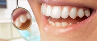
An “attractive smile” means not only straight and snow-white teeth, but also the correct contours of the tissues that frame the dental crowns. Unfortunately, their outlines are far from always ideal. If you are unhappy with your gum contour, contact your dentist for a plastic surgery procedure, which has become quite common in modern practice.
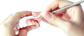
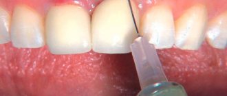
To perform gum surgery, local anesthesia is sufficient. The doctor can use either a scalpel or a laser. What exactly would be the optimal solution in your case, the specialist will decide during the examination. As a rule, to outline the desired result, the doctor draws the future gum line on the teeth.
The operation is performed by a periodontist. Before doing this, be sure to make sure that the specialist is qualified and look at examples of work he has already carried out.
Why is the tooth root exposed?
The reasons for the development of gum recession can be different, and in some cases there is a combination of several factors at the same time.
- aggressive brushing with a toothbrush - there is such a thing as brush gum recession. They occur when there is excessive pressure on the toothbrush when brushing, or when using a brush of unsuitable stiffness (hard). If you have increased tooth sensitivity when brushing, if your toothbrush becomes deformed over time (the villi move in different directions), you should consult a dentist about proper self-hygiene.
An example of brush gum recession on the upper jaw.
- incorrect position of the tooth in the dentition. Sometimes even a small bite problem can lead to gum pathology. This occurs because the tooth bears excess load when chewing or when teeth are closed, which over time leads to atrophy of the tissues of the supporting apparatus of the tooth, including the gums. In addition, excessive protrusion of the tooth root from the dentition also leads to gum atrophy.
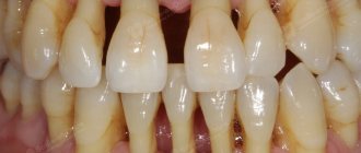
Multiple gum recessions caused by malocclusion.
- caries - in cases of gingival caries, gum loss often occurs due to the presence of an infectious focus in the immediate vicinity of the gingival margin.
Gum recession caused by tooth decay in the immediate vicinity of the gum margin.
- Tartar can also cause root exposure.
- anatomical features - for example, a thin biotype of gingival tissue in combination with other factors can lead to recession. Shallow vestibule of the oral cavity (in which the muscles of the lips attach very close to the gingival margin and gradually “pull back” it, exposing the roots of the teeth). In addition, improper attachment of the lip and tongue frenulum can contribute to gum recession.
The frenulum of the lower lip, a thin tissue biotype, caused the exposure of the lower incisor root
- bad or professional habits - for example, chewing a pen/pencil, biting threads (for a seamstress), pinching metal objects with teeth, etc.
- lip or tongue piercing - constantly touching the gums and regularly injuring it, metal jewelry can lead to exposure of the roots of the teeth.
- periodontitis - with inflammatory diseases of the gums, the roots are often exposed due to the resorption of the tissues surrounding the tooth, including gum tissue. Unlike “normal” gum recession, periodontitis is treated differently and the prognosis for such recessions is somewhat worse (as a rule, it is impossible to restore the volume of lost gum by completely covering the exposed root).
Types of surgery
At the preparatory stage, professional teeth cleaning is required. This is necessary to destroy plaque and food debris trapped in periodontal pockets. Immediately before the operation, local anesthesia is administered. The dentist performs a series of sequential actions in accordance with the chosen method. There are three main ways to perform a gingivectomy.
Simple means:
- Wave-like dissection of the gums and periosteal tissue at the site of inflammation.
- A similar cut is made from the inside.
- At the ends of each of the incisions, vertical cuts are made.
- The gum is separated and needs to be eliminated.
- Curettage of the bone cavity and wound surface is performed.
- A bandage is applied, which the patient wears for up to 48 hours.
Gentle - almost the same as the previous one. The difference is that in this case, not the entire wall of the pocket between the tooth and the gum is cut, but only part of it (no more than 3 mm). Then curettage is also carried out on the remaining part of the gum tissue.
Radical - refers to complex surgical procedures, since the tissue of the alveolar arch of the jaw is involved in the process. In this case, wave-shaped incisions are also made, after which the gum edge is removed along with epithelial cords, granulation tissue and affected areas of bone. At the last stage, a gum bandage is applied to promote rapid healing.
How to treat gum recession?
To cover the exposed root, a small operation is performed in the gum area.
There are many methods of gum plastic surgery, but they are fundamentally divided into 2 types:
- gum plastic surgery with local tissues (when the existing gum is “stretched” or moved to the root in a certain way so that the exposed area is closed, and fixed with small sutures, which are removed after healing).
- gum plastic surgery using a “patch” (the patch can be a special collagen-based material or the patient’s own tissue from the hard palate or the area of the far upper tooth - gum transplantation).
The choice of method is at the discretion of the doctor, according to each specific situation.
Indications
The operation may be prescribed in the following cases:
- “Short teeth” due to too wide a band of gum tissue.
- An uneven edge that looks unsightly.
- The gap between the gum and tooth (pocket) is too large.
- Inflammatory processes (periodontitis, gingivitis), which serve as an obstacle to fixation of the crown.
- Damage to gum tissue with the risk of spreading to neighboring areas.
There are a number of indications for the operation.
In these cases, tissue must be removed not only for aesthetic reasons, but also due to the fact that the gap between the teeth and gums is a place where bacteria accumulate, which can lead to the development of inflammatory processes.
The operation is not performed if there are contraindications , which include:
- decompensated diabetes mellitus;
- blood diseases;
- cardiovascular diseases in the stage of decompensation;
- infectious diseases in the acute stage;
- immune pathologies.
In addition, surgery is not indicated if the inflammation has already affected the bone tissue.
Is gum grafting painful?
No . Operations to eliminate gum recession are performed under local anesthesia; the surgical intervention is 100% painless. In the postoperative period, especially with gum plastic surgery using local tissues, patients often do not experience pain at all. If pain occurs, it is usually only on the day of surgery, a couple of hours after the intervention, when the effect of the local anesthetic wears off. During this period, the pain is perfectly eliminated with painkillers prescribed by the doctor. During the entire rehabilitation period, on average, 1-2 painkiller tablets are required.
In cases of gum plastic surgery using a “patch,” unpleasant sensations may be added due to the presence of a donor zone, which, according to patients, resembles the feeling “as if you grabbed hot tea,” but this does not always occur. The presence/absence of pain is determined by the area from which the “patch” is taken. If anatomical conditions allow, doctors can remove the graft from the donor area absolutely painlessly.
How to identify the problem?
If the flushing space corresponds to the norm, then:
- the prosthesis is fixed correctly;
- the gap between the bridge and the crown allows hygiene procedures to be carried out without any difficulties;
- the risk of developing periodontal pathologies is minimal;
- The device works normally for a long time.
You should consult a doctor as soon as possible if:
- after installation of the prosthesis there is noticeable discomfort or pain;
- there is a change in the location of the apparatus in the oral cavity;
- increased sensitivity to cold/hot;
- there is a burning sensation and other obvious signs of deterioration in the condition of the oral cavity.
Solution
If the gums rise or fall above the coronal part, you must consult a dentist. The doctor must take measures to restore the normal fit of the prosthesis, taking into account the distance between the structure and the surface of the oral cavity.
It is important to understand that the presence of any foreign object in the mouth is initially accompanied by discomfort, which goes away within a few days. If negative symptoms do not go away longer, you should immediately inform your doctor about this so that appropriate correction can be made.
Correction methods
- Local treatment with anti-inflammatory and antiseptic drugs (performed after removal of the orthopedic device).
- Correction of an existing prosthesis or production of a new one (the decision is made based on the results of an orthopantomogram).
- Fitting the prosthesis using the grinding method.
- Measures to strengthen supporting teeth with repeated prosthetics.
- In rare cases of the bridge coming off, which increases the risk of displacement of the supporting units, the prosthesis is reinstalled. To prevent this, it is important to consult a doctor immediately after identifying the problem.
What are the features and complications after gum recession surgery?
With a mild course of the rehabilitation period, patients do not note any peculiarities . But sometimes it is possible:
- the appearance of white plaque on the gums (this is normal, one of the healing options after gum surgery).
- swelling of the soft tissues of the face (from minor swelling to significant) depending on the extent of the surgical intervention (number of teeth involved), the size of the recession, the method of its closure, anatomical features and the patient’s body.
- hematomas on the facial skin (rarely encountered with gum surgery).
- increase in body temperature.
- pain (relieved by painkillers).
Consequences if the problem is left unattended
- Acute pain syndrome when exposed to teeth in the gingival margin area
- Severe inflammation of the gums or, conversely, the acquisition of an unnaturally white tint
- Significant deterioration in smile aesthetics, lengthening of teeth, formation of gaps
- Active development of caries, high risk of wedge-shaped defects
- Excessive accumulation of plaque at the base of the teeth, formation of periodontal pockets
- Spread of atrophic processes to periodontal and bone tissues with subsequent loss of teeth
What are the recommendations and restrictions after gum recession surgery?
- Do not disturb the wound surface (with tongue, food, foreign objects, etc.) this is the main condition for good healing.
- Do not bite (food eaten should be soft or finely chopped).
- Do not brush your teeth in the surgical area with a toothbrush (hygiene in this area should be maintained only as permitted by the attending physician).
- Do not eat or drink hot foods.
- Avoid physical activity.
- Follow the recommendations of your doctor and take prescribed medications.
Alternative options
- Cleaning teeth using ultrasound . Tartar is destroyed, the integrity of the unit is preserved.
- Drug treatment using antiseptics. The best option is Chlorhexidine in the form of rinses or compresses.
- A course of antibiotics . Allows you to quickly relieve the inflammatory process and alleviate the human condition.
Conservative treatment is not always effective. If characteristic symptoms reappear or the situation worsens, gingivectomy becomes mandatory.
What happens if gum recession is not treated?
- root caries (the root, unlike the crown of the tooth, is not covered with enamel - a dense “pearl” shell, and therefore is more vulnerable). Normally, the root is covered by the gum and is not exposed to the aggressive environment of the oral cavity (acids, humidity, microorganisms, enzymes). With gum recession, the exposed root is susceptible to destruction.
- abrasion of root tissues (again, due to the lack of enamel, tooth root tissues are not resistant to abrasion).
- increased sensitivity of teeth.
- the transition of a recession to a class that is not subject to complete closure.
- more plaque (the surface of the root is rougher than the surface of the tooth crown, which means that plaque is retained more when the roots are exposed).
Large gum recession, caries of the exposed tooth root, an abundance of soft plaque on the root surface.
Reasons for changing the size of the flushing hole
The gap may increase for the following reasons:
- tissue inflammation, accompanied by periodontal destruction (can occur when the immune system is weakened, as well as along with allergic manifestations and oral infections);
- violation of the sealing of the fixing elements of the orthopedic structure;
- damage to the integrity of the bridge at the junction with the crown (even minor damage to the prosthesis leads to its displacement and subsequent impact on the tissue around the tooth);
- poor oral hygiene and inflammation of the gums leads to their enlargement, due to which the rinsing hole is reduced;
- improper preparation of the oral cavity (poor sanitation, etc.) leads to damage to the enamel, due to which the gums rise above the coronal part;
- periodontal diseases, which entails a change in the original position of individual elements of the dentition.
Errors by the dentist or non-compliance with the technological process when installing a prosthesis can result in either an increase in the rinsing space or its complete absence.
Reference! Fixing the bridge too tightly increases the risk of injury to the gums in all parts of the oral cavity. Insufficient fit of the prosthesis, in turn, can lead to impaired speech functions, cause difficulties while eating and even difficulty breathing.
Deviation of the planting density from the norm is fraught with rapid damage to the structure beyond the possibility of repair.
Types of gum loss by severity and prevalence
The treatment regimen depends on the causes of the pathology, the severity of the lesion, and the volume of atrophied tissue.
Types by severity:
- Mild - loss of mucous membrane up to 3 mm. Conservative methods are usually sufficient.
- Average - 3-5 mm. In addition to therapeutic methods, there is a possibility of using surgical ones.
- Severe - more than 6 mm. It cannot be done without surgical intervention.
Depending on the volume of affected tissue:
- Localized Reduction in mucosal volume in the area of several teeth. As a rule, it develops as a result of injury and affects the tissues on the vestibular (outer) side of the teeth. Conservative treatment is often sufficient for mild tissue loss.
- Generalized The pathological process affects almost the entire dentition. Occurs in periodontal diseases, as well as in older people as a result of natural aging. There is a high probability of surgical treatment with significant atrophy.
The optimal treatment plan is drawn up by our periodontist after a detailed diagnosis.
Medvedeva Tatyana Alexandrovna
Dentist-periodontist, 6 years of experience
Specialist in the diagnosis and treatment of gum diseases. Conservative, surgical, regenerative treatment. Microsurgical operations without pain under sedation.
More about the doctor
Indications for plastic surgery in the oral cavity
The most common cause of gum loss is prolonged inflammation and treatment of periodontal disease or periodontitis. In these cases, there is a need to treat gum recession, which has noticeably decreased in volume and exposed the roots of the teeth. Other indications include:
- peeling of the gums from the surface of the dental crowns with the formation of deep periodontal pockets due to periodontitis or periodontal disease;
- uneven gingival margin, disrupting aesthetics;
- a defect called the “shark smile” and manifested in overhanging mucosa and visual shortening of the crowns;
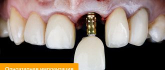
- the need to form an aesthetic gum margin during dental implantation;
- the need to maintain gum aesthetics after flap surgery;
- a short or powerful frenulum of the tongue or lip.
Effectiveness of events
The result of treatment directly depends on the severity of the recession
- At the initial stage , with a slight decrease in the gingival margin, the prognosis is favorable; you can quickly get rid of the defect 100% once and for all .
- The middle stage , at which exposure of tooth roots is already observed, requires an integrated approach. By combining therapeutic and surgical techniques, recession closure can be achieved .
- In severe forms , it is not always possible to completely eliminate the defect .
Timely treatment improves the chances of a favorable prognosis. When the first symptoms appear, we recommend not to self-medicate, but to contact a specialized specialist - a periodontist.
Where does treatment begin?
Before proceeding directly to restoring aesthetics, it is necessary to eliminate provoking factors - replace crowns, bridges, removable dentures, redo fillings, and perhaps even correct the bite.
Engaging in gum restoration without eliminating the root cause is useless and ineffective . If we immediately move on to closing the recession, the problem will remain and will only get worse.
If a loss of mucous membrane is observed against the background of bite defects, it is logical to first carry out orthodontic treatment, even before performing surgical correction of the gingival contour. If you correct your bite after plastic surgery, the scars remaining after the operation may prevent the teeth from moving in the desired direction. Therefore, the first step is to restore a physiologically correct bite using braces, aligners or other structures.
In addition, if the recession was insignificant before the installation of corrective devices, after correcting the bite, an independent rise in the gum level is possible. Surgery may not be required at all .
