Polyodontia is an abnormal number of teeth.
In medicine, this disease is often called hyperdontia, and “extra” dental elements are called supernumerary teeth. Research is still being conducted into why this pathology occurs. Most scientists associate it with disturbances in the formation of tooth germs.
Nature provides that a person grows no more than 20 milk teeth and 32 permanent teeth in a lifetime, but exceptions occur, and in our time quite often. According to statistics, on average, dental anomalies occur in 2% of the world's population, most often in men.
In 2014 alone, two operations were performed, in one of which 80 teeth were removed, and in the other, a record 232 teeth. Until this time, the maximum figure was 37 teeth.
The most common hyperdontia (anomaly in the number of teeth) is an anomaly of the upper incisors. Supernumerary teeth are less common among the lower incisors and in other parts of the jaw. They can come in a wide variety of shapes and sizes. These are usually small, cone-shaped teeth.
Extra teeth lead to deformation of the dentition, so it is recommended to remove supernumerary elements. Another reason for removal is that most patients with this pathology have a lisp.
The formation of extra teeth is quite common today. According to statistics, 70% of patients have only one extra incisor, in 25% of cases – 2 supernumerary elements, and only 5% of all patients have 3 or more teeth during examination.
How many implants need to be placed?
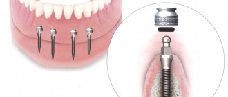
In the absence of three adjacent units of the dentition, the following are placed:
- 2 implants - are introduced along the edges of the defect, serve as support for a permanent bridge prosthesis consisting of three welded crowns (two of them are fixed to titanium roots, and the central one is hinged);
- 3 implants - replace each lost tooth and, after healing, are covered with separate crowns.
Which option will be chosen and which orthopedic structures will be used depends on the location of the defect.
Front teeth
Implantation of three adjacent incisors has a number of features due to increased aesthetic requirements:
- white zirconium abutments are used that are not visible through the gum;
- if necessary, plastic surgery of soft tissues is performed;
- smaller implants are selected for installation in the narrow bone of the frontal jaw;
- Crowns made of lightweight materials with bite relief are placed as temporary prostheses.
If three teeth in a row are missing from the front, both restoration options are suitable (2 and 3 titanium roots), however, the installation of three implants followed by prosthetics with single crowns is preferable. In this way, identity to natural teeth is achieved, an even gingival contour is formed, which guarantees maximum aesthetics.
Chewing teeth
When replacing three adjacent units of the lateral jaw, high demands are placed on the functionality of the structures. They must be securely fixed, withstand severe chewing loads, and prevent atrophic processes.
In such cases, we choose the option of installing three implants with single crowns, since a bridge on two titanium roots does not provide uniform pressure on the bone and leads to its resorption.
When implanting several teeth in a row, three or more, it is important to assess the condition of adjacent units. If they previously served as supports for a bridge, then, as a result, they are destroyed and cannot be restored - they will have to be removed and work with a more extensive defect.
Implantation of 3 teeth, E.max ceramic crowns
Attending doctor
Strigin Vladimir Igorevich
Find out the price
Causes of polyodontia
Medicine has not yet found an exact answer to the question of what are the causes of supernumerary teeth. Scientists put forward several hypotheses:
- Atavism. Supernumerary teeth are explained by the fact that the dental system strives to return to the original number of elements laid down by nature. There is evidence that our ancestors had 6 incisors on both the lower and upper jaws. As a result, many doctors consider atavism to be the cause of the development of polyodontia in humans.
- Splitting of the tooth germ. Even in the embryonic period, the activity of the dental plate is disrupted in the child, as a result of which hyperdontia is formed. Violations can be caused by viruses, poor ecology, drugs, medications prohibited during pregnancy, alcohol and other factors. This hypothesis is increasingly supported today, because recently the disease has been rapidly progressing due to bad habits and poor ecology.
The causes of hyperdontia continue to be researched. Scientists cannot give an exact explanation for this anomaly, but most of them are inclined to the second hypothesis - the splitting of the tooth germ at the embryonic stage.
What does polyodontia look like?
Quite often, extra teeth are almost indistinguishable from normal ones. It is not uncommon for them to grow in the form of a drop or a thorn. These dental elements can appear either individually or fused with permanent ones. They can form tooth-like formations and entire arrays of teeth.
Also in medical practice, there are cases where polyodontia was hidden and was detected only by radiography. There are many different cases of abnormal development of the number of teeth, and if you notice symptoms, you should definitely contact the dentist.
Types of polyodontia disease
Polyodontia in the oral cavity manifests itself in different ways. By studying the statistics, signs and symptoms of the disease, dentists were able to classify the types of this anomaly.
Depending on the origin, the disease is divided into two types:
- False polyodontia. Provides for a baby tooth that does not fall out, regardless of the person’s age. At the same time, it fulfills its functions, does not create discomfort to the bite, and is firmly fixed in the patient’s jaw. In addition, teeth fused together and other anomalies are classified as a false type of disease.
- True polyodontia. It can be caused by genetic predisposition, as well as terogenic factors. At the same time, extra molars begin to form in the human jaw.
As for the placement of extra teeth, dentists distinguish the following types of disease:
- Typical hyperdontia. Applies to those patients in whom extra teeth appear only in the dentition and do not extend beyond it. Many scientists are confident that this is simply heredity, because our ancestors had a more developed dental system than modern people.
- Atypical hyperdontia. It occurs much less frequently and is characterized by the appearance of teeth outside the dentition.
In case of anomalies with baby teeth, the latter pose almost no threat. On the contrary, such a tooth can last a lifetime. But the permanent molars, over which the supernumeraries grow, should be removed, if only because it is not aesthetically pleasing.
Often, the patient grows extra fangs or incisors, or even several front teeth at once. In addition to a ruined smile, the disease can cause serious complications if the necessary measures are not taken in time.
Second lower molar
Average age of teething: 11-13 years
Average age of root formation: 14-15 years
Average length: 19.8 mm
The crown is somewhat smaller in size than the first molar, and the more symmetrical second lower molar is characterized by a close arrangement of roots. The roots gradually converge distally and have closely spaced apices.
Exposure is made at the mesial portion of the crown with access extending only slightly distal to the central sulcus. After trephination of the cavity with a fissure bur with a cutting apex, an elongated ball-shaped bur is used to expand it until free access is achieved. The distal root angles often allow for a smaller opening than the lower first molar.
Particular attention should be paid to the shape of the mouth of the distal canal. A narrow oval orifice indicates the presence of a slit-like lumen in the distal canal, which requires more careful treatment.
All carious tissue, leaking fillings and denticles should be removed and replaced with a suitable temporary filling prior to endodontic treatment.
The lower second molar is most susceptible to vertical fractures. After preparing the access, but before starting endodontic treatment, the clinician should examine the floor of the pulp cavity using fiber optics. For mesial-distal crown or root fractures, the prognosis is extremely unfavorable.
To avoid vertical fractures after endodontic treatment, it is necessary to completely cover the cusps with a restorative structure.
Symptoms of the disease in children
The first supernumerary teeth in children appear before birth or in the first six months of life. The main inconvenience they cause is difficulty in feeding.
Polyodontia of primary teeth in older children occurs with symptoms similar to the eruption of regular teeth. In this case it is observed:
- temperature increase;
- swelling of the gums in the place where the tooth should erupt;
- pain;
- excessive salivation;
- swelling of the nasal mucosa;
- loose stool.
Symptoms are especially severe when extra teeth appear in the upper palate.
If hyperdontia makes itself felt in a two-year-old child, this can interfere with the formation of normal speech. In turn, due to injury to the tongue and mucous membranes, some kind of inflammation constantly appears in the oral cavity.
When supernumerary teeth appear in very noticeable places in school-age children, ridicule towards the patient may occur, which is fraught with the development of psychological problems and complexes in the future.
The recipe is ready
That's all, in short, what is needed to create teeth. Thus, the recipe for creating a tooth looks something like this:
- Stem cells - assorted
- Scaffold is a natural product
- Growth factors - to taste
Regenerative medicine technologies are progressing incredibly quickly. And now, probably, the most basic provisions for dental tissue engineering have already been developed. They all stem from our knowledge of the cellular and molecular basis of tooth development. We understand that the best result in tooth bioengineering can be achieved only in the presence of two types of cells, and not one: these are both epithelial cells and mesenchymal cells (where would we be without them?) [28]. However, you cannot build a tooth on cells alone. Thus, the role of growth factors and extracellular matrix cannot be excluded here. Fortunately, science does not stand still, and new provisions are being actively developed. Perhaps, in the near future, the treasury of knowledge called “dental tissue engineering” will be replenished with another equally valuable “coin”.
But, despite all the promising potential of tissue engineering in dentistry, there are still problems to be solved related to the conduct of clinical trials, the innervation and blood supply of the bioengineered tooth, its ligamentous apparatus, the timing of its eruption, as well as the choice of a pool of stem cells and technology for working with them , and a number of other equally pressing tasks [10], [29].
As for the most basic thing, namely stem cells: in the experiments performed (it is worth noting that almost all of them were carried out on mice), they mainly used embryonic stem cells. But in the clinic their use is sharply limited, including by law. Therefore, only postnatal stem cells remain (not counting iPSCs, where things are also not calm), and here we face the following snag: unlike mice, humans lack a niche of dental stem cells, which is why our teeth do not have the ability to constantly grow. Those MSCs that are suitable for use cannot be obtained without damaging the tooth, or even more so if the tooth was previously treated endodontically, that is, with pulp removal. Those to which access is open do not have odontogenic competence, for example, gingival MSCs. This is just one of the dilemmas that remain to be resolved (Fig. 9).
Figure 9. The fight for healthy teeth for humanity.
website accuratedentistry.net
Symptoms of hyperdontia in adults
Polyodontia affects permanent teeth more often than baby teeth. An adult usually develops dystopic and impacted supernumerary teeth.
Dystopic teeth are those that appear outside the dental arch. Most often they erupt on the lingual surface of the gums and in the palate. With this form of the disease, the patient typically:
- poor pronunciation of sounds;
- noticeable malocclusion;
- change in the usual arrangement of teeth: curvature of the angle at which they grow, as well as their rotation
- around its axis;
- frequent injury to the oral mucosa and, as a result, its inflammation;
- disruption of chewing processes, resulting in digestive problems.
Among other things, dystopic teeth often cause psychological problems. Due to a non-aesthetic, and sometimes completely unattractive smile, the patient becomes withdrawn and uncommunicative. Psychological problems, in turn, cause chronic diseases of the endocrine, digestive and nervous systems.
Impacted supernumerary teeth are teeth that do not erupt, but continue to remain in the bone tissue of the human jaw. Often they hardly make themselves felt until complications begin. Dentists diagnose this anomaly during a routine examination of the patient.
This abnormality in the number of teeth is accompanied by the following symptoms:
- normal teeth begin to loosen (the condition is considered pathological);
- the bone begins to protrude (if the impacted tooth is too close to the edge of the jaw);
- Aching pains appear periodically.
One of the most difficult situations is when extra teeth grow in place of impacted third molars. Wisdom teeth cannot grow and begin to negatively affect the roots of other teeth, which in turn can lead to serious complications.
Non-Hong Kong "Triad"
Now that we know so much about the origin and development of the tooth, we can move directly to the topic of interest to us - tissue engineering.
Tissue engineering is a set of methods and procedures aimed at the regeneration of biological tissues. It includes a triad of main elements (Fig. 4): stem cells, extracellular matrix or scaffold, growth factors and signaling pathways [10].
Figure 4. Tissue engineering triad. The basis of the tissue engineering triad is stem cells, growth factors and extracellular matrix.
[10]
The goal of tissue engineering is to replace lost cells, tissues and organs, or promote their regeneration, or simply restore impaired function.
Today we hear and read a lot about stem cells. This is a hotly debated branch of science. The information that goes out to consumers, as a rule, is not always objective. What exactly are stem cells, and how and which of them can be used in dental tissue engineering?
Let's get acquainted: stem cells are undifferentiated embryonic or adult (postnatal) cells that are capable of going through a huge number of cell divisions while in an undifferentiated state, as well as forming intermediate cell types - precursors that can differentiate into various cells and create full-fledged tissues and organs (Fig. 5) [10], [11].
Figure 5. Classification of stem cells according to their ability to differentiate. Based on the scale of differentiation, stem cells are divided into totipotent, pluripotent, multipotent and unipotent. Totipotent cells are capable of differentiating into any cell type of an adult organism. Pluripotent cells can produce specialized cells of the three germ layers (ectoderm, endoderm and mesoderm), but not the entire organism. Multipotent cells produce a limited range of cell types. Unipotent cells are capable of differentiation into only one type of cell [13].
[11]
The first cell line of embryonic stem cells was isolated back in 1998 [12]. In fact, not so long ago, and from the point of view of the course of history one can say quite recently, but the progress is colossal [10].
Embryonic stem cells are isolated from the blastocyst during embryonic development. They give rise to three germ layers: ecto-, endo- and mesoderm. These cells are totipotent, meaning they can develop into each of the more than 200 cell types in the adult body [10].
There are currently 3 known sources of mammalian embryonic stem cells: cells isolated from the inner cell mass of the blastocyst; teratoma cells and primary germ cells of the embryo [10].
As was previously mentioned, stem cells are not only embryonic, but also postnatal. As for “adult” stem cells, they exist in the body in various tissues, including bone marrow, blood vessels, liver, skin, adipose tissue and dental tissue. They are localized in special niches where their proliferation, migration and life span are regulated. Postnatal stem cells are multipotent, meaning they give rise to only one type of cell.
Dental stem cells are a population of postnatal mesenchymal stem cells (MSCs) that have the ability to self-renew and differentiate [4], [14]. Depending on the location of the MSC depot (Fig. 6) [15], they are divided into:
- pulp stem cells;
- apical papilla stem cells;
- stem cells from extracted baby teeth;
- dental follicle progenitor cells;
- periodontal ligament stem cells;
- MSCs obtained from the alveolar process;
- MSCs of the gums;
- progenitor cells (MSCs aimed at differentiation only into a certain type of cell) of the tooth germ.
Figure 6. Dental stem cells. Schematic representation of sources of dental stem cells. For an explanation of the abbreviations, see the box below.
[15]
Abbreviations
WHO World Health Organization MSCs mesenchymal stem cells ECM extracellular matrix ABMSCs alveolar bone-derived mesenchymal stem cells BMP bone morphogenetic protein DFPCs dental follicle progenitor cells DPSCs dental pulp stem cells FGF fibroblast growth factor GMSCs gingival mesenchymal stem cells iPSCs induced pluripotent stem cells PDGF platelet derived growth factor PDLSCs periodontal ligament stem cells SCAP stem cells from the apical part of the human dental papilla SHEDs stem cells from human exfoliated deciduous teeth TGPCs tooth germ progenitor cells germ progenitor cells)
Let's look at some of them.
Pulp stem cells can be quite easily isolated from the pulp of extracted teeth. They represent a very attractive and promising source of autologous stem cells and can be used both for the regeneration of dentin, pulp and cement, and for the restoration of bone tissue [15]. In addition, they exhibit strong neuroregenerative activity, which is of particular value in the treatment of spinal cord injuries: pulp MSCs, in addition to suppressing the early inflammatory response, inhibit the apoptosis of neurons, astrocytes and oligodendrocytes after injury, which leads to the preservation of the nerve fiber and myelin sheath. They have also been found to promote the regeneration of severed axons. Thus, scientists hypothesize that pulp MSCs could provide significant therapeutic benefits in the treatment of spinal cord injury [16].
Stem cells from extracted primary teeth are a postnatal population of stem cells with high proliferative capacity, high viability, and the potential for multilineage differentiation (e.g., into osteoblasts, neuronal cells, and odontoblasts) [15].
Gum mesenchymal stem cells are ideal for restoring damaged periodontal tissue, muscles and even tendons. But it is not yet entirely clear whether they are capable of forming dentin and pulp cells [15].
Tooth germ progenitor cells are a relatively new population of stem cells that were discovered in the mesenchyme of the third molar germ at the bell stage. They show the same multilevel differentiation as other dental MSCs, including the ability to differentiate into adipocytes, osteoblasts, odontoblasts, chondrocytes and neurons, and can also differentiate into cells with the morphological, phenotypic and functional characteristics of hepatocytes. Hence, it is assumed that this type of stem cells can be used in the future to treat liver diseases [15].
Thus, each type of dental stem cells has its own characteristics and areas of application not only in dentistry, but also in other areas of medicine.
In addition to the MSCs described above, induced pluripotent stem cells (iPSCs) derived from somatic cells are also used in tissue engineering. They were first discussed in 2006, when Japanese scientists Kazutoshi Takahashi and Shinya Yamanaka showed that somatic cells can be reprogrammed into iPSCs by increasing the expression of certain transcription factors (Oct3/4, Sox2 and Klf4) [17], [18]. These cells themselves are immunologically neutral and, just as importantly, do not raise the same ethical controversy as embryonic stem cells. However, viral agents were used to reprogram them, which could lead to the formation of neoplasms [19]. There were attempts to use chemical molecules instead of viruses [20], but, unfortunately, the percentage of successful reprogramming turned out to be small. New methods for obtaining iPSCs are now being developed, since their application looks quite attractive and very promising.
Consequences of polyodontia disease
Polyodontia in humans can often be the cause of retention. This is a phenomenon in which normal teeth are unable to erupt due to the interference of supernumerary teeth. The former may remain in the jaw or take an abnormal position.
In addition, even if the complete incisor grows before the supernumerary one, the latter will be able to displace it. This will lead to the person being unable to chew food normally. And if several extra incisors grow at once, they can cause the loss of permanent teeth.
Lateral lower incisor
Average age of teething: 7-8 years
Average age of root formation: 10 years
Average length: 21.1 mm
Very similar to the lower central incisor, so access cavity preparation follows the same principles.
Their similarity can cause rare but serious errors. Hasty installation of a rubber dam, identical fillings and carelessness can lead to preparation of the access cavity on the wrong tooth. This error can be prevented by marking the vestibular surface of the tooth with a felt-tip pen before applying the rubber dam.
Trauma, periodontal disease, carious lesions and malocclusion can lead to obliteration of the canal. When moving apically to identify the orifice, great care must be taken to prevent unnecessary destruction of the crown and root. Labial perforations were discussed in Illustration IX. If the bur is not oriented along the long axis of the tooth, there is a risk of lateral perforation. The situation becomes more complicated with the traumatic loss of an anatomical crown. Without anatomical landmarks, a lateral perforation can be easily made when moving in a coronal direction. To prevent this, access is carried out without a rubber dam so that the root can be palpated.
Lateral perforation with endodontic files and Gates-Glidden burs is facilitated by the presence of slit-shaped canals with a narrow, hourglass-shaped cross-section. To avoid vertical fracture along the approximal root wall, minimal expansion and preparation of the space for the abutment pin is indicated.
Apical curves and accessory canals are common in the lower incisors.
Diagnostics
Examining supernumerary teeth during an x-ray is not as easy as it seems. They can be superimposed along the contour onto the permanent ones and remain invisible. In such cases, patients are recommended to undergo a computed tomography scan, which shows a more accurate picture of the disease.
If the extra dental elements have already erupted, the dentist can easily detect them. In practice, the patients themselves find the erupted supernumerary teeth and already at the initial appointment with the dentist they complain about the pathology.
"Eights" have long lost their functionality
In ancient times, people ate raw meat, roots, and they needed massive molars to chew solid food. Over time, people began to consume thermally processed, softer foods, and the urgent need for the most distant units disappeared. However, you should not completely discount them.
Despite the fact that they themselves do not take much part in chewing food, “eights” restrain the loosening of the teeth, which bear the main chewing load. And in old age they can serve as a support for a bridge.
Symptom relief
Most often, in adults, extra teeth erupt without any symptoms, but for children this can become a problem that needs to be addressed.
Supernumerary teeth erupt with the same symptoms as regular teeth, so the treatment for them is the same.
- To lower the temperature, it is recommended to give your baby Paracetamol or Ibuprofen. If the child is very small, these drugs can be used in the form of suspensions or rectal suppositories. In addition to lowering the temperature, these medications do an excellent job of treating pain and inflammation.
- To relieve gum pain, local anesthetics are used - ointments and gels (for example, Kalgel, Dentinox, Solokoseryl). These remedies cope well with painful sensations and slightly relieve inflammation.
- Adults and children over 2 years of age can be treated with folk remedies: propolis, honey, decoctions of calendula, chamomile and lemon balm. Some decoctions help reduce pain and relieve inflammation. Traditional methods of treatment should be used only after consultation with your doctor.
- If primary supernumerary teeth have partially erupted, stimulation of eruption is prescribed. For this purpose, vibration and electrical stimulation, as well as special massage, are used.
Mandibular canine
Average age of teething: 9-10 years
Average age of root formation: 13 years
Average length: 25.6 mm
The canine of the lower jaw is more powerful and significantly wider than the incisors in the mesial-distal direction. It rarely causes treatment problems. The atypical form with two roots can be problematic, but is rare.
The access cavity is oval and can be expanded in the incisal direction to facilitate vestibular-lingual access. In the cervical region the canal is oval, in the middle third it is rounded. To completely clean its walls, directed instrumental action is necessary.
If there are two roots, one of them will always be easier to instrument. The other canal must also be opened and funnel-shaped in accordance with the first to prevent dentine filings from entering it and impairing access. Pre-bending the instruments during the initial approach will allow the clinician to walk along the walls of the buccal or lingual root until the tip of the instrument enters the orifice. Once a difficult-to-reach canal has been identified, every effort must be made to shape and create a funnel-shaped orifice to keep access open.
Normal deletion
If the dentist decides that in a particular case, polyodontia can only be treated by removing an extra tooth, the patient should count on the following procedures:
- First of all, the patient should be sent for radiography. This is necessary in order to determine the size and number of roots, as well as the ratio of supernumerary and normal teeth.
- After collecting research, the doctor gives the patient anesthesia and removes excess teeth.
- In some cases, soft tissue sutures may be necessary after surgery.
Removal of impacted teeth
In order for the operation to be successful and polyodontia to be cured without any complications, the doctor must fully examine the patient and plan his further actions.
- To begin with, X-rays and/or computed tomography are performed to determine the exact topography of the anomaly.
- Removal is performed under local anesthesia, but there are cases when general anesthesia can be used on the patient.
- First, the mucous membrane is peeled off, then the bone tissue is opened and the root and crown parts of the tooth are removed.
- If necessary, bone defects are covered with osteoplastic material, and the mucous membrane is sutured.
After tooth extraction, the patient continues treatment at home: takes antibiotics (if prescribed by the attending physician), rinses the oral cavity with antiseptic solutions.
Until the wound heals after surgery, it is not recommended to eat too hot, hard or spicy food. You should also brush your teeth carefully, especially on the operated side.
Removing "eights" is always difficult
If the unit is located correctly and does not have serious pathologies, it will not be difficult to remove it. To carry out the removal without the risk of complications, before the procedure the patient is prescribed an X-ray of the tooth.
However, most often the third molar grows incorrectly, partially erupts or becomes intertwined with neighboring roots. In this case, the intervention will be carried out in a complex way. The final decision on the choice of method is made by the doctor after examination and obtaining the results of an x-ray of the tooth.
When using a complex method, manipulations are performed in the following sequence:
- The gum is cut and peeled away from the bone.
- To access the roots, a hole is drilled into the bone.
- The unit is extracted (in whole or in parts).
- The wound is treated and stitches are applied.
The procedure can take from 20 minutes to 2 hours. Everything is individual and only an experienced doctor can professionally perform the removal. Therefore, you should be careful when choosing a clinic and specialist.
After surgery, it is recommended to eat soft foods and refrain from physical activity for several days. Antibacterial therapy may be prescribed for prophylactic purposes. Pain and swelling persist for 3-4 days and then gradually disappear.
Visiting the dentist immediately after the figure eight begins to erupt will save you from many serious complications.
Complex dentistry "Sanident" invites all residents of Ivanteevka and Shchelkovo to use the services of experienced dentists who can cope with any dental disease.