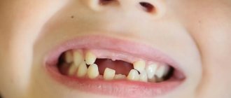
A dystopic tooth, or dystopia for short, is a tooth that has grown in the wrong place. In this case, the tooth is incorrectly positioned relative to the jaw arch and its specific socket in particular. People under 20 years of age most often complain about dystopia (this phenomenon is often associated with the growth of wisdom teeth), as well as children whose molars are growing.
Dystopic wisdom tooth: what is it?
A dystopic wisdom tooth is a third molar, also known as a “figure eight”, which is positioned incorrectly relative to the rest of the dentition. Such a tooth almost always needs to be removed.
A special case of a dystopic wisdom tooth is an unerupted, but fully formed figure eight, which turns out to be turned parallel to the gum. In this case, the tooth is there, although it is not visible, and it is in an incorrect position, so we can talk about dystopia.
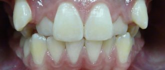
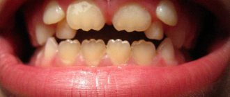
Two examples of dental dystopia
Therapy
Treatment for dystopia may be different in each case. Consider your options.
If a wisdom tooth is found to be dystopic, and when chewed it is of less value, then dentists, as a rule, recommend an extraction procedure. Its extraction is especially indicated if the “eight” injures the neighboring one.
It is important to know that the removal of a dystopic tooth can lead to complications, such as dislocation of an adjacent premolar, jaw dislocation, and bleeding.
Teeth must be removed in the presence of pulpitis, periodontitis, cysts, or periostitis . Sometimes the procedure can only take place in a hospital, since this requires a full-fledged surgical intervention with further rehabilitation of the patient.
When there is enough space in the dental arch for normal placement, dentists use braces as a treatment. If the position of the canine is incorrect, most often they try to save it, since it is involved in chewing activities.
The adjacent premolar is removed to make room for the canine tooth. Treatment with braces takes the longest, as retainers will subsequently be required. This is necessary to ensure that the dystopic tooth does not return to its original position.
The stages of treatment are illustrated in the video:
If a tooth is injured, they try to restore it using special splints and wire ligatures. When restoration is not possible, removal is performed, and then prosthetics or implantation.
If the tooth does not provoke any functional problem and does not cause discomfort, then the procedure of grinding sharp cusps is sufficient.
Causes of dystopic wisdom teeth
Very often, dental dystopia is associated with genetic reasons. A predisposition to shifting dentition is inherited along with small jaw size and other aspects of individual appearance that can cause teeth to shift. Doctors call other factors that provoke improper growth of molars, incisors and fangs:
- atypical formation of the tooth germ in the embryonic period;
- macrodentia, partial adentia, supernumerary teeth and other diseases;
- improper eruption (can be caused by early removal of the primary bite);
- small jaw size;
- mechanical damage, dental trauma;
- bad habits such as chewing foreign objects, thumb sucking (affect the development of malocclusion);
- late eruption of canines, for which there is no longer room in the dentition.
Modern dentistry successfully cures the patient, regardless of the causes of tooth dystopia and its position in the gum. The main thing is to seek help in time.
Why does retention occur?
Most often, wisdom teeth cannot fully erupt. This is due to the peculiarities of their location. In the process of evolution, eights have lost their meaning. Some people don't have them at all. In others, they often “sit” under the gum and contribute to the displacement of neighboring units, which causes the bite to change.
Retention is caused by:
- heredity;
- premature removal of baby teeth, which leaves little room for constant sweat;
- excessively thick gum walls (very rare);
- malocclusion, severe crowding.
Types of wisdom teeth dystopia
Wisdom tooth dystopia is classified depending on which direction the crown is shifted. The following types of disease are distinguished:
- vestibular (forward displacement);
- oral (teeth located behind the entire row);
- mesial (forward bend);
- distal (backward tilt);
- with supraposition (above the dentition);
- with infraposition (below the dentition);
- with tortoposition (the tooth is rotated around its axis);
- with transposition (the tooth is in the place of another tooth).
In the most severe cases, you can observe the appearance of a tooth outside the oral cavity: for example, inside the nose. This is very dangerous and requires immediate treatment.
In what cases is dental surgery difficult?
Complex manipulations regarding tooth extraction include the following situations:
- with disconnected roots that are excessively curved inward or directed in different directions;
- when the problem tooth is located in the affected bone;
- in the complete absence of a crown, when there is nothing to grab onto with forceps;
- if a filling was previously installed in the tooth, which can crack under the mechanical influence of forceps;
- when the tooth is impacted or dystopic;
- removing the "eight"
These are the main clinical features in which it is impossible to get rid of a diseased tooth using a standard simple procedure. Moreover, in each specific case the reasons for complex extraction may be individual.
Symptoms of dystopia
Symptoms of dental dystopia are most often visual and easily noticeable. It is enough to look at the position of all the elements of the dentition to see that some of them are in the wrong places or are simply growing unevenly. Other symptoms:
- there is no tooth in the bite, and the period of eruption has already passed;
- inflammation and pain are felt at the site of the wisdom tooth;
- The wisdom tooth did not appear on time
It is interesting that in a number of cases there is no wisdom tooth at all - it does not form and does not erupt. Dentists call this option a variation of the norm.
Classification of impacted teeth
Taking into account how deep the unit is under the gum tissue, retention is usually classified into:
- partial (part of the impacted crown rises above the surface of the gum and is clearly visible to the naked eye);
- complete (the entire crown is covered by gingival tissue or is located deep in the jaw bone).
Doctors also take into account where the tooth is located. If in bone tissue, then they speak of retention with immersion, if above it - without immersion.
Impacted dystopic molars, incisors, and canines are also found. They grow out of place or at the wrong angles. Then there is a significant displacement of the dental axis.
Consequences of dystopia
Although dental dystopia does not seem like a terrible disease, it can create many problems. This is why wisdom teeth that have grown incorrectly are most often removed. Among the most unpleasant consequences of the disease:
- disruption of the entire dentition, because due to one incorrect element, the rest move or cannot erupt correctly;
- injury to the tissues of the cheeks, tongue, lips, which can cause ulcers and even provoke the development of tumors;
- complication of oral hygiene, which leads to inflammation, caries, both on the dystopic and adjacent teeth;
- the appearance of gingival pockets where bacteria accumulate, leading to pericoronitis;
- impaired chewing function, which in turn reduces the quality of food and can lead to deterioration of the gastrointestinal tract;
- difficulty pronouncing words, poor diction;
- violation of the facial skeleton, unaesthetic appearance of the lower part of the face.
Almost always, dystopia leads to unpleasant aesthetic consequences, which causes complexes and rejection of one’s appearance in the child, and then in the adult. Of course, this can be worked out with a psychologist, but it is much wiser and more correct to deal with the cause than to deal with the consequences.
Treatment of dystopic upper canines
The upper canines are critically important teeth for the dentition, given the characteristics of their morphology, position, protective function in the structure of occlusal interactions, as well as the volume of visualization in the smile profile. The proper position of the canines in the dental arch is one of the criteria for the predicted stability of the results of dental treatment.
Dystopia and tooth retention can develop due to physical blocking by other teeth, thickening of bone tissue or mucosa due to the presence of supernumerary teeth, odontoma or some other tumor. The third molars of the lower jaw are characterized by the highest prevalence of cases of dystopia and retention, while the second place is occupied by the canines of the upper jaw. During eruption, the upper canines must travel a significant distance from the lower edge of the orbit to the edge of the bony ridge when all other anterior teeth have already erupted. The prevalence of dystopia and retention of the upper canines is about 2%, and among women this pathology occurs 2-3 times more often than among men. About 66% of cases of dystopia and retention of the upper canines are situations with their displacement in the projection of the palate, while the remaining 33% fall on dystopia in the vestibular position. Bilateral dystopia is noted with an 8% prevalence rate. In Latin American countries, according to publications by authors from Colombia, Mexico and Brazil, the prevalence of canine dystopia is almost the same as in Europe or the USA. However, countries such as Greece or Turkey, for example, are characterized by a slightly higher level of registration of cases of the above-mentioned pathology (at a rate of approximately 4%).
Dystopia of the upper canines can cause displacement of other teeth, cyst formation, resorption of the roots of the lateral incisors (especially when their roots are oriented palatally), the development of localized and widespread pain, as well as inflammatory disorders. Clinicians should be aware that there may be a difference of up to 6 months between the child's chronological age and the age of canine eruption to ensure that there is no developmental or eruption disorder. Only the clinical sign of the absence of a canine tooth in the dentition at the age of 12 years is not a dominant criterion for making any diagnosis. Timely diagnosis and appropriate interventions allow a more optimal approach to the issue of proper occlusal correction. If early interventions are unsuccessful, a multidisciplinary approach to treatment may be necessary, which involves the implementation of surgical and orthopedic phases of intervention.
Causes of dystopia
Potential causes of dystopia suggested by Becker and Chaushu include: increased size of the tooth germ, odontoma, the presence of an unerupted tooth in the path of the problematic unit of the dentition, delayed eruption of adjacent teeth, and the presence or absence of lateral incisors. Three main criteria must be considered to properly evaluate dystopic maxillary canines: diagnostic principles, treatment plan, and biomechanical principles. To adequately assess the existing disorder, it is necessary to use both clinical and radiological diagnostic methods. The treatment plan for canine dystopia often involves close collaboration between the orthodontist and the oral surgeon. The implementation of biomechanical principles must take into account the possibilities of effective application and orientation of orthodontic force vectors for therapeutic purposes.
Bishara conditionally divided all the causes of dystopia into local and general. Among the common causes, the author identified disorders in the structure of the thyroid gland, hypovitaminosis of vitamins A and D, infectious diseases, radiation, and several syndromes, including Crouzon syndrome and Down syndrome. Local causes of dystopia include the presence of supernumerary teeth, odontomas, trauma at an early age, cleft lip or palate. Other potential causes are associated with abnormalities in the morphology or position of the tooth germ, disturbances in the eruption path of the canines, ankylosis of the periodontal ligament, prolonged retention or, conversely, too rapid loss of primary canines, iatrogenic causes, bifurcated roots, or the influence of idiopathic factors.
It is generally accepted that the specific cause of buccal retention of the maxillary canines is a lack of space in the dentition or insufficient length of the dental arch. Tomographic studies have demonstrated a relationship between narrow maxillary shape and buccal canine retention. But similar associations were not found for palatal dystopia and maxillary canine retention. Jacobs reported the influence of tooth size discrepancy on the possibility of developing impaction of canines, with canines being more prone to such problems due to their significantly longer eruption path compared to other teeth. Becker suggested that palatal retention of the canines may be caused by a lack of direction from the roots of the lateral incisors (in the absence or morphological deformation of the latter). Other authors have cited genetic causes behind canine impaction, including microdontia of the lateral incisors, enamel hypoplasia, defective eruption of primary molars, or distally disrupted eruption patterns of second premolars. Peck et al associate palatal dystopia and canine impaction with a broader set of chromosomal abnormalities.
Planning of surgical/orthodontic interventions and algorithm for selecting them
To help clinicians determine the optimal treatment method for maxillary canine impaction, an algorithm for selecting surgical and orthodontic types of interventions will be presented below.
Diagnosis of canine retention
The diagnostic process should follow a logical sequence and include a detailed analysis of personal and family history of existing dental disorders, the results of a comprehensive clinical examination, which in turn includes: palpation, X-ray screening through orthopantomography, lateral cephalography and radiography in the superior occlusal projection. It is also recommended to obtain additional targeted images using the Clark method, although from the point of view of cost-effectiveness, orthopantomography is a more reasoned diagnostic approach. For a more detailed analysis, you can use the method of cone beam computed tomography. The latter makes it possible to diagnose root resorption of adjacent teeth, possible transposition between the lateral incisors and canines, as well as the trajectory of the eruption of the canines in cases of its projection over the apical areas of the lateral incisors. In addition, CBCT can identify root splitting and signs of ankylotic changes. According to Ericson and Kurol, CBCT data confirmed evidence of root resorption in 38% of maxillary lateral incisors and 9% of maxillary central incisors due to canine impaction (Figures 1 and 2).
Photo 1. Scan of palatally impacted canines (front view).
Photo 2. Scan of palatally impacted canines (occlusal view).
CBCT method and canine retention
As a valuable X-ray diagnostic tool, the use of CBCT is indicated depending on the amount and nature of the information that the doctor received during the clinical examination. The use of CBCT must be justified taking into account the individual needs of each individual patient. This means that CBCT should not be considered as the first imaging modality in the absence of clinical suspicion of maxillary canine impaction. Localized CBCT imaging is only warranted after careful clinical evaluation and in cases where other radiographic diagnostic modalities do not provide the necessary data to identify or differentiate the pathology. Although it is intuitive to assume that 3-dimensional imaging is certainly superior to 2-dimensional imaging, this assumption has not been completely unambiguous in cases of impacted canines. Systematic reviews comparing diagnostic approaches to the verification and assessment of canine impaction using CBCT and traditional imaging methods have shown that, although CBCT leads to greater consistency in diagnosis, the choice of different treatments for the same pathology by different clinicians - dentists continues to vary. In other words, CBCT does not eliminate differences in existing personal treatment preferences among dentists. Therefore, orthopantomography in many cases remains the most appropriate method of radiological diagnosis. A similar conclusion was reached in studies of angulation and position of impacted canines: although CBCT allows a more accurate assessment of the above-mentioned parameters, the treatment plan for this disorder varies significantly among different dentists, that is, the level of patient-centered effectiveness of impaction treatment canines remains quite low, despite the fact that the tomographic method is used. In summary, there is no convincing evidence that CBCT should be considered a first-line diagnostic modality for assessing maxillary canine impaction, although its use is indicated in cases where 2D diagnostic results do not provide adequate diagnostic information.
Open or closed surgical technique?
In a systematic review of the literature examining treatment options for high palate impacted canines, Parkin et al concluded that the available evidence does not support the conclusion that closed versus open surgical techniques are superior when compared. None of the above-mentioned techniques was characterized by a better treatment outcome in terms of gingival aesthetics, dental health and patient comfort. The authors recommended more detailed randomized trials. In another systematic review, Sampaziotis et al compared the effectiveness of open and closed operative approaches for the management of impacted maxillary canine teeth. The researchers also concluded that the difference in periodontal and esthetic outcomes between the two treatment methods was not significant. The level of postoperative discomfort in patients of both comparison groups was similar. The only difference was that the open surgical technique required less time, although this conclusion was based on the results of only two studies analyzed. In a systematic review with meta-analysis, Cassina et al concluded that the open surgical approach has a lower risk of ankylosis and a shorter required traction period for impacted canines. It should be noted that this systematic review analyzed only a small number of clinical studies. To formulate targeted clinical recommendations regarding the choice of a particular treatment method, the researchers recommended conducting controlled randomized trials. The disadvantage of the method of closed induced eruption of canines is that if the orthodontic appliances used lose stability, additional surgical interventions are required. In addition, since the tooth is located inside the bone tissue, it is much more difficult to control the force vector, therefore, traction is essentially a blind movement. In addition, in cases of closed type interventions, the risk of developing ankylosis increases. This is why many doctors prefer the technique of open canine movement, although no consensus decision on this matter has yet been formed.
Consideration of location early in the treatment of impacted canines
Removing a temporary canine is the simplest method of preventing the development of retention of permanent canines. This manipulation can be performed in isolation, or in combination with the use of a place holder, such as a Nance button or a maxillo-palatal arch. In some cases, a device can be used to expand the dental arch. Jacobs emphasized the importance of correcting the lack of arch space in the upper jaw, and Baccetti, in turn, noted the importance of early expansion of the palate during the mixed dentition period to prevent the development of early signs of retention. Palate expansion is an effective and cost-effective method of restoring arch width to avoid future bite problems. Pediatric dentists should recommend orthopantomographic examination for patients aged 7 to 11 years. According to orthopantomography data, it is possible to objectively assess the proximity of the canine crown in relation to the roots of the lateral and central incisors. After removal of temporary canines, the clinical situation can significantly improve even with signs of resorption of the roots of the lateral incisors. However, you need to understand that early extraction of primary canines does not always work.
In a randomized clinical controlled trial, Naoumova and Kjellberg provided a clear algorithm for the temporary removal of primary canines. When potentially impacted permanent canines are located in sector 2 (between the distal surface of the lateral incisor and the midline of the lateral incisor) or in sector 3 (between the midline of the lateral incisor and the distal side of the central incisor), and their conventional inclination to the vertical plane is 20-30 degrees , removal of temporary fangs is indicated for prophylactic purposes. In cases where the inclination is less than 20 degrees or more than 30 degrees, the extraction of temporary canines will not significantly affect the result of retention of permanent teeth. The same applies to permanent canines impacted in sectors 1 (deciduous canine), 4 (distal central incisor to midline of the central incisor), or 5 (midline of the central incisor to midline of the maxillary arch). The exposure and direction of impacted canines from the surfaces of nearby tooth roots is carried out surgically. The authors proposed to advance the canine first palatally or buccally, depending on the situation, and then distally. In patients with severe crowding, it is recommended to evaluate the response of the impacted canine to traction before deciding whether to remove a premolar.
Recommendations for anchoring impacted teeth
For orthodontic pulling of impacted teeth, a stainless steel arch with a rectangular cross-section or a thick transpalatal beam of a sufficient level of rigidity is used. The use of these traction materials is extremely important in cases of palatal impaction of canines. In the process of orthodontic treatment of these teeth, it is necessary to abandon the use of flexible arches, since their action can provoke the development of undesirable side effects in the area of adjacent teeth during the process of canine traction. Sufficient intra-arch anchorage can be achieved by using 0.019 x 0.025 orthodontic wire in a 0.022 slot. This approach prevents arch deformation, open bite changes, intrusion of adjacent teeth and ensures the prevention of associated complications. Swing gate expanders, transpalatal arches, Hass expanders, quad coils, Hyrax expanders and orthodontic implants can be used as external elements as alternative fixation options. In cases where a transpalatal beam is used, its mechanism of action is to use cantilever extensors to pull the affected canine into the projection of the palate. After this, the canine is directly pulled out and its position is normalized using positional arches (photo 3-4).
Photo 3. A swinging gate-type device for direct traction of palatally impacted canines during the implementation of the open surgical method.
Photo 4. Exposure of a palatally impacted canine. A transpalatal bar was used as an anchor with an additional handle to provide traction on the canine.
Recommendations for activating impacted canines
The vectors of force acting on the canines should ensure their displacement away from the roots of adjacent teeth, especially in cases where impacted teeth are adjacent to the roots of the lateral incisors. With deep palatal retention, activation of the canines must occur first occlusally and then distally to achieve the required position in the dental arch. Elastic chains or threads, nickel titanium springs, extrusion assist springs, ballista springs, cantilever arms, and orthodontic implants may be used to enhance traction. If the development of ankylosis is suspected, the traction force should be reduced and the patient should be referred to a periodontist. In such cases, it is recommended to obtain a plain periapical x-ray to evaluate the presence of osteoid tissue to ensure that the periodontal tissue follows the tooth as it advances.
Treatment time for impacted canines
In assessing the duration of orthodontic treatment, Fink and Smith reviewed six private orthodontic practices and 118 case reports of patients without evidence of canine impaction and concluded that the average orthodontic treatment time was approximately 23.1 months, with a range of 19.4 to 27.9 months. . Another study evaluated the duration of treatment in adolescents with palatal impacted canines using closed orthodontic eruption. In cases of unilateral retention, the average treatment time was 25.8 months, while in cases of bilateral retention it increased to 32.3 months. A similar study was conducted among adults with palatally impacted canines who were treated with the same protocol. The treatment success rate in adult patients was found to be 69.5%, compared to the 100% success rate reported among adolescents. Another interesting discovery was that in all cases of unsuccessful treatment for non-eruptive canine pathology, the age of the patients exceeded 30 years.
The orthodontist is often faced with the problem of creating sufficient space in the dentition before pulling out the canines. This preparatory stage of treatment can take from 2 to 4 months. The position of problematic canines can be assessed by palpation, radiography, or transmucosal probing. Palpation of the unerupted canine allows the doctor to feel a hard, well-defined area that can be used to determine the location of the impacted tooth. If this bulge cannot be detected, an x-ray may be necessary. For this purpose, lateral, occlusal, panoramic and periapical imaging techniques are used. During radiographic assessment, the SLOB rule (“same-lingual, opposite-buccal” - same-lingual, opposite-buccal) is often used: on two images of the same area, taken at different angles of the tube, it is determined in which direction visually “ the problematic tooth moves. For the purpose described above, the method of cone-beam computed tomography can also be used for greater diagnostic accuracy.
Intervention techniques for vestibular retention of canines
Approximately a third of all clinical cases of canine retention occur due to their vestibular transposition. In such cases, use one of the two intervention techniques described above:
1. Gingivectomy, which is essentially the cutting out of gum tissue. In this case, the tip of the canine should be at or below the mucogingival junction. This procedure is indicated when there is a wide area of keratinized gum tissue. It is performed with a special Kirkland knife or a 15th blade with an external bevel. If the canine tip is located coronal to the cemento-enamel junction of the lateral incisor, this is another indication for gingivectomy. At the end of the procedure, at least 3 mm of keratinized gingiva should be accessible apical to the level of the cemento-enamel junction. To place a bracket, at least two-thirds of the surface of the canine crown must be exposed. The disadvantage of this intervention is that the procedure may need to be repeated when the gum tissue is restored, or if there is not enough keratinized gum.
2. The method of creating an apically repositioned flap is an intervention option for a deficiency of keratinized gingival tissue, and is preferable in cases where the canine is localized mesial to the lateral incisor. The flap must be secured and adapted to the tooth. Contraindications to an apically repositioned flap include the risk of gingival recession and irregular gingival margins, as well as the potential need for extensive bone grafting. The primary incision is made using a 15 c ridge projection blade in order to obtain the maximum volume of keratinized gums. After this, vertical incisions are made and the flap is moved in a lateral or apical direction. The design of the flap provides for the same width of the base and the coronal part of the flap, or the base can be slightly narrower; The flap thickness should be 4-5 mm to ensure proper width in the mesiodistal direction, extending to 1.5 mm beyond the corner of the tooth (Figure 5-6). The bone covering the impacted tooth must be removed with a curette or bur to expose the crown surface. The flap is then positioned at the cemento-enamel junction and secured with sutures to ensure proper stability. Depending on the level of retention, a periodontal bandage can be used to prevent overgrowth of soft tissues. The bracket is installed during the flap formation procedure, or after 10 days. If the impacted canine is too apical, a closed orthodontic eruption technique is used. The orthodontic phase in this case begins 4-6 weeks after surgical exposure. In cases where the canine is surrounded by a wide follicle, the flap incision must be made wider than the size of the follicle in order to achieve proper subsequent adaptation of the flap to the tooth and bone tissue. If the flap is adequately adapted, it will not move when the lip moves.
Photo 5. Application of an apically displaced flap during the treatment of bilateral canine impaction. During the manipulation, the median frenulum was cut out and braces were installed.
Photo 6. After the orthodontic phase of treatment, the canines were positioned in the dentition.
Surgical methods for treating palatal impaction of canines
Two surgical approaches can be used to expose palatally impacted canines: an open approach with a trapezius or lunate flap, or a closed approach.
Open surgical method. A semilunar oblique incision is made with the 15th blade or 15c blade from the mesio-palatal side of the tooth, continuing it to the distal palatal side. The incision is made to the entire depth of the tissue to the bone, after which the full-tissue flap is separated using a periosteal elevator. The bone covering the canine is removed using a curette or rotary instrument. The follicle is cut out and scraped to completely expose the fang, creating conditions for fixing the bracket. A ligature is attached to the bracket for traction. Hemostasis is ensured by local anesthesia, bone wax, or a sponge soaked in an anesthetic solution with an adrenaline concentration of 1:50,000. To fenestrate the gum tissue in the projection of the bracket, a new 15c blade is used, thus, as if forming a window for access to the bracket through a flap that is fixed with sutures. A periodontal dressing is used if necessary. In cases where it is necessary to apply direct traction, orthodontic force is applied 2 weeks after the operation; before that, the tooth is given a chance to erupt independently in the direction of the occlusal plane, after which its position in the dentition can be corrected (photo 7-8).
Photo 7. Spontaneous eruption of impacted canines 7 months after surgical exposure.
Figure 8. After the canines had erupted to the level of the occlusal plane, they were placed into the dentition using orthodontic traction. The total duration of treatment was 12 months.
Closed surgical method. When performing this manipulation, an intrasulcular or crestal incision is made with the 15th blade or 15c blade from the premolar to the midline. The full-tissue flap is separated using a periosteal elevator to expose the impacted tooth. Any residual tissue that interferes with the eruption of the canine is removed by curettage. After fixing the bracket and installing the ligature, the flap is fixed into place. The advantage of this approach is the preservation of gingival aesthetics, but the disadvantage is the possible peeling of the bracket due to the lack of dryness of the working field during its installation. Consequently, in some cases there is a need for additional surgical intervention.
Complications during surgical/orthodontic treatment of impacted teeth
Complications that orthodontists may encounter when treating impacted teeth include devitalization, the need for re-exposure, ankylosis, damage to adjacent teeth, and external root resorption. An adequate treatment protocol preserves the integrity of the periodontal attachment. To date, there are no studies explaining the difference in treatment outcomes for impacted canine teeth between adults and adolescents. It has been suggested that the periodontal ligament around unerupted canines atrophies in older patients, thereby slowing their ambulation and making it less predictable. The use of CBCT allows a comprehensive assessment of the morphology of the tooth and root, as well as the surrounding periodontal complex. In cases where there is no ligamentous space or with a hook-shaped apex, more careful planning of the intervention is necessary to avoid the development of ankylosis. The prevalence of ankylosis increases with age, therefore the possibility of successful treatment of impacted canines decreases. Patients should be informed about the risks and benefits of treatment for ankylosed canines so that they can choose between orthodontic treatment and possible extraction or further implantation.
Adverse reactions from the periodontium
An appropriate surgical approach is critical to predict the soft tissue and periodontal condition during the treatment of impacted canine teeth. Rapid movement, heavy loads and poor oral hygiene are most responsible for a possible negative reaction to periodontal treatment. Marginal bone loss, gum recession, and sensitivity are complications of long-term orthodontic treatment. To optimize treatment time, some doctors suggest using approaches with the formation of perforations and tunnels, although other specialists consider these approaches to be periodontally compromising. After all, if during treatment it is nevertheless possible to position the canine into the required position, then the patient will be left with a periodontal defect that requires augmentation intervention to restore the integrity of the tissues.
Root resorption of impacted canine and/or adjacent teeth
In conventional orthodontic treatment of non-impacted teeth, the risk of root resorption is directly related to the duration of treatment. At the same time, the anterior teeth of the upper and lower jaws are more sensitive to the risk of developing root resorption. Other factors associated with a greater likelihood of resorptive damage are anterior retraction, extraction of teeth, significant orthodontic forces and irregularities in the shape of the apical region of the teeth. Remington et al followed a large sample of patients for 10 years after completing their treatment. They concluded that if the development of resorption is associated with treatment, then stopping the movement of teeth under the influence of orthodontic traction will most likely stop the resorptive process.
Ericson et al reported that the likelihood of lateral incisor root resorption was 3 to 4 times higher in women than in men treated with impacted canines. In addition, the diagnosis of resorptive pathology in women using CBCT is more accurate than in men. In general, CBCT can identify 50% more cases of root resorption than 2D x-rays. It is still not completely clear what the mechanism of resorption of the roots of the lateral and central incisors is in cases of treatment of canine retention. Perhaps it is caused directly by the movement of the fang and its pressure on adjacent structures; on the other hand, the cause of the pathology may be an enlarged tooth follicle. Yan et al reported a correlation between the development of root resorption of adjacent teeth and a distance of less than 1 mm between the root resorption and the impacted canine. Dağsuyu's studies could not find any associations between the size of the canine follicle and the resorption of the roots of the lateral incisors. However, the authors reported that lateral incisors with signs of resorption were in close contact with more asymmetrical and enlarged canine follicles. Chaushu stated that when the canine follicle size is greater than 2 mm, the risk of developing root resorption of adjacent teeth significantly increases. If there are signs of resorptive damage to the root of the lateral incisors (even in conditions of only half the formation of the canine root) in patients aged 9-10 years, it is recommended to expose the canines and shift them away from the adjacent teeth. After such distalization of the canines, no orthodontic forces act on them for some time in order to create conditions for the full development of the root. Movement of problematic canines away from incisors with signs of root resorption may promote restoration of cementum over exposed dentin. If there is resorption of the buccal or palatal surface of the root of the lateral incisors, it should be diagnosed before treatment. Pre-existing resorptive lesions rarely cause loss of central or lateral incisors, but require special attention. Vermette found that the roots of impacted canines, after they had fully moved into the dentition, were shorter than normal ones. The most pronounced resorptive signs were found on canines impacted in the middle of the bone crest. In many cases, root resorption can be avoided by physiological movement of the lateral incisors during canine traction.
Summary
Although impaction of the maxillary canines is relatively uncommon among the general population, the frequency of registration of this pathology among dental patients is quite high. Canine impaction requires an integrated approach to diagnosis and treatment. Some clinical cases of canine retention can be solved by removing their deciduous counterparts and using “place holders” - special orthodontic appliances. If it is not possible to achieve an effective result through minimally invasive interventions, then complex surgical and orthodontic treatment is resorted to. This article describes protocols for surgical and orthodontic treatment techniques for cases of palatal and vestibular canine impaction. The doctor must independently choose the most optimal approach, based on the characteristics of the clinical situation and the evidence base. In case of buccal retention, special attention when choosing a treatment method should be paid to the position of the canine relative to the mucogingival border. To correct such clinical cases, gingivectomy, apically positioned flap, or closed surgical technique can be used. For palatal retention of canines, the open surgical method has many advantages over the closed surgical method. However, the latter also has its own specific indications. The use of proper orthodontic traction techniques and consideration of basic biomechanical principles significantly reduces the risk of developing iatrogenic complications. The following three factors are critical to orthodontic success: adequate anchorage in the maxilla, use of an effective bonding protocol, and application of adequately targeted forces of optimal magnitude to provide traction. Possible complications that may be noted during the treatment of impacted canines include the development of ankylosis, resorption of the roots of adjacent teeth, disruption of the gingival aesthetics in the projection of the canines, and the potential need for excessively prolonged treatment.
Authors: Miguel Hirschhaut, DDS Nelson Leon, DDS Howard Gross, DDS, MS Carlos Flores-Mir, DDS, DSc
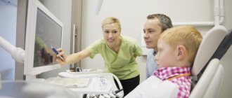
Diagnosis of dystopia
An orthodontist or even a dental therapist can easily determine tooth dystopia during a routine examination if the anomaly is not complex. In some cases, when an element of the dentition remains in the gum, ends up in the palate or another place, during the examination one can only suspect a problem.
If the doctor has not found one of the teeth that should have already erupted in a child (or an adult, if we are talking about eights), he will refer the patient for an x-ray examination. The picture clearly shows all the irregularities and you can see the formed teeth in the soft tissues. To clarify the position and for subsequent treatment, arthopantomography, creation of plaster models of the jaws, and teleradiography are used.
Preparing for dental surgery
If a patient is faced with the removal of a complex tooth, then an experienced doctor will not perform such manipulations without prior preparation. An x-ray is required before the operation. This is necessary to clarify the location of the tooth in the bone, as well as to evaluate the tissues surrounding it.
When a patient has an inflammatory process, it is first cured by taking antibiotics.
Complications after pulling out problem teeth rarely occur, but to avoid them, the procedure should be trusted to a highly qualified doctor.
When is the best time to treat dental dystopia?
Like any dental disorder, dystopia should be treated immediately after the problem is discovered. The easiest time to change the position of teeth is during childhood and adolescence (up to about 18 years of age). This is due to the time of formation of the jaw bones. Until adulthood, the bone is still quite soft, which makes it easy to align the bite.
Sometimes, although rarely, trainers are used to treat dystopia. Read more about them in the article: Trainers for teeth straightening: description, varieties, tips for use
If we are talking about dystopic molars , then they are almost always removed. The third molars themselves are considered an atavism and an optional element in the dentition; moreover, their treatment is associated with problems and difficulties. Therefore, doctors do not try to save these teeth.
Sometimes treatment is associated not with removal, but with grinding of the tooth. This decision is made if the dystopic element does not interfere with the chewing function and does not violate the aesthetics of the oral cavity. In this case, it is recommended to be constantly monitored by a doctor, since such elements of the dentition are easily affected by caries. Moreover, the consequences are much more serious than in ordinary cases: incorrect tooth position stimulates the development of bacteria, inflammation, and complications.
Installation of braces for the treatment of canine dystopia
Content:
- Why does retention occur?
- Classification of impacted teeth
- Is it necessary to remove an impacted tooth?
- How impacted units are removed
- Recovery after removal of an impacted tooth
Retained are units that cannot independently erupt and take the place allocated for them.
They remain inside the jaw bone or are located directly under the soft tissues, mucous membranes of the oral cavity. Removing an impacted tooth is a complex operation for which doctors always prepare in advance. It is not carried out on an emergency basis.
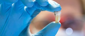
Removal of dystopic wisdom tooth
Removing a dystopic wisdom tooth is a fairly serious operation that requires professionalism and care from the doctor. Incorrect actions can lead to dislocation of adjacent teeth, lower jaw, injury to the mandibular canal and other problems. Therefore, it is very important to contact experienced specialists who are familiar with the specifics of removing complex teeth.
Indications for removing a dystopic tooth
Clear indications for removal:
- detection of a jaw cyst;
- traumatic dystopia;
- diseases of the jaw caused by this tooth;
- difficulties in treating caries in adjacent teeth;
- the appearance of pulpitis and periodontitis.
The practice is that the decision to remove a child is made taking into account all these factors. In an adult, it is more likely to decide whether to keep a tooth or not – removal is considered a universal solution, the simplest and safest. They resort to it in almost all cases.
Stages of removing a dystopic wisdom tooth
The removal operation is carried out in several stages. Compliance with technology allows you to protect the patient and simplify the operation for the doctor. Also, before starting work, detailed studies must be carried out: a full x-ray of the jaw is taken, all available caries is cured, etc.
During tooth extraction, the surgeon performs the following actions:
- Introduction of anesthetic. This may be local anesthesia or general anesthesia if several teeth are to be removed at once.
- Complete exposure of the tooth. To do this, a flap of mucous membrane and periosteum are peeled off.
- I sawed off the walls using a drill. This is necessary to align the element and make it easier to remove.
- Tooth extraction. The entire molar is removed using forceps. If the tooth was previously destroyed, its fragments are removed. It is important that the doctor removes all the fragments, otherwise inflammation will occur.
- Application of antiseptics to prevent the development of wound infection.
- Returning the mucosal flap to its place.
- Applying seams.
Once the wound is stitched, the operation is over, but the treatment is not. After a week, you should definitely see a doctor to have the stitches removed. It is also advisable to be observed by a specialist for 1-2 months to make sure that the removal had no consequences.
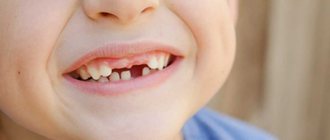
Removal of a dystopic tooth in pictures
How impacted units are removed
The operation to remove an impacted tooth is always difficult. It can only be performed successfully by an experienced and highly qualified dental surgeon. Unerupted wisdom teeth are especially difficult to pull out.
On average, surgery takes about one and a half to two hours. It includes the main steps:
Introduction of anesthesia. This may be local anesthesia, sedation or general anesthesia - it all depends on the location and depth of the unit. The doctor also takes into account the individual wishes of the patient and his state of health.- Excision of the gum and gaining access to the bone tissue in the area of the impacted tooth. A laser or scalpel is used. The first option is more preferable, as it makes the operation less invasive and eliminates the risk of bleeding.
- Preparation of exposed bone to gain access to a submerged tooth. Boron is used here.
- Removing the impacted unit with forceps - an elevator. If the tooth contains several roots and is large, it is first sawed and then removed in pieces.
- Performing plastic surgery of hard and soft tissues, suturing (if necessary).
- Treating the surgical wound with antiseptics and anti-inflammatory solutions.
After the extraction of an impacted tooth, the patient spends some time in the maxillofacial surgery hospital. He is prescribed daily wound treatment and various physical procedures. These measures can reduce the likelihood of developing postoperative complications.
Forecast of dystopia
The appearance of a dystopic tooth in a child is not such a serious problem (especially if there is only one). Before a person’s facial skeleton stops growing, the position of the main teeth can be normalized quite quickly. Therefore, it is recommended to begin treatment immediately after identifying the problem. In this case, all the teeth will most likely fall into place and in the future, in adulthood, will no longer cause concern.
If correction of dystopia in an adult is required, this almost always causes many difficulties. After 18-20 years, teeth are no longer so mobile, so correcting their position requires many preparatory interventions. It is much easier to remove a problematic tooth than to try to put it back in place.
Being cured in a timely manner, dystopia has virtually no negative impact on a person’s future life. Most often, the correct bite is maintained for a long time and does not require supervision by a doctor.
Prevention of dystopia
Dentists are confident that if you carefully monitor your child’s dental health from early childhood, you can avoid most bite problems. Dystopic teeth are no exception, because their appearance is not always associated with hereditary factors. To prevent dystopia, doctors recommend the following:
- proper nutrition, adherence to the daily routine of a pregnant mother;
- prevention of jaw injuries from an early age;
- regular visits to the dentist, wearing devices to correct malocclusion;
- giving up bad habits (including weaning off the pacifier after a year and from thumb sucking in sleep), etc.
If you and your child regularly visit a dentist, he will give recommendations on what exactly to do in your case.
Reasons for development
The causes of such a phenomenon as dystopia can be genetic diseases, embryonic, hereditary, and external factors :
- a genetic disease due to which the child has an underdeveloped jaw and at the same time large teeth;
- deviation during embryonic development, which leads to improper formation of primordia;
- development of teeth beyond normal;
- the formation of molars ahead of schedule or their uneven appearance;
- bad habits (sucking a pacifier or finger for a long time), various bruises, injuries to the oral mucosa;
If the fangs do not appear until the age of 9-12, then there is no place for them and as a result they take someone else’s place and a problem naturally arises.
“Eights” can be dystopic due to the fact that they do not have predecessors in the primary occlusion and they have to independently make their way through the bone tissue.
Conclusions. Expert advice
Dystopia is the incorrect position of a tooth in the jaw: its displacement forward or backward, down or up, as well as rotation around its axis or tilt. The appearance of such an anomaly is associated both with hereditary factors and with bad habits or mechanical damage to the jaw. The sooner dystopia is detected, the easier it is to cure. In adults, the wisdom tooth most often found to be dystopic is the third molar, because there may simply not be enough space in the jaw for it to erupt.
The consequences of such anomalies include general malocclusion, frequent inflammatory diseases, impaired chewing function, and much more. Depending on the complexity of the case, dystopia is treated either by tooth extraction or with the help of braces. If the decision is made to remove it, it is important to carry out the operation carefully and correctly. If treatment is started on time, the prognosis for the disease is positive.
The healing process of the hole after a complex removal
Basically, tissue healing occurs after 7-10 days. It should be noted that this period is quite difficult for the patient. As soon as the effect of the anesthetics stops, aching pain immediately occurs. To eliminate it, you should take pain medication.
The swelling and redness that appears will decrease every day. If there is slight itching in the socket area, this indicates intensive tissue restoration. Removal of sutures after healing is not required since they “dissolve” on their own.
During the regeneration period after removal, it is very important not to disrupt the integrity of the blood clot formed in the socket. To do this, you need to follow a few simple rules:
- during the first 2-3 hours after surgery, do not consume any drinks, much less food;
- Under no circumstances should the well be heated, cleaned or washed;
- do not rinse your mouth for 24 hours after removal;
- When eating food, you need to chew it on the opposite side in relation to the hole.
If you follow these recommendations, the tissue will soon recover without complications.