Vladimir Lutsenko to talk about 3D scanning of teeth and intraoral/intraoral scanners
- founder and head of the Star Smile company, the leader of the Russian aligner market, providing digital 3D dental modeling services in more than 25 countries around the world.
If we talk about intraoral dental scanning, the first intraoral scanners appeared in the early 2000s. The first companies to sell intraoral scanners on an industrial scale were Sirona (Germany) and OrthoCAD from Israel, which sold iTero intraoral scanners in America.
Computer modeling in dental prosthetics - what is it?
Computer modeling, as is already clear from the very name of this technology, allows you to use a regular computer/tablet for dental prosthetics, but, of course, with special “software” (that is, software). Using it, a three-dimensional digital model of the patient’s future smile is created, taking into account all the anatomical features - the shape and color of the teeth, their size and combination with the contours of the face, skin color, gum level, size of the lips and nose, and the width of the smile.
The use of a computer for smile modeling not only speeds up the process of prosthetics, but also optimizes the relationship between the dentist and the patient, as well as the dentist and the dental technician. Patients especially like that they actively participate in the modeling process - together with the doctor they choose the color and shape of their future teeth.
Complex on 4 OSSTEM implants with delayed loading - from RUB 170,000.
Complex implantation Osstem (South Korea) with delayed loading after 4-6 months.
Guarantee for the doctor’s work - unlimited Call now or order a call
Opening hours: 24 hours a day - seven days a week
For computer smile modeling in dentistry, a concept called Digital Smile Design, or DSD for short, is used - which translates to “digital smile design.” It was developed in 2007 by Brazilian dental technician Christian Coachman, and since then it has gained great popularity among the global dental community. By the way, DSD models the future smile, which becomes a kind of aesthetic standard for the prosthesis, but does not model the prosthesis itself - for this they use other solutions, which we will discuss below.
A few words about the shortcomings
3D implantation has a huge number of advantages, but it also has disadvantages. In particular, no software will help an unprofessional doctor who does not thoroughly know the anatomy of the jaw system and does not have computer modeling skills. In addition, this approach requires the presence of special equipment in the clinic - a scanner for performing computed tomography (although the best option would be to contact specialized medical centers to conduct a more in-depth diagnosis on a tomograph with better image detail), as well as computer software itself - a program for planning the process implantation
Three-dimensional dental implantation allows you to obtain an almost accurate prediction of the treatment outcome. Naturally, a computer cannot completely replace a person, but advanced high technologies significantly simplify and speed up the treatment process and, of course, make it better. Naturally, provided that the work is carried out by a professional doctor with high qualifications and extensive experience.
Tasks of computer smile modeling
- select the shape and color of crowns/veneers in accordance with the patient’s requests,
- see your future smile with new teeth in advance and “try on” it: this happens on a computer screen or on a tablet,
- assess the performance of the prosthesis in advance: how the jaws will close, whether the bite relationship will be disrupted,
- rejuvenate the face through Dental face lifting and the application of the principles of the “golden” section and “beauty mask”: these parameters are taken into account during digital modeling (colors and shapes of crowns) - as a result, the face looks more attractive and younger,
- form and study a model of the patient’s jaw before implantation.
Benefits of 3D implantation
Careful planning of dental implantation has a huge number of advantages:
- visualization of the result of implantation: the patient will be able to see on the computer not only how dental implantation will be carried out, but also the result of treatment with a demonstration of a renewed smile,
- maximum precision when installing implants and reducing the risk of errors even when installing structures with pronounced bone tissue atrophy,
- minimal tissue trauma when installing implants due to the use of templates,
- shortened treatment and rehabilitation periods.
Indications and contraindications for digital modeling
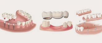
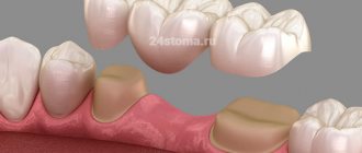
The scope of computer smile design in dentistry is quite wide. Digital smile design is suitable for patients who need prosthetics with restorative inlays, veneers (as well as their variations - ultraneers, nanoeers, etc.), crowns, bridges, and, less commonly, removable dentures.
The DSD concept is used in the development of implant prostheses. Moreover, these can also be either single crowns on implants or extended bridge-like structures with acrylic gum (its color and shape are also digitally modeled).
Don't know what type of prosthetics to choose?
We will help in the selection, advise where to read more information and compare types of prosthetics.
Consultation with an orthopedic doctor in Moscow clinics is free! Call now or request a call
Working hours: from 9:00 to 21:00 - seven days a week
The only contraindications to computer modeling are contraindications to the installation of dentures. This includes caries, the presence of serious inflammation in the oral cavity and massive deposits of tartar. For example, untreated swelling of the gums or installing a filling after the “smile parameters” have been removed will lead to the orthopedic design being uncomfortable. Therefore, before creating a prosthesis, even a virtual one, existing dental diseases should be cured.
Navigation implantology today
Today, the “discover-saw-perform” approach of implant surgeons has undergone major changes. Previously, the implant was installed based on visual inspection of the bone crest, without taking into account the convenience of prosthetics. Since the active introduction of the CT method into dentistry, the risks of errors in choosing the length of the implant have decreased.
The method made it possible to preliminary estimate the diameter of the implant and plan its location in the bone. However, even a brilliant surgeon is unable to do this manually, without special navigation tools. Even laboratory surgical templates that set the drilling point in the bone do not solve the problem.
The situation has changed with the advent of 3D guide template manufacturing technologies in dentistry. The principle is simple. First, the doctor conducts 3D diagnostics and designs the position of the implant on virtual models. Then, using computer technology, a guiding surgical template is made, which is clearly fixed on the prosthetic bed and sets the diameter, direction, and depth of bone drilling for further installation of the implant.
For which restoration method is DSD suitable?
Dental prosthetics is carried out using two methods – direct and indirect. But digital smile design is used only in one case, read about it further.
Direct method - the prosthesis is created in the patient’s mouth
The direct method most often involves applying a liquid filling directly to the patient's teeth while the patient is in the dental chair. This is how they restore small chips, cracks, and darkening on the front teeth, and restore chipped enamel of the lateral teeth. Before the filling hardens, the dentist can give the patient a mirror to evaluate the result and tell him whether he is satisfied or not. There is no need for computer modeling here because all the work is done by hand (not in a laboratory).
Indirect method - the prosthesis is created in the laboratory
Dentures created in a dental laboratory are of much higher quality than those produced by the direct method. For the indirect manufacturing method, in addition to the skills of specialists, equipment is required - various forms for real models, firing and baking ovens, milling machines, presses and modern high-quality materials (ceramics, glass ceramics, zirconium dioxide).
Here, the use of 3D modeling of a future smile helps the dental technician better navigate the work being performed, because DSD data is combined with special programs that model the prosthesis itself and control its production. Of course, not all clinics use the original Digital Smile Design program (because they don’t want to purchase it from the developers), and digital modeling, in principle, works the old fashioned way manually. But as practice shows, it is this concept that helps create the most beautiful, comfortable and durable designs.
On-site 3D scanning from Star Smile company
While on-site scanning
operates in Moscow and St. Petersburg. Thanks to this service, all our clients - clinics or doctors - can call a Star Smile employee with an intraoral scanner, who, by agreement, will come to your clinic at the appointed time and scan the patient.
How much does on-site 3D scanning cost?
By the way, the on-site scanning service is free when ordering Star Smile aligners. It increases the accuracy of aligners, speeds up the process of their production, and eliminates the possibility of errors when taking impressions. And as an added bonus: your clinic will look more advanced, high-tech to your patients.
For now, this service is available in Moscow and St. Petersburg. Since we Star Smile operates in more than 70 cities of Russia, we are already considering other cities in which on-site scanning will be in demand by orthodontists and dentists.
Stages of computer modeling and prosthetics
The process of dental restoration includes several stages that follow one after another. Let's look at them further and tell you when computer smile modeling is used and what initial data is needed for it.
Taking impressions or 3D scanning of the oral cavity
Now computerization can accompany the prosthetics process from start to finish. For example, dentists often abandon the usual casts or impressions of the dentition and use digital “casts” (using a 3D oral scanner).
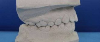
But the usual physical impressions can also be used. True, in this case you will have to transfer them to the laboratory, make a plaster model1 and scan it - in order to create a digital version for its correction in Digital Smile Design. This route takes longer (2-7 days).
Photo and video session, assessment of bite parameters
Photographs (as well as short videos) are taken in order to evaluate the patient’s smile parameters, their relationship with other facial parameters, and also to understand how the facial muscles work. Filming is carried out when the patient is smiling, calm or deliberately “angry”, when he speaks or is silent. All photos and videos, and there can be several dozen of them, are loaded into the program and combined with digital impressions.
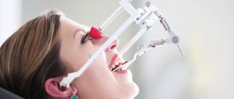
Also at this stage, the dentist uses various jaw movement analyzers (articulators, HIP plane analyzer, face bows). After all, in a future prosthesis, not only beauty is important, but also how it will relate to the existing bite.
Virtual smile modeling
All received data (imprints, photos, etc.) are entered into a computer smile modeling program. Then the parameters are processed, combined, and a three-dimensional projection is displayed on the screen that fully corresponds to the patient’s natural dentition. Next, adjustments are made to improve your smile. Moreover, you can immediately make several options for a new smile - with different shapes or heights, widths of teeth, with different shades of enamel, for example. And then the best option is selected, both from the dentist’s point of view and based on the patient’s wishes.
By the way, such a virtual projection can be combined with digital computed tomography of the patient and begin to develop a prosthesis, incl. and for placement on implants. Popular programs for creating prostheses are CAD/CAM systems CEREC and inLab from Dentsply Sirona, NobelProcera from Nobel Biocare.
Wax modeling and fitting of the prosthesis blank
In some cases, patients who need veneers, extended bridges or removable orthopedic structures should go through one more stage - fitting of the prosthesis blank. This is necessary to assess the comfort of the new teeth and their relationship with the neighboring ones. Here, after developing a model, the dental technician makes a “trial” prosthesis (from silicone, plastic, etc.), and then attaches it to a wax model, after which he hands it over to the orthopedic dentist, who tries it on the patient. If necessary, adjustments are made and the layout is sent back to the laboratory.
Creation of the prosthesis and its final installation
The approved version of the prosthesis goes through all stages of manufacturing - milling on a robotic machine, baking, glazing, polishing, etc. The production technology depends on the type of prosthesis, the properties of the material and its volume, which is needed to create each specific structure.
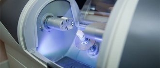
Previously, all production machines were manually adjusted by a person, using measurements also taken by hand. Therefore, there was no need to talk about accuracy, especially the first time. Today, dental devices (namely CAD/CAM systems) are configured largely automatically, in accordance with the parameters set by the computer. Therefore, inaccuracies and human errors are excluded here.
Today, dental crowns, bridges and the thinnest veneers are cut automatically by machines - quickly and the first time. As a result, the doctor simply fixes the prosthetic structure, without first trying it on and subsequent adjustment.
Virtual articulator
The virtual articulator is an effective tool for achieving optimal results in orthopedic treatment. It can be used to control dynamic occlusion when modeling prostheses. The position of the models inside the real articulator is accurately transferred to the program via the scanner.
First, the models are plastered in the usual way. They are then mounted on a special scanner stand with a mounting plate. The stand ensures the positioning of the plaster model in relation to the hinge axis of the virtual space.
Rules for caring for a new smile
Despite the fact that computer modeling allows you to create comfortable, beautiful and durable dentures, this does not mean that new teeth do not need to be cared for. On the contrary, proper care will only extend the life of the restoration and preserve its integrity and aesthetics. However, there is nothing difficult about care. Restorations need to be cleaned 2-3 times a day with a brush and paste (only without abrasive particles); you should not bite nuts and candies, chew chewing gum or toffee. And if veneers are installed, then you should not bite off food with your front teeth, especially hard food (carrots, apples, etc.).
Why is 3D modeling used?
3D modeling directly affects two mandatory stages of implantation:
- diagnostics;
- planning.
Diagnostics.
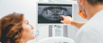
With the help of three-dimensional modeling, it is possible to increase the success of diagnosing dental pathology by 2/3. Conventional images (sighted x-ray and orthopantomogram) give an idea of only 25-30 percent of the tissues shown in one projection. This does not allow timely recognition of problems and may reduce the success of implantation.
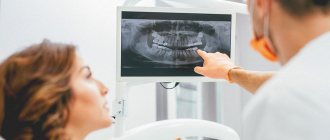
The use of 3D tomography makes it possible to see teeth from all sides, evaluate the tissues surrounding them, and see what is inside the teeth without opening them. Also, three-dimensional modeling allows you to assess the topography of the mandibular nerve, blood vessels, the condition of the joints, sinuses, and assess the height and volume of the upper and lower jaw.
This is especially important in relation to the alveolar ridge. Implantation in this location may be associated with problems caused by insufficient tissue height. Implants can pass through the bone and exit into the maxillary sinus. This complication of implantation is a common cause of odontogenic sinusitis.
Planning.
With the help of three-dimensional scanning it is possible to achieve high implantation efficiency. The plan of the operation, the type of placement of the implants - all this is carefully thought out at the initial stage. Based on the planning results, a special template is created from acrylic or other materials. It is placed on the jaw during implantation to make punctures and implant implants exactly in the places where they are needed.
The result of three-dimensional modeling is 100% compliance of the implantation result with the originally planned one. And this scheme really works.
How much does computer smile modeling cost?
It is worth noting that this type of creation of prostheses increases the cost of overall treatment, and modeling is not available in every clinic. However, many dentists are able to competently build a pricing policy in such a way as to minimize the cost of additional manipulations and equipment used. That is, in one clinic a patient may be charged an additional 10-15 thousand rubles for computer modeling of a smile. And in some places it is already included in the turnkey price and increases the final price of the prosthesis by only 1000-2000 rubles, or does not change it at all.
Patient Questions
QUESTION: I read about trying on a smile on a computer and wanted to clarify whether it is possible to do this if you only need 2 veneers? I have a gap between my front teeth and would like to somehow see the result in advance. And then suddenly I don’t like what the doctor does. Elena
ANSWER: Hello, Elena. Yes, computer smile modeling can be used for any number of necessary prostheses - be it 1-2 veneers or a whole complex for the front teeth. This concept really allows you to “try on” a new smile virtually - the doctor will show suitable options on the computer screen, and you will choose the one you like best. You can also select the color of the veneers so that it matches perfectly with the adjacent teeth and meets your expectations.
- Haug S. Correct modeling, 2006.
Author: Sambuev B. S. (Thank you for your help in writing the article and the information provided)
Modeling dentures - the possibilities of modern technologies in orthopedic dentistry
Modeling a smile and a future orthopedic system is an integral stage of prosthetics. And today, for this purpose, good dental clinics use advanced 3D planning technologies, which make it possible to work out all the smallest details of the future prosthesis using special computer programs. This progress has allowed specialists to significantly improve the quality of services provided. Thanks to him, modern prostheses are as comfortable as possible, reliable, durable and at the same time highly aesthetic. In addition, the use of such modern technologies can significantly reduce the time required to restore missing teeth. Read more about computer modeling of dentures further in this article.
Modeling a smile and a future orthopedic system is an integral stage of prosthetics
New teeth for patients protected by computer technology
As you know, medicine has undergone significant changes in recent years, which allows patients to receive all their guarantees regarding safety, efficiency, reliability, comfort and good treatment results. Such positive trends are also occurring in the dental industry, and in particular in dental implantation.
Today, everyone can safely decide to transform their smile with the help of 3D implantation, or rather, thanks to 3D technologies in dentistry. In the article below we will take a closer look at what “secrets” are available to professional doctors and how new teeth can now be obtained in just a few days.
When 3D models are needed - tasks and scope
The most widespread use of comprehensive digital planning is for single-stage dental implantation protocols. These are methods with immediate loading: all-on-3, all-on-4, all-on-6, basal complex, zygomatic and basal implantation. Without 3D technologies, it is difficult to imagine their use, since high-precision diagnostics and elaboration of the operation are needed in advance.
All of these methods are suitable for patients with complex cases of bone atrophy, periodontitis and periodontal disease. At the same time, they allow you to avoid complex surgery to restore bone tissue volume - augmentation, or osteoplasty. But for implantation to be successful, diagnostics and detailed planning are instead required. This is what digital technologies provide, becoming a solution to the problem of atrophy. The doctor, examining the patient, collects all the necessary information about the volume and quality of bone tissue, the condition of the gums and remaining teeth, and can reliably determine the places where implants are attached to bypass atrophy.
Read more about tooth restoration in case of bone tissue loss in the material “Implantation for jaw bone atrophy.”
Special programs cover the entire cycle[1] of dental implantation procedures, which includes:
- high-precision diagnostics,
- planning dental implantation,
- process visualization,
- step-by-step study of surgical intervention (similar to a simulator),
- selection of implants and modeling of future prostheses,
- preliminary assessment of the result obtained.
“My sister is a dentist. Of course, she doesn’t install implants, but treats caries. But their clinic is good and she sometimes tells us what innovations they have now. So, it turns out that in order to place implants, you first need to plan the entire operation on a computer. So, he says, there are practically no complications afterwards, the patients are satisfied. A few years ago, I myself had a dental implant done in a clinic, even without an x-ray. Then I suffered from inflammation and had to undergo procedures that were not very pleasant. And now I don’t like the way this implant looks.”
Julia P., review from gidpozubam.ru
Possibilities of modern orthopedic dentistry
As can be seen from all of the above, today orthopedic dentists have at their disposal significantly more technological capabilities for creating high-quality, aesthetic and durable prostheses than 10-15 years ago. Thus, to create modern orthopedic devices, advanced programs for computer 3D modeling are used, as well as the CEREC system based on CAD/CAM technology. All these innovations are now used to create high-quality inlays, veneers, crowns and bridges.
On a note! Any removable denture allows you to restore the functionality of the dental system by only 60-65%1. Therefore, even modern new generation models still cause certain inconveniences, especially at the adaptation stage. In addition, they cannot stop atrophy in the jawbone in the area of missing teeth. Coping with this problem and ensuring maximum comfort after prosthetics is possible only through implantation. And if it is necessary to restore a large number of teeth at once, today complex implantation is carried out using one-stage protocols.
In the field of implantology, the main emphasis is now on the use of one-stage treatment protocols, which allow for complex restoration of the jaws quickly and with minimal risks of complications, even in conditions of jaw bone resorption without the need to first build up atrophied areas of bone tissue. In this case, a fully functional adaptive and sometimes permanent prosthesis can be secured within a week after implantation of titanium roots.
To create orthopedic devices, 3D modeling programs are used, as well as the CEREC system based on CAD/CAM technology
If we talk about removable prosthetics, then in this area we can highlight modern clasp dentures, as well as models of the new generation “Quadrotti” and “Acri-Free” - these are reliable and durable systems, moderately elastic, but at the same time durable, practically indestructible. causing discomfort even at the addiction stage. As for permanent crowns, bridges and veneers, experts recommend giving preference to such modern materials as zirconium dioxide and glass ceramics. Products made from them are not only strong and durable, but also extremely aesthetic.
It is obvious that dentistry as an independent field of medicine is progressing quite quickly. New technologies and materials are constantly appearing in the arsenal of orthopedic dentists and dental technicians, allowing them to continually improve dentures, find new, more effective and reliable solutions to give their patients beautiful smiles for many years.
1Kosorukov, N.V. Diseases of the mucous membrane of the prosthetic bed in persons using removable dental structures, 2007.
