What is needed to make a diagnosis for an orthopedic patient?
At the first visit to an orthopedic doctor, the patient is examined. Dentistry and orthopedics provides basic and additional examination methods.
The main examination methods include:
- Collection of complaints: at this point, the doctor asks the patient what the patient is complaining about right now. Accordingly, the doctor clarifies the problems that, from the patient’s point of view, need to be solved: difficulties in chewing food, mobile teeth, sensitivity in certain areas, pain when chewing in the temporomandibular joint, bleeding, pain in teeth covered with orthopedic structures and rubbing or poor fixation denture.
- Collecting an anamnesis of the disease: here the doctor clarifies the time frame - how long ago and how intensely certain complaints are bothering you.
- Collection of general anamnesis:
Past diseases: the general condition of the body, all diseases that the patient suffers from and data on allergic reactions are specified.
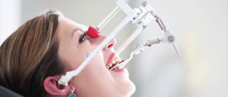
Visual inspection. The doctor examines the patient externally - the skin, the condition of the lymph nodes, whether the mouth opens freely and any external defects.
- The condition of the TMJ is determined without additional examination methods, that is, they pay attention to the degree of opening, uniformity of movement, the presence of clicks, crunching and pain when moving the joint, and also note the condition of the masticatory muscles.
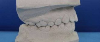
- Next, the oral cavity is examined directly and a dental formula is drawn up. A dental formula is a short table in which, opposite each tooth, the doctor makes a note about caries or other disease, whether this tooth is covered with a crown, whether it is damaged or missing, and the degree of mobility is also noted. Under the table, a note is made about the presence, nature and location of dental plaque.
- Condition of the oral mucosa – the color of the mucosa and its condition are noted: degree of moisture, identified pathologies.
- Next, the doctor focuses directly on examining the teeth that the patient is complaining about - their condition, position in the dentition, abrasion, mobility, whether the roots of the teeth are exposed and whether there is sensitivity during probing or from temperature stimuli, as well as the presence and size of periodontal gum pockets.
They also separately describe the condition of fillings or carious defects, orthopedic structures - both removable and non-removable: the condition of inlays, crowns, removable dentures - the integrity of the prosthesis, fixation of the prosthesis, how well the prosthesis corresponds to the boundaries.
- The doctor also pays attention to the bite - the way the patient closes his teeth. This can provide a lot of data to determine the cause of the pathology.
The procedure for examining a patient and filling out a medical history during orthopedic treatment
- home
- For patients
- Articles
- The procedure for examining a patient and filling out a medical history during orthopedic treatment
- 1. Last name First name Patronymic (of the patient).
- 10. Type of bite.
- 2. Current complaints.
- 11. Language.
- 3. History of the disease.
- 12. Additional examination methods.
- 4. General condition.
- 13. Diagnosis.
- 5. External inspection.
- 14. Plan for preparing the oral cavity for prosthetics.
- 6. Research of TMJ.
- 15. Orthopedic treatment plan.
- 7. Objective data.
- 16. Diary.
- 8. Inspection of the system.
- 17. Dispensary observation.
- 9. Examination of teeth and dentition.
- 18. Epicrisis.
Last name First name Patronymic (of the patient).
Current complaints: impaired chewing, aesthetics, tooth mobility, increased sensitivity of teeth, pain in the TMJ, pain under the base of a removable denture, poor fixation, pain in the tooth under the crown, bleeding gums, bad breath, suppuration, swelling of soft tissues , taste disturbances, etc. Complaints about parafunctions of the masticatory muscles (is there a habit of biting your lips or clenching your teeth while watching a movie, reading books or working, is there a habit of grinding your teeth, complaints from the masticatory muscles (pain, fatigue)).
History of the disease: when did certain complaints appear, what is it associated with (caries, periodontitis, periodontal disease, trauma, surgery, etc.), whether treatment or prosthetics were previously carried out (how long ago?).
General condition: presence or absence of bad habits (smoking, drinking alcohol), concomitant diseases (chronic diseases; hepatitis, tuberculosis, syphilis, HIV), allergy history, whether anesthesia was previously performed during treatment, tooth extraction, its effectiveness.
External examination: skin color, presence of defects, symmetry and type of face (conical, obverse-conical, square, round), height of the lower third of the face, protrusion of the chin, pattern of lip closure, severity of nasolabial and chin folds.
Research of the TMJ: opening of the mouth (intermittent, free, the nature of the movement of the lower jaw is smooth or jerky, displacement of the lower jaw to the side), the presence of crunching, clicking, noise in the TMJ when moving the lower jaw.
The state of the tone of the masticatory muscles and their soreness on palpation, the state of the submandibular lymph nodes, their soreness and size on palpation.
Objective data
| 8 | 7 | 6 | 5 | 4 | 3 | 2 | 1 | 1 | 2 | 3 | 4 | 5 | 6 | 7 | 8 |
Inspection of the mucous membranes: color and moisture content of the mucous membranes, color and shape of the gingival papillae.
Examination of teeth and dentition:
- condition of intact teeth (size, anomalies of position, shape, color of hard tissues, mobility, exposure of necks or abrasion of teeth (degree), sensitivity (type: hot, cold, sour, sweet, etc.), size of periodontal pockets);
- condition of teeth with defects of hard tissues (size and topography of the defect, condition of fillings is satisfactory or not, IROPD, mobility, exposure of necks or abrasion of teeth (degree), sensitivity (percussion, probing, temperature reaction), size of periodontal pockets);
- condition of inlays, veneers, pin structures (size, topography, condition, correspondence to the bite, mobility of the abutment tooth or prosthesis, exposure of the necks (degree), sensitivity), crowns, bridges (size, topography, condition, correspondence to the bite, mobility of the abutment teeth, exposure of the necks of the supporting teeth, sensitivity, abrasion of the chewing or other surface of the dentures), the size of the periodontal pockets of the supporting teeth;
- condition of removable dentures (type, integrity of the base and prosthesis fixation systems, service life, fixation of the denture in the oral cavity, balance, compliance with boundaries).
Type of bite: fixed, not fixed; orthognathic, direct, biprognathic, prognathic, progenic, cross, deep, open.
Condition of the dentition: shape, size and topography (classification) of defects, secondary deformations and movements of teeth.
Condition of toothless jaws: classification of the upper jaw (Doinikov, Kurlyansky, Oksman, Schroeder), lower jaw (Doinikov, Keller, Kurlyansky, Oksman); degree of mucosal compliance according to Supple; attachment of the frenulum of the upper and lower lips, buccal-gingival cords.
Tongue: size (normal, microglossia, macroglossia), shape (round, oval, V-shaped), attachment of the frenulum of the tongue.
Additional examination methods: description of x-ray examination (intraoral x-ray, panoramic x-ray, orthopantomography, targeted x-ray of teeth), description of diagnostic models, study of loss of chewing efficiency according to I.M. Oksman
| teeth | 1 | 2 | 3 | 4 | 5 | 6 | 7 | 8 | Total |
| V/H | 1 | 1 | 2 | 3 | 3 | 6 | 5 | 4 | 25% |
| LF | 2 | 1 | 2 | 3 | 3 | 6 | 5 | 3 | 25% |
Diagnosis is a logical conclusion, a synthesis of the obtained subjective and objective research data. The diagnosis in orthopedic dentistry should reflect the size and topography of defects in the hard tissues of teeth, dentition, the condition of the oral mucosa, as well as concomitant diseases of the dental system and complications.
For example:
- a defect in the hard tissues of the tooth (necessarily which one?), carious, non-carious or traumatic origin (non-carious diseases include: enamel hypoplasia, wedge-shaped defects, fluorosis, acid necrosis and pathological abrasion; trauma - acute and chronic), the degree of destruction of the coronal part of the tooth must be indicated .
- partial edentia (which jaw?) according to Kennedy: bilateral end (I class), unilateral end (P class), included in the area of the lateral teeth (W class), isolated included in the frontal region (1U class). Complications: traumatic occlusion, decreasing bite, secondary deformation (Godon-Popov phenomenon).
- complete adentia: degree of atrophy according to I.M. Oksman, pliability of the mucous membrane according to Supple.
Plan for preparing the oral cavity for prosthetics: sanitation of the oral cavity (removal of dental plaque, dental treatment, removal of teeth or roots); special preparation (depulpation of teeth, elimination of occlusal disorders, orthodontic preparation, alveolotomy, excision of scars, strands of the mucous membrane, deepening of the vestibule or floor of the oral cavity).
Orthopedic treatment plan:
- Inlay, veneer from what material and for what tooth;
- Pin structure (single-root, dismountable, how it is made, temporary, permanent) on the abutment tooth;
- Single crown (from what material) on the abutment tooth;
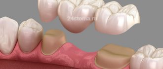
- The bridge prosthesis is made of what material, supported by what teeth;
- Partially removable laminar denture on the upper part, lower part, with which teeth (plastic, ceramic, porcelain), clasps on which teeth;
- Clasp prosthesis (splinting clasp prosthesis) indicating the fixation system (cast clasps, type of attachments, telescopic crowns) and on which teeth;
- Other types of structures are possible, indicating the type of materials, manufacturing method and supporting teeth.
Diary: displays the date of patient admission, the amount of work performed and must be certified by the signature of the immediate supervisor.
Dispensary observation: if necessary, the date of examination (year, month) of the subsequent visit for the following diseases is noted: pathological abrasion, periodontal tissue diseases, complete edentia, etc.
Epicrisis: describes the scope of orthopedic treatment (aesthetics, anatomical shape of teeth, integrity of dentition, height of the lower third of the face, tooth mobility), indicates the extent to which chewing efficiency has been restored (according to I.M. Oksman), and provides recommendations for oral care and the use of prostheses.
ILLUSTRATIVE EXAMPLE
“Maintaining an outpatient card for a dental patient”
Last name I.O.: Ivanov V.P.
Year of birth: 1991.
Current complaints: disturbances in chewing and aesthetics.
History of the disease: a day ago, the crown of the central tooth of the upper jaw broke off while eating. The tooth was treated three years ago due to caries complications.
General condition: bad habits – smokes; concomitant diseases – no; hepatitis, tuberculosis, syphilis, HIV – denies; allergy history - not burdened, anesthesia was previously carried out, effective, without pathologies.
External examination: skin color – clear; face – symmetrical; face type – conical; the height of the lower third of the face is not changed; the chin does not protrude; lips close without tension; nasolabial and chin folds are moderately pronounced; mouth opening – free, painless; movements of the lower jaw are smooth, there are no displacements during movement.
Examination of the TMJ: the presence of crunching, clicking, noise in the TMJ during movement of the lower jaw is not recorded, the masticatory muscles are painless on palpation, the submandibular lymph nodes are painless on palpation and are not enlarged.
Objective data:
| P | R | P | P | ||||||||||||
| 8 | 7 | 6 | 5 | 4 | 3 | 2 | 1 | 1 | 2 | 3 | 4 | 5 | 6 | 7 | 8 |
| P | P | P |
Examination of the oral mucosa: the mucous membrane is pale pink, moderate humidity, gingival papillae are normal.
Examination of teeth and dentition:
- intact teeth without pathology, not mobile, percussion and probing are painless, temperature reaction is negative, periodontal pockets are 0.1 mm.
- The fillings are in satisfactory condition, correspond to the bite, the marginal fit is tight.
The teeth are not mobile, percussion probing is painless, the temperature reaction is negative. IROPZ 16; 25; 26; 36; 44; 45 – 0.5.
Type of bite: fixed, straight.
Condition of the dentition: the shape of the dentition is ellipsoidal in the upper jaw, parabolic in the lower jaw. There are no secondary deformations.
Tongue: normal size, oval shape, normal frenulum.
Additional examination methods: from 07/05/2010. On an intraoral radiograph, there are 21 teeth in the periapical tissues without pathological changes, the canal is sealed to the physiological aperx, along its entire length.
or according to I.M. Oksman
| teeth | 1 | 2 | 3 | 4 | 5 | 6 | 7 | 8 | Total |
| V/H | 1 | 1 | 2 | 3 | 3 | 6 | 5 | 4 | 25% |
| LF | 2 | 1 | 2 | 3 | 3 | 6 | 5 | 3 | 25% |
Loss of chewing efficiency – 3% according to I.M. Oksman.
Diagnosis – defect of hard dental tissues as a result of caries, destruction of crown 21 on 1/2 of the surface, IROPD 16; 25; 26; 36; 44; 45 – 0.5; loss of chewing efficiency 3% according to I.M. Oksman.
Plan for preparing the oral cavity for prosthetics: not carried out.
Orthopedic treatment plan: stump pin design for 21 teeth; single metal-ceramic crown for 21 teeth.
Diary:
| date | Volume of work performed | Manager's signature |
| 5.07.2010 | Inspection, documentation. Preparation of the root of the 21st tooth for a core post structure, modeling the post structure in the oral cavity with Lavax wax, applying a temporary filling - dentin paste. | |
| 6.07.2010 | Checking and fitting the metal stump pin structure to the root of the 21st tooth. Honey. treatment of the root and canal of the 21st tooth with 3% hydrogen peroxide and liquid for degreasing the canals, air; metal stump pin structure - 95% alcohol, air. Fixation of a metal stump pin structure into the root of the 21st tooth on a Fuji. | |
| 7.07.2010 | Under application anesthesia with Ludoxor spray, insertion of a retraction thread into the periodontal sulcus of the 21st tooth, preparation of the 21st tooth for a metal-ceramic crown. Taking a double impression from the upper jaw “Spidex”, from the lower jaw – an alginate impression “Hydrogum soft”. | |
| 8.07.2010 | Determination and fixation of central occlusion. | |
| 10.07.2010 | Checking and fitting the cast frame of a metal-ceramic crown for 21 teeth. Determination of the color of ceramic cladding: Ivoclar – 4 A. | |
| 12.07.2010 | Checking and fitting a metal-ceramic crown for 21 teeth. Honey. treatment of the stump of 21 teeth with 3% hydrogen peroxide and air; metal-ceramic crown - 95% alcohol, air. Fixation of a metal-ceramic crown for 21 teeth on “Fuji”. Advice and recommendations on oral care and denture care are given. |
Dispensary observation: the patient does not need a preventive examination of the oral cavity once every six months.
Epicrisis: as a result of orthopedic treatment of a defect in the hard tissues of the 21st tooth, the anatomical shape, aesthetics, integrity of the dentition of the upper jaw, and chewing efficiency were restored in full (100%).
Patient V.P. Ivanov, born in 1991, was given the necessary recommendations for caring for the oral cavity and the metal-ceramic crown on the 21st tooth.
Additional examination methods in orthopedic dentistry include:
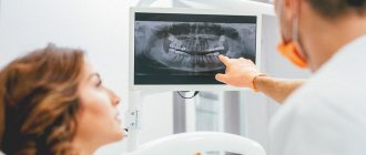
- X-ray image. Typically, orthopedic patients undergo an orthopantomogram, or OPTG as it is called for short. This is a large shot of both jaws and all the teeth. It allows you to see the overall picture and understand what additional examination methods will be needed. To clarify data on specific teeth, targeted intraoral radiographs are taken - small pictures that fit 1-3 teeth. In such photographs, the image of the tooth is larger and clearer, which allows you to see what is invisible in a large panoramic photograph.
- CT scan. This is essentially an advanced X-ray image - thanks to CT, you can get more than just one angle, but immediately look from all sides and see what is not visible on a planar image. To plan large-scale orthopedic work, a CT examination is simply necessary.
As a result, after carrying out all the necessary examinations and examinations, the doctor makes a diagnosis of orthopedics and dentistry based on the data obtained. Next, depending on the diagnosis, a treatment plan is drawn up - each diagnosis has a treatment protocol and objective requirements for the situation in the oral cavity. In orthopedics and dentistry, diagnoses are made only after a thorough examination.
Reliable diagnosis is the key to successful treatment
Dear readers, in this article we will try to tell you as comprehensively and in “simple” language as possible about what diagnostics of dental diseases is, how and why it is carried out, how a plan for the necessary treatment and dental prosthetics is drawn up.
Each clinical case is individual, as well as the required treatment individually. There are no two completely identical people, just like no two identical teeth. Each person in need of restoration of the dental system always needs an individual treatment plan, which can be drawn up based on the examination and diagnosis. One of the keys to successful treatment is diagnosis and careful planning in each specific case. The quality, durability and beauty of your teeth will depend on the correct implementation of these important factors...
As a rule, after the first introductory consultation and filling out medical documentation, diagnostic measures are carried out. The scale and type of diagnosis depends on the complexity and severity of a particular clinical case.
Many diseases that patients had no idea about are detected at various stages of diagnosis, which allows the necessary measures to be taken in time. For example, a person who went to the dentist about a chipped or worn-out tooth would not even think that this was due to an incorrectly formed bite or incorrectly made restorations. Many factors that at first glance are not directly related to the patient’s complaints are, in fact, often the result of another disease or destruction that may not appear immediately; the patient simply does not notice them. If you do not identify the problem, or do not diagnose it at all, but immediately begin to restore this tooth (which was the purpose of the patient’s visit), then the quality of such treatment will be in doubt, moreover, the situation will worsen over time and lead to even greater destruction. The task of any doctor is to diagnose the clinical situation in a timely and detailed manner before carrying out therapeutic or restorative measures.
So, the first diagnostic event is a survey.
Based on your complaints, the dentist collects information about previously suffered diseases, the timing of their occurrence, and their course. Determines your psychological status and readiness for dental treatment. During the interview, try to answer the doctor’s questions in as much detail as possible, listen carefully and do not hesitate to ask if something is not clear. This way you can help the dentist solve your problem. Even during the interview, the dentist pays attention to your face, evaluates symmetry, identifies edema and swelling, studies your articulation and much more, and so begins the next stage of diagnosis - examination.
After the external examination, the dentist begins to examine the oral cavity. In addition to the usual mirror and probe, there are quite a few modern additional examination methods for identifying diseases and making a correct diagnosis. It is believed that a “standard” dental examination of a patient (examination using a dental probe and mirror) will reveal only 30% of the necessary information. Therefore, high-quality diagnostics in modern dentistry is unthinkable without additional examination methods.
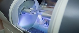
The most important and informative methods include visiographic and tomographic studies in dentistry. You can read more about these types of diagnostics in the section on computed tomography.
In addition to identifying various pathologies of the dentofacial area, radiovisiography and computed tomography are an integral part of modern planning of dental prosthetics.
The most accurate calculations for dental implant placement are made using 3D tomographic images. Calculating the exact location of the implant plays a vital role in its functioning and the correct placement of the crown on it. This is a real breakthrough in dentistry. Today it is unthinkable to install implants without preliminary tomographic diagnostics. But tomography is not the entire arsenal of modern diagnostics, which allows you to correctly install an implant, filling or crown.
In addition to successfully identifying and treating dental disease, it is necessary to correctly restore the shape of a tooth or an entire row of teeth. After all, after you have had root canal treatment on your tooth, it still needs to be restored. And if you have a tooth removed, then you need to install another one in its place.
Everyone knows that a tooth has a strictly defined shape: cusps, fissures, various ridges and pits. All these elements together determine its shape. Only a properly shaped tooth can ensure proper functioning and each, even the most inconspicuous element on the tooth performs its strictly defined task. If such an element is lost or not created during dental restoration, the entire tooth will suffer, then, like a chain reaction, this will affect the bite, which can subsequently negatively affect the temporomandibular joint, the symmetry of the face, and much more. Each tooth must occupy its specific position in the dental arch and in the bite with an accuracy of fractions of a millimeter.
Let's look at, for example, the destruction of a chewing tooth in the lower jaw
The patient came with complaints about the crown of tooth 46 chipping while eating; the patient does not experience any pain, but he is bothered by the sharp edge of the tooth, which scratches his tongue. The tooth chipped the day before.
The patient denies the presence of chronic diseases, HIV, hepatitis and allergic reactions.
An external examination, examination of the condition of the temporomandibular joint and the condition of the oral mucosa revealed no pathologies. In the oral cavity, the condition of the teeth is satisfactory; no pathologies other than a chipped tooth 46 were identified. The plaque is scanty - the patient had professional oral hygiene a month ago.
The patient's bite is correct and orthognathic.
When examining the chipped tooth 46, we note that the tooth is depulped, while the seal is preserved - an insulating gasket is in place - it protects the filling material of the tooth root canals from infection by oral bacteria. The chip occurred to approximately half the height of the tooth crown on the lingual and distal (back) sides. In this case, the size of the defect in the coronal part of the tooth is more than half the size of the crown.
Next, we move on to additional research methods - we will do an OPTG to identify possible defects on other teeth and assess the condition of neighboring teeth and teeth opposite on the upper jaw and a CT scan in the area of tooth 46 to determine the quality of canal filling and the presence of inflammation in the root area 46.
In the photographs we see that the canals are sealed tightly, up to the apical foramen (that is, to the end) and there are no clearings to suspect the presence of inflammation. The adjacent teeth are vital and do not require treatment.
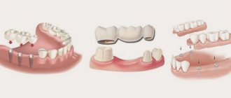
What are the dangers of missing teeth?
Losing teeth, as it may seem at first glance, causes not only psychological discomfort or an aesthetic defect. The loss of even one tooth can lead to deformation of the dentition, curvature of the remaining teeth, malocclusion, bone tissue atrophy, impaired diction, and the loss of several anatomical units leads to disruptions in the functioning of the entire body. The patient has difficulty chewing food, which leads to problems in the digestive system.
Therefore, it is important to restore the integrity of the dentition as soon as possible, especially since modern orthopedic dentistry offers many treatment options.
Dentists at the multidisciplinary department of orthopedic dentistry in St. Petersburg provide a range of services for dental prosthetics and restoration of the integrity of the dentition. We install ceramic inlays, temporary and permanent crowns on implants and natural teeth, Maryland Visio Ling bridges, clasp dentures, Ti beam structures, veneers, etc. You can make an appointment with a doctor at the Department of Orthopedic Dentistry by calling: 8 (812) 456-15-15 or using the online registration form on the website.
Setting goals
Goals of orthodontic treatment:
- achieving the correct jaw ratio;
- elimination of dental distortions;
- restoration of chewing and speech functions;
- elimination of facial asymmetry and other defects.
Planning includes the following activities:
- initial examination and consultation;
- complex of diagnostic studies;
- development of a treatment protocol and agreement with the patient.
Most clinical cases allow for several correction options. The doctor conducts a consultation on each so that the patient can choose the most optimal option.
Classification of dentition defects
There are several types of classification. Dentists mainly use three of them:
- Kennedy classification:
- Group 1 - bilateral absence of chewing teeth;
- Group 2 - unilateral absence of a molar;
- Group 3 - unilateral absence of lateral teeth with preservation of distal support;
- Group 4 - defect of the anterior jaw.
- Beltman classification:
- Class 1 - the defect includes edentulous chewing teeth.
a) dentition with unilateral terminal defects;
b) dentition with bilateral terminal defects.
- Class 2 - there is one or more included defects (if the terminal units are present, there may be no teeth in other parts of the row).
a) the number of missing units does not exceed three;
b) the number of missing units is three or more.
- Classification according to the Gavrilov system:
- Group 1 - dentition with terminal one- and two-sided defects;
- Group 2 - dentition with anterior and lateral one- and two-sided defects;
- Group 3 - combined defects;
- Group 4 - single preserved dental units.