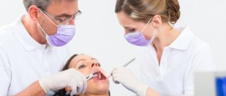
A. A. Glotova , periodontist, dental clinic “DENTiK LUX”, member of the Russian Periodontal Association RPA (Krasnodar)
E. N. Shastin Professor of RAE, Head of the dental clinic “DENTiK LUX” (Krasnodar)
The article describes the etiology, pathogenesis, and clinical cases of peri-implant tissue disease. Data are presented indicating a connection between periodontal diseases and dental implants. The need for comprehensive treatment of periodontal diseases prior to implantation is described.
Dental implants have revolutionized dentistry, providing new hope for millions of people with missing teeth. Since the debut of implants in the 1960s, we have made great strides in understanding the biology of osseointegration. However, with an increase in the number of implants installed, the number of late complications also increases: mucositis, peri-implantitis, complete disintegration.
According to epidemiological studies, after 10 years, peri-implantitis develops in approximately 40% of patients who receive implants, while mucositis in the implant area occurs in approximately 80% of patients. ( Berglund at al 2015 Clin Periodontolog & Implant Dentistry, Eke at al. J Dent Res 2012, Kassebaum at ol Dent Rec 2014).
In almost every book on implantation you will find that the success rate of this treatment method today, with modern equipment and the latest technologies, is 98% or higher, and the survival rate of implants for 10 years is 95–97%.
The Heterburg Study of the Swedish Population
This study (Journal on Dental Researelu 2015 Effectiveness of Implant Therapy analyzed in a Swedish population) is by far the largest on implant survival. Of the 25,000 patients with implants installed in 2003, 4,716 patients were selected. Early implant loss was 4.4% (121 patients) on the patient side and 1.4% (154 implants) on the implant side. Nine years after implant treatment, 596 patients with 2367 implants were evaluated. Late loss was 4.2% on the patient side (25 patients), and 2% on the implant side (47 implants). Overall loss: patients 7.6%, implants 3%.
When assessing the prevalence of caries or periodontitis, the number of patients with these diseases is considered, and not the number of affected teeth, so implantation failure should be assessed in terms of the number of patients, not implants. Thus, the survival rate of implants over 10 years is 93%, and not 97%, these data allow a more realistic assessment of the result and improve clinical work.
CONTENT
- 1 Biomechanical considerations 1.1 Biological width
- 1.2 Tip effect
- 1.3 Crown to root relationship
- 1.4 Treatment planning
- 2.1 Apically repositioned flap with bone recontouring (resection) [13] 2.1.1 Indications
- 2.2.1 Indications
Biological width and periodontal complex
In the implant field, biological width is also defined as the distance from the most prominent coronal projection of the connective epithelium to the alveolar bone, including the epithelium (approximately 1 mm) and the connective tissue attachment (approximately 1 mm). By analogy with a tooth, the dentogingival complex additionally contains a section of the implant-gingival sulcus (approximately 1 mm). If this distance is less, resorption of the bone ridge surrounding the implant should be expected (Clin Oral Implants Res. 1991 Apr-Jun; 2(2):81-90 “The soft tissue barrier at implants and teeth” Berglundh T., Lindhe J., Ericsson I., Marinello C.P., Liljenberg B., Thomsen P.).
Coronal lengthening
Lengthening the coronal part is a procedure that can minimize the risk of violation of the biological width. When considering the applicability of this procedure to a specific case, one should calculate the feasibility of its implementation in general. Preparation for surgery should include determining the biotype of gingival tissue, assessing its condition, and high-quality x-rays. In some cases, such as severe abrasion/wear of teeth, a sensitivity test should also be performed. A wax up, a wax-up model, is often helpful in discussing the treatment plan with the patient.
Etiology
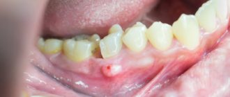
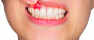
According to the report of the sixth European Workshop on Periodontology in 2008, mucositis and peri-implantitis are recognized as infectious diseases caused by bacteria (Cindy & Meyle, 2008, Consensus Report EP6). Mucositis is an inflammatory lesion that is limited to the mucosa around the implant (Fig. 1 a, b, c). Peri-implantitis is a pathological condition that occurs in the tissues surrounding dental implants, caused by microorganisms and characterized by tissue inflammation and bone loss around the implant (Fig. 2, 3 a, b).
Rice. 1 a. Level of bone tissue at the time of fixation of the metal-ceramic bridge.
Rice. 1 b. Peri-implant mucositis. The patient, 5 years after fixing the bridge, complained of discomfort in the area of the implants.
Rice. 1st century Level of bone tissue 5 years after fixation of a metal-ceramic bridge. There is no pathological resorption in the implant area, the diagnosis of mucositis is confirmed.
Rice. 2. Peri-implantitis. Bone resorption for 2/3 of the implant length 3 years after implant placement.
Rice. 3 a. Hygiene is unsatisfactory. 6 months after permanent prosthetics, peri-implantitis was diagnosed.
Rice. 3 b. Hygiene is unsatisfactory. 6 months after permanent prosthetics, peri-implantitis was diagnosed.
In recent years, numerous experimental and clinical studies have established that the formation of bacterial biofilm is the main etiological factor in the development and progression of peri-implantitis. The main types of subgingival bacteria include Peptostreptococcus micros, Fusobacterium nucleatum, Prevotella intermedia.
Inflammation of the gums and bone around implants develops under the influence of gram-negative anaerobic microflora, which is also associated with periodontitis. With peri-implantitis, a large number of periodontal pathogens are found, in particular red complex bacteria (Porphyromonas gingivalis, Tannerella Forsythia, Treponema denticola), A. Actinimycetemcomitans, Prevotella intermedia. It is assumed that the microflora of the oral cavity at the time of implantation influences the formation of biofilm on the surface of the implant. Accordingly, existing periodontal pockets in the area of natural teeth serve as reservoirs of pathogenic microorganisms that can colonize installed implants.
Periodontal and peri-implant pockets at the same probing depth in the same patient have a similar microbiological composition. Thus, rationally, periodontal diseases must be pre-treated before implantation as part of a comprehensive periodontal therapy protocol.
Risk factors
Risk factors are of great importance for the occurrence of diseases around implants. Local factors include anatomical and clinical conditions that favor the colonization of pathogenic bacteria: for example, poor oral hygiene, deep gingival pockets, unfavorable denture design (Fig. 4).
Rice. 4. The patient undergoes a routine examination 1 year after total prosthetics. Hygiene is unsatisfactory, food remains, soft plaque. The gums are hyperemic and swollen both in the area of the teeth and in the area of the implants.
Rice. 4. The patient undergoes a routine examination 1 year after total prosthetics. Hygiene is unsatisfactory, food remains, soft plaque. The gums are hyperemic and swollen both in the area of the teeth and in the area of the implants.
General risk factors determine the response of the macroorganism to the microbial load - for example, the patient’s general health, bad habits. A combination of risk factors can lead to a severe response to inflammation resulting from bacterial infection, which will increase the rate of bone resorption in peri-implantitis.
Oral microflora colonizes the surfaces of implants and prosthetics in the same way as traditional fixed dentures, so good oral hygiene is critical to the long-term success of implant treatment. The accumulation of bacterial plaque on the gingival surface of the prosthesis leads to the development of mucositis, which is clinically manifested by redness, swelling of the mucous membrane and bleeding when probing the gingival sulcus. Lack of timely treatment leads to further progression of the inflammatory process and its spread to bone tissue (Fig. 5).
Rice. 5. Implants in the area of the 32nd, 42nd, 43rd teeth. Condition of the gums 2 months after permanent prosthetics. Unsatisfactory hygiene in the area of metal-ceramic prosthesis on implants. Bleeding on probing.
When planning implant treatment, one must take into account the need to create optimal conditions for self-hygiene in the implant area. It should be remembered that in the absence of proper care, peri-implantitis develops very quickly, even if the patient has no history of periodontitis.
Removing all teeth does not completely eliminate periodontal pathogenic bacteria, since they are able to penetrate soft tissues; also, if there are family members diagnosed with periodontitis, there is a constant supply of periodontopathogenic microflora from the outside (through hygiene items, dishes, kisses).
The integration of soft tissues to the intragingival elements of orthopedic structures is important. Titanium and ceramic abutments promote the formation of a mucosal attachment strip 1.5 mm wide. There is little or no attachment to gold and veneering ceramics, which increases the likelihood of developing deep pockets. In addition, there is better adhesion of soft tissues to relatively rough surfaces. However, when a rougher surface in the mouth is exposed, bacterial plaque accumulates on it more quickly. Thus, when choosing an implant system, the dentist faces a certain dilemma. On the one hand, it is necessary to achieve better attachment of soft tissues to the intragingival structures of the structure, and on the other, to avoid excessive accumulation of bacterial plaque when exposed in the oral cavity.
It is believed that excessive occlusal load can cause bone loss around implants. Many authors have studied the relationship between implant load and marginal bone resorption. Most of these studies have been conducted in animal models over a period of 12–15 years. It turned out that the load on the implants is not accompanied by bone resorption around them. On the contrary, the degree of mineralization of the bone in direct contact with the implant increases compared to an implant that does not experience such stress. Thus, excessive loading of implants in the long term can lead solely to biomechanical damage (screw loosening, crown decementation, abutment fracture, implant fracture), and bone resorption around integrated implants is associated only with bacterial infection.
Cigarette smoking should be considered as a potential risk factor, since tobacco smoke directly affects local immunity in the oral cavity. There is a decrease in chemotaxis and phagocytosis of neutrophils, stimulation of anti-inflammatory cytokines, activation of subgingival anaerobes, all of which can cause early and late complications after implant installation.
The professionalism, theoretical knowledge and manual skills of the doctor play a big role in the success of implant treatment. Unfortunately, due to the popularization of implantation, doctors have formed the opinion that installing implants is a fairly simple manipulation that does not require much theoretical knowledge and manual skills. According to recent studies in the field of peri-implantitis, in more than 90% of cases, this disease is caused by poor planning and suboptimal treatment.
Pathogenesis
Direct contact between the accumulation of microorganisms and the soft tissues surrounding the implant leads to inflammation and an increase in probing depth, similar to the processes occurring in the area of natural teeth. Experimental studies confirm a similar inflammatory response around teeth and implants in response to bacterial irritants. In both cases, the lesion is localized in the area of the gum edge. The progression of the inflammatory process and tissue infiltration by large lymphocytes leads to bone resorption. Elastase-producing cells are found in greater numbers in peri-implantitis than in periodontitis, indicating a more rapid progression of peri-implant bone damage.
As plaque accumulates, the tissue damage around the implant spreads in the apical direction. The inflammatory infiltrate includes plasma cells, macrophages, lymphocytes and a large number of polymorphonuclear leukocytes. Moreover, it is the increase in the number of polymorphonuclear leukocytes that may explain the increase in elastase concentration in peri-implantitis compared to periodontitis. The inflammatory infiltrate during peri-implantitis directly extends to the alveolar bone and penetrates into the medullary spaces. This circumstance distinguishes the nature of inflammation in peri-implantitis, since in periodontitis the inflammatory infiltrate is separated from the bone tissue by a dense connective layer about 1 mm thick. In peri-implantitis and periodontitis, cytokines are found that increase the activity of osteoclasts, however, in peri-implantitis, interleukin-1A predominates, while in periodontitis, tumor necrotizing factor alpha has a greater influence. The absence of a periodontal gap and ligament in the implant is important in the pathogenesis of the development of the process. The tooth has excellent blood supply and strong immunity; therefore, the body is able to resist infection and periodontitis lasts for years - from the moment a subgingival infection occurs until the complete loss of the tissues surrounding the tooth, it can take 15–20 years. For an implant, due to the differences in structure and pathogenesis described above, complete disintegration can occur within 3–5 years from the onset of peri-implantitis (Fig. 6).
Rice. 6 a. Level of bone tissue at the time of permanent fixation.
Rice. 6 b. The patient has no complaints. A routine examination with X-ray control 2 years after fixation revealed a saucer-shaped loss of bone tissue.
Disturbance = inflammation
Any disruption of the periodontal ligament and restoration is likely to cause inflammation. Any inflammation in this area can negatively affect the result of the entire work. Understanding the function and location of the ligament increases the likelihood of successful restoration. In particular, factors such as
- position of the gum edge,
- marginal fit,
- contact areas,
- prosthesis shape,
- Iatrogenic damage.