The procedure for removing the upper teeth can be carried out planned or emergency. In the first case, the indications are: significant or complete destruction of the supragingival part of the tooth and the inability to use it for prosthetics, chronic periodontitis that cannot be treated conservatively, periodontal diseases with tooth mobility of the third degree, and others.
Urgent removal of the upper tooth is required for purulent periodontitis, periostitis, acute osteomyelitis, lymphadenitis (if the tooth is the source of the disease), sinusitis, deep fracture of the crown exposing the pulp, as well as some other conditions and diseases.
Before the operation to remove the upper tooth, an examination is carried out and, if necessary, an x-ray is taken. As a rule, the operation is done under local anesthesia. Before surgery, the dentist thoroughly cleans the teeth and mucous membranes to prevent infection of the socket.
Depending on the type of tooth, the doctor uses various devices:
- removal of the front teeth (incisors and canines) is carried out using straight forceps;
- for extraction of premolars you need bayonet-shaped and S-shaped forceps, a straight elevator;
- large molars are removed using coronal S-shaped forceps equipped with spikes.
In some cases, it is necessary to saw the tooth into pieces or cut out part of the bone tissue. This mainly applies not to the removal of front teeth, but to the extraction of molars (especially wisdom teeth).
What instruments are used to remove teeth?
Dental forceps have been and remain tools for removing teeth. For certain groups of teeth, a different type of forceps is used, since our teeth have different structures and are located differently in the dentition. For example, to remove the upper anterior tooth and the maxillary canine, there are straight forceps, and the remaining upper teeth are removed using S-shaped ones. The incisors of the lower jaw are pulled out using forceps curved at 90º with narrow cheeks (the part of the forceps that grasps the crown or root of the tooth being removed). The fangs and the two teeth following them are pulled with forceps, on the contrary, with wide cheeks. To remove large molars of the lower jaw, forceps with spikes that go between the roots are used.
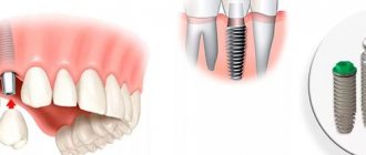
Let's start implanting the upper front teeth
a highly accurate surgical template
, individually made based on the CBCT image and using the 3D Shape intraoral scanner . Under the Straumann Bone Level SLActive implantation system, as part of a complete protocol for implantation of the upper anterior incisors.
Here you can observe the result of one-stage implantation of the front teeth; Straumann implants were installed. A bed for the implants was formed, which were installed in a strictly necessary position so that we could do aesthetic work not only on implantation, but also on further prosthetics of the upper anterior incisors.
You can see Straumann implants installed in exactly the right position
, as we planned. They are presented with the template removed. Bone biomaterial was placed in the sockets, which prevents the socket from collapsing in order to preserve the outer contour of the gum. In addition, in the area of the tooth that had a fistulous tract (tooth 11), in the photo it is on the right, a collagen membrane was sutured. The membrane was tunneled and secured with a single suture to allow bone to form and mature normally in the socket.
Here you see the same implants, but in front view. You can clearly see how the collagen membrane was fixed, and it should be noted that in order for the work to be aesthetic on the one hand, and minimally traumatic on the other, mucosal detachment was carried out using the tunnel method
with further installation of the membrane.
What is the typical tooth extraction process like?
When teeth are removed, local anesthesia is first administered. The doctor then removes about half a centimeter of gum tissue from the tooth. Then forceps are applied to the crown of the tooth being removed. When removing teeth in the upper jaw, the doctor presses on the forceps with his entire right hand. When removing teeth on the lower jaw, pressure is applied with the thumb of the right hand. The tooth is then dislocated to destroy the tissue that holds it in place. To remove single-rooted teeth, such as front teeth, rotational or pendulum-like movements are performed. When removing molars, pendulum-like movements are performed. The culmination of this action is the tooth extracted from the hole.
Healing time
The dentists of our clinic understand each patient very well. Any procedure associated with surgery is stressful for a person. And in the period after graduation, you will certainly want to have a snack, relieve stress with hot tea, smoke, and perhaps even drink something stronger. We ask: read the leaflet carefully and try to follow all the recommendations. As the tissues recover, the pain will disappear, and after 12-14 days the hole will be completely filled with epithelium. Take care of yourself, follow the doctors' recommendations and you will reduce the time spent on treatment!
How is a complex tooth extraction performed?
Complex wisdom tooth extraction is considered to be a case when the tooth cannot be removed with the simple application of forceps. As a rule, in such situations, access to the root of the tooth being removed is first created by cutting the mucous membrane and periosteum. Complex tooth extraction with an oblique or horizontal position is carried out in parts, for which a laser or a special saw is often used. You should not be afraid of this, since cutting a hard-to-reach tooth only shortens the time of its removal. After the procedure, the doctor smoothes the sharp edges of the bone wound, washes it with hydrogen peroxide or furatsilin, the mucoperiosteal flap is placed in place and fixed with sutures.
In complex cases, tooth extraction surgery does not have a uniform technique. How the doctor will act depends on the specific case.
Operation procedure
The standard (simple) scheme for removing the upper tooth includes the following measures:
- a medical history examination, during which the surgeon makes sure that the patient has no contraindications to the operation;
- referring the patient for an X-ray examination and subsequent study of the resulting images;
- injection of local anesthetics;
- detachment of the gums adjacent to the enamel using a smoothing iron or a rasp;
- grasping the diseased tooth with the cheeks of dental forceps and dislocating it by rocking or rotating around an axis;
- extraction of teeth and movable bone fragments from the alveolus;
- sanitation of the alveolar socket (tissue treatment with antiseptic and anti-inflammatory agents, removal of cystic neoplasms, granulomas, etc.);
- suturing (if necessary).
Simple extraction is carried out in cases where the dental crown is well visualized, and the surgeon has the ability to securely grasp it with the cheeks of dental forceps. In case of incomplete eruption of the tooth, its increased fragility, the absence of its crown part, as well as significant curvature of the tooth roots, surgical (complex) removal is performed. In this case, the doctor can cut the gums, drill out bone tissue, divide the crown into lobes and take other measures designed to facilitate the procedure of removing the upper incisor, canine, molar or premolar from the alveolus.
Contraindications to the procedure
You should temporarily avoid deleting:
- with a decrease in blood clotting, which was caused by taking medications or the onset of menstruation;
- after suffering a hypertensive crisis, heart attack, infectious diseases;
- with severe heart rhythm disturbances;
- during gestation (if there are serious indications, surgical intervention is allowed in the second trimester).
If a patient has hemophilia or mental disorders, the operation is performed only in an inpatient setting.
Postoperative care
After the operation is completed, the surgeon books the patient for a preventive examination and dressing, prescribes anti-inflammatory, antihistamine, antibacterial and painkillers. In addition, the doctor explains to the patient how to properly care for the postoperative wound. In particular, the surgeon may recommend:
- refuse food for 3 hours after surgery;
- ensure that the wound surface is not injured when chewing food, rinsing the mouth, brushing the teeth with a brush, floss or toothpick, etc.;
- refrain from hot drinks, alcohol, cigarettes, excessively salty, sweet, hard or spicy foods for 3 days after the procedure;
- Avoid physical activity, visiting a bathhouse or solarium for a week.
The appearance of heavy bleeding or purulent discharge from the alveolar socket, the development of inflammatory processes and severe pain in the postoperative wound area are signs of complications of the procedure. If they are identified, it is necessary to contact the surgeon who performed the operation and undergo comprehensive treatment according to the scheme drawn up by him.
When is complex tooth extraction indicated?
Tooth extraction is considered difficult due to tumor or edema, periodontitis, periodontitis, abscess and gumboil. The presence of a cyst and a fistulous tract in the tooth also complicates the removal procedure. Impacted (unerupted) teeth are also indications for surgical tooth extraction. Complex cases include the removal of a dystopic wisdom tooth located outside the dentition; removal of 4 teeth to correct malocclusion; removal of baby teeth in children at an early age. Severe curvature of the roots and fracture of the apical part of the root are also indications for surgery. Please note that complex tooth extraction is not performed during pregnancy.
The method in which your tooth will be removed depends on the individual case. Only a specialist can determine the removal strategy. In any case, you should not be afraid of this procedure. A competent doctor will perform the removal correctly, and all you have to do is say “thank you”
Now - the stage of prosthetics of the upper front teeth
In this photo you can see the fixation of temporary crowns
on previously installed Straumann implants. It can be seen that the crowns have settled exactly as they were originally. As you remember, we decided to use the patient's old crowns as temporary crowns.
screw-retained
temporary crowns
. The shafts extend exactly to the back surface of the teeth, as pre-planned in the computer program.
You can see what the patient received on the day of the operation, namely:
on the day of the operation the patient left with her teeth!
And almost no one around you will notice that any surgical procedures have been performed. The patient looks exactly the same as she came BEFORE the immediate implantation surgery.
Temporary crowns for the front teeth, as you can see in the photo, we left the old ones.
Since the accuracy of implantation was very high, i.e. Straumann implants were positioned in the bone, I would say, with filigree precision, so there was no need to make additional temporary prostheses. Here I would just like to note to readers of the article
, those who are planning implantation and surgery - do not worry about the fact that if your front teeth are implanted in the frontal area, you will have to walk without teeth for some time.
No, that's not true. After the operation, you will leave with your teeth on the same day
.
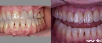
Final picture of the result of implantation and prosthetics
You can clearly see how the Straumann Bone Level implants stand and how deep they are.
Both implants and temporary structures (crowns) cost exactly as needed. They were delivered exactly as planned, which gives a positive result. And it gives us all the prerequisites that the result of a one-step implantation of the front teeth, performed by doctors of the German Implantology Center, will be highly aesthetic and durable. And it will delight our patient for many, many years.
Indications and contraindications
Indications for removal are:
- tooth root fractures;
- severe destruction;
- chronic form of periodontitis;
- atypical structure, inclusion or dystonia, leading to pathologies (difficulty opening the mouth, diction disorders, etc.);
- advanced caries;
- strong mobility;
- crowded teeth;
- neoplasm on soft tissues near the root, large cysts.
Contraindications:
- canine removal (treatment recommended);
- pregnancy;
- mental disorders;
- traumatic brain injuries;
- epileptic seizures;
- diabetes;
- diseases of the nervous and respiratory systems.
Before removal, the doctor conducts a thorough examination, determines whether the tooth can be saved and what measures need to be taken to achieve this. In most cases, doctors prefer treatment, as this allows the functionality of the row and aesthetics to be preserved.