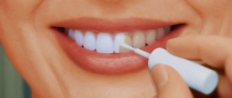
Radiovisiographic examination is a method of computer radiographic examination in dentistry. Its goal is to obtain an image of the tooth and tissues around it with minimal radiation exposure. It has proven itself well in diagnosing hidden carious processes, inflammation of the pulp or periodontium, and identifying foreign objects in the tooth canal. Thanks to him, the dentist has the opportunity to control the quality and effectiveness of the treatment. During the procedure, a detector is inserted into the oral cavity, which is aimed at the area under study: its image is displayed on the monitor.
You can undergo radiovisiography in Moscow at the dental department of CELT. Our clinic has been operating in the capital's paid medical services market for almost three decades. We have a modern radiovisiograph, which allows us to accurately and quickly make diagnoses, identifying diseases in the early stages of their development. Our surgeons, orthodontists, therapists, and orthopedists have decades of experience in scientific and practical work. They constantly improve their professional knowledge and skills and use effective and gentle treatment methods. You can find out the price of the study by going to the “Services and Prices” tab. To avoid misunderstandings, check the numbers with the information line operators or at your doctor’s appointment.
Consultation with a dentist-therapist - 1,000 rubles.
Radiovisiographic examination - 370 rubles.
At CELT you can get advice from a dental specialist.
- The cost of a consultation with a dentist-therapist is 1,000
Make an appointment
Radiovisiography in dentistry
Without radiovisiographic examination, diagnostics in dentistry would be impossible. This is computer radiography, which replaced the outdated film radiography. Thanks to it, you can get a clear image in just a few seconds, while the patient’s radiation exposure will be minimal - ten times less than when using a film device.
A digital visiograph is equipment that should be in any modern dental clinic, since today it is the only method that fully complies with international standards. Its operation is based on the same principle as a standard device, but the signal is sent not to the film, but to the CCD sensor (or, as it is also called, the matrix). The latter has greater light sensitivity than conventional film, so minimal exposure to radiation is sufficient to obtain a clear image.
The visiograph sensor is connected to a converter, which reads its signals, transforming them into a digital image. The latter is sent to the monitor where the doctor can see it. The digital format also has many advantages: it does not take up much space on your hard drive, where it can be stored for a long time, it can be transferred to a USB drive or sent by email.
Advantages of computer radiovisiography
- Safety. The patient receives 93% less radiation than with a traditional X-ray procedure. This technique is used even in pediatric dentistry.
- High speed of examination. Radiovisiography does not require much time. The photograph is immediately digitized and displayed on a computer screen; there is no need to develop the film.
- Best image quality. Ready-made materials allow you to analyze the condition of the maxillofacial area. A specialist can adjust brightness, contrast, and increase the scale to conduct a better analysis.
- Collection and storage of information. All data can be saved electronically, which greatly simplifies the process of subsequent endodontic treatment and orthodontic intervention.
- Minimum contraindications. This examination has virtually no contraindications. It is applicable for most patients, including children.
One of the disadvantages of the procedure is that the spatial resolution is 1.5–2 times lower than with traditional X-ray diagnostics. However, this disadvantage can be corrected by changing the contrast of the digital image.
Radiovisiography allows examination to be carried out at a high level, painlessly and safely for the patient. Full control of the therapeutic intervention makes it possible to avoid complications and guarantee high-quality dental treatment.
Dentists at the AlfaDent clinic perform similar procedures every day. Contacting experienced specialists guarantees effective therapy. Detection of pathologies at an early stage using a visiograph will allow you to avoid expensive treatment.
Indications and contraindications for dental radiovisiography
The study can be used for targeted study of dental units, as well as for identifying hidden carious processes and inflammation of surrounding structures.
Indications
- Diagnosis of caries, inflammatory processes of pulp and periodontium;
- Monitoring their treatment (quality of endodontic treatment, in particular, filling of canals, correct placement of pins, tightness of seal to the walls of the tooth);
- Preparation for surgery to increase bone tissue to determine the degree of its atrophy and the characteristics of the process;
- Suspicions of neoplasms in the thickness of the soft tissues of the oral cavity;
- Cystic formations and granulomas at the roots of teeth;
- Determining the location of fragments of broken dental instruments in the root canal;
- Traumatic damage to a tooth (dislocation, bruise, fracture);
- Control of the implant after installation;
- Before sealing the fissures of teeth in order to ensure the absence of caries.
Contraindications
Absolute contraindications:
- The patient has serious mental disorders;
- Pathologies of a neurological nature;
- Depressive states.
Due to the minimal level of radiation, radiovisiography of the maxillofacial area can be performed on a child, provided that he can sit still for several seconds.
If indicated, the study can be performed on women during pregnancy and lactation. It is optimal to do this in the second trimester, after warning the diagnostician about pregnancy. This is necessary so that she is put on a special apron, which provides additional protection from x-rays.
Indications for the procedure
There are quite a few indications for radiovisiography, although it can also be done for preventive purposes, that is, without obvious dental problems.
Caries
Carious damage to dental tissue is quite clearly visible on visiograph images. You can even see lesions in the interdental space or natural recesses of the teeth (fissures). Also available for such diagnostics is root caries, which will most likely go unnoticed during the initial dental examination of the oral cavity and dentition.
Inflammation of tooth tissue - pulpitis
Inside the dental canal is the pulp, which contains the nerve. The most common cause of toothache is pulpitis, that is, inflammation of the pulp. To make the right decision whether to remove the nerve or whether it is enough to perform treatment and filling, the dentist prescribes radiovisiography to the patient. The pictures will clearly show which tooth is affected and to what extent. After all, sometimes it happens that one tooth hurts, but it is felt in a slightly different area.
Periodontitis
Periodontitis is a hidden disease, as it affects the ends of the tooth root. The patient may feel pain, but only a targeted X-ray image taken with modern equipment will help to recognize the cause of its occurrence.
Foreign bodies in the canal
In dental practice, there are cases where inflammation in the tooth and pain occurred due to the presence of a foreign body in the dental canal. Of course, it does not appear there by itself; it is a consequence of improper treatment and leaving particles of instruments (for example, used to clean the pulp), cotton swabs, and so on in the tooth.
Neoplasms in the oral cavity
Using computed x-ray images, you can recognize oral pathologies such as cysts, tumors, fibromas, and granulomas. Similar neoplasms may have similar symptoms, but it is important to accurately determine the diagnosis in order to prescribe effective treatment.
Advantages of radiovisiographic examination
This research method is considered the best option both for a dental office and for a large clinic in which several specialists work at once. Its advantages:
- Efficiency and the ability to receive an image instantly, without waiting;
- High clarity and information content of the image, in which you can see the condition of the teeth, surrounding tissues, and implants;
- Minimum number of restrictions (possibility of use for children and pregnant patients);
- Convenient digital format, the ability to print the image on paper.
Do not forget that visiography is an environmentally friendly and harmless research method. It does not require the use of film or aggressive chemicals for development. The radiation dose received during filming of the lower jaw is 2 microsieverts, the upper – 5 microsieverts. For comparison, when using a film apparatus, it is equal to 30 and 80 microsieverts, respectively.
The resulting images clearly show:
- Carious lesions, cystic formations, granulomas and inflammatory processes. They are transmitted in black;
- Fillings, inlays, pins, dentures – white areas;
- Connective and bone tissue are gray areas.
Advantages
- Safety. The radiation dose is 90% lower than with conventional x-rays: radiovisiography is no more dangerous than a digital camera or an office scanner. Not contraindicated for children, nursing mothers and even pregnant women - but not during the second trimester and strictly according to indications.
- Save time. You don't have to wait under the cabinet for 10-15 minutes while the film is developed. The image will immediately appear on the monitor in the doctor’s office.
- Elimination of the darkroom process. No equipment is needed to develop the film and print the image.
- Creation of a patient database. The images are “linked” to medical records, and the dentist can review them at any time. Image processing. Changing brightness and contrast, comparing negatives and positives, zooming in on images, and creating panoramas allow doctors to analyze in detail the structures shown in the image.
Manufacturers of radiovisiographs guarantee that up to 60 procedures per year are allowed without any harm to health.
This will not exceed the norm of annual radiation acceptable for humans - up to 1000 microsieverts (µSv). So, when taking an image of the lower teeth, the radiation load on the body is 2 μSv, the upper ones - 5. Note: versus 7-14 μSv with film X-rays.
Reviews about our dental therapists
I would like to express my gratitude to the dentist Elena Nikolaevna Kiseleva and her assistant Svetlana - they are real specialists and at the same time sensitive, not burnt out by years of practice.
Thanks to them, I have been coming back here for many years. Thanks to the management for such doctors! Read full review Svetlana Nikolaevna
13.08.2021
I am very grateful to Evgeniy Borisovich Antiukhin for removing my three eights. Especially considering that the lower tooth was not the simplest (it was located in an embrace with a nerve). The removal took place in 2 stages, one tooth under local anesthesia, two under general anesthesia. I had no idea that wisdom teeth could be... Read full review
Sofia
28.12.2020
Radiovisiography technique
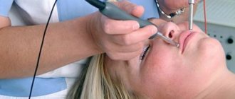
The procedure is painless, with a minimum of discomfort. The patient is seated in a chair, covered with a lead apron, given a CCD sensor in a disposable cover to bite on, and asked not to move. To take a picture, 0.06-0.08 seconds is enough.
When examining the maxillofacial area in the area of the distant chewing teeth, a gag reflex may occur. The first time the image may not be obtained in patients with current anesthesia - due to loss of sensitivity, it is difficult to hold the tongue motionless.
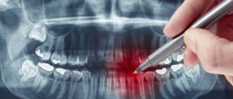
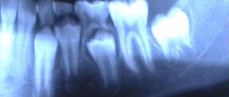
Radiovisiographic image
How is dental radiovisiography performed?
The procedure is very simple and absolutely painless and does not require preparation. The patient is asked to take a sitting position on a chair near the device, after which a sensor is applied to the area under study and the patient is asked to gently bite the sensor with his teeth. This prevents its displacement. After this, the tube of the device is directed to a certain area of the jaw. The diagnostician starts it, and X-rays begin to illuminate the tissue. The signals are captured by the sensor, then sent to the converter and (already in the form of a graphic image) to the monitor.
Execution technique
Radiovisiography of a tooth with pathology (or a regular x-ray if the dental clinic does not have computer equipment) is a mandatory procedure before starting treatment. It is carried out in order to make an accurate diagnosis and assess the condition of the diseased tooth. Follow-up images may be required during treatment. For example, after cleaning the canal, the doctor must use an image to ensure that the nerve has been completely removed and the tooth has been prepared for filling.
An image of the teeth may also be required after treatment to assess its effectiveness.
Radiovisiography does not require special training.
The patient sits on a chair and gets as close to the device as possible. To protect the body from exposure to x-rays, a special apron is worn. Next, the patient covers the digital sensor with his mouth and clenches his teeth. The sensor is located as close as possible to the area where the pathology occurs. A picture is taken, which takes a few seconds (during this you cannot move or breathe).
Difference between radiovisiography, OPTG and computed tomography
Unlike visiography, OPTG - orthopantogram (or, as it is also called, panoramic image) covers not one or three teeth, but the entire dental system. An orthopantomograph is used to obtain images. Most often, such diagnostics are prescribed to young patients in order to assess the condition of the rudiments of permanent teeth. As for computed tomography, it provides not a two-dimensional, but a three-dimensional image of the patients’ jaw system. For this reason, it is carried out before implantation and bone augmentation operations that precede it, in order to plan them in detail.
You can make an appointment with dentists and undergo radiovisiography at CELT online or by contacting our information line operators: +7 (495) 788‑33‑88.
Make an appointment through the application or by calling +7 +7 We work every day:
- Monday—Friday: 8.00—20.00
- Saturday: 8.00–18.00
- Sunday is a day off
The nearest metro and MCC stations to the clinic:
- Highway of Enthusiasts or Perovo
- Partisan
- Enthusiast Highway
Driving directions
Advantages of radiovisiography
Dental diagnostics using a radiovisiograph has the following advantages.
- Survey speed. Thanks to visual imaging, you can get an image 10 times faster than using outdated X-ray models.
- High quality image. The equipment allows you to display an enlarged image on the monitor, which can be processed to clarify details.
- Ability to save pictures. Images can be archived in the patient's electronic medical record.
- Digital image processing. The specialist has the ability to change the brightness, contrast and size of the image. The device’s software allows the dentist to obtain data for the manufacture of implants and dentures.
Types of X-ray
In addition to dental radiovisiography, there are other diagnostic methods.
- Extraoral radiography. It is used if, for a number of reasons, it is not possible to conduct intraoral diagnostics.
- Bitewing, or intraoral, examination. To carry out the procedure, a special film is used, which is clamped between the teeth to take an image.
- CT scan. A tomograph is required for the study. Such diagnostics are highly informative.
- Teleradiogram. The procedure is carried out before operations or installation of braces. As a result, the doctor will receive a picture of the entire skull.
- Orthopantomogram. It is a panoramic photograph of the dentition, allowing you to see unerupted units.
Advantages of digital radiovisiography for the dentist
A few years ago, specialists used only film X-rays, but now more and more dentists are resorting to digital research methods. Filmless diagnostics are good because the received data is instantly processed by the program and displayed on the display (medical workers will no longer need to use dangerous chemical compounds to develop photos).
Today, if necessary, it is very easy for dentists to look at any image taken several weeks ago, since the results of the study are stored in the PC memory. The resulting photo can be sent via the Internet to any corner of the world, for example, if the doctor is on vacation or away for work.
The advantage of the visiographic method is that the photo can be enlarged several times, the contrast can be adjusted and the teeth and roots can be examined in detail. Using the program, the dentist can examine the texture of bone tissue, alveolar processes, tooth roots or periodontal tissue.
What does it have to do with x-rays?
Digitized image.
Many people mistakenly believe that a visiograph is the same as an x-ray. It has nothing in common with x-rays. A visiograph is a digital equipment that does not require chemical reagents or X-ray film to obtain an image. This directly affects the exposure time, because the digital output method provides instantaneous image acquisition on a computer monitor. Therefore, a special room is not required to place the visiograph. It can be placed next to the dental unit, in close proximity. The patient does not need to leave the dental chair to take the image. Thus, the patient's appointment time is significantly reduced.