The clinics of the German Implantology Center are equipped with the most modern diagnostic equipment necessary to prepare the patient for dental implantation. According to dental market experts, both clinics of the Research Center, located in the Ramenki and Dorogomilovo districts of Moscow, occupy the TOP 1 rating for 2017-2019.
private dental clinics in Moscow and Russia.
Let's see what diagnostics are carried out before implantation in our clinics, without what equipment, procedures and diagnostic data it is currently impossible to prepare qualitatively
for implantation surgery.
The chief physician of the Research Center, implant surgeon Dakhkilgov Magomed Umatgireevich answers these questions:
As an implantologist with more than 20 years of experience
Magomed Umatgireevich, who has made a serious practical contribution to the development of this area in Russia, makes great efforts to ensure that the equipment of clinics meets
leading world standards.
He brought clinics to the TOP1 ratings in Moscow and Russia, which today makes it possible to successfully perform dental implantation even in the most complex clinical cases.
Please note that some of the material is accompanied by a video from our YouTube channel, we recommend spending a little of your precious time and watching the videos, it will be useful. More than 1,000 patients use the recommendations of medical specialists from the Scientific Research Center on YouTube every day
.
What examinations and tests need to be completed before implantation?
Speaking about tests before dental implantation, it should be noted that tests are needed, but not in every case. In cases where:
- the patient does not have chronic diseases, and he knows about it,
- the patient has no complaints about any organs or systems,
- he has not suffered from any serious illnesses recently,
- there have been no major organ surgeries,
is NO SPECIAL need to take tests
.
But, nevertheless, if there were any problems before, if the patient is being treated by specialists, then he must first undergo tests depending on the organ that is bothering him.
ATTENTION!
The patient needs to approach diagnosis and preparation with the utmost seriousness, since any problem that was undetected or ignored can subsequently lead to tissue inflammation - peri-implantitis, even to rejection or removal of the implant.
What's the difference in price?
The difference in price is the cost of producing radiopaque and implantation templates. For assessment, for one jaw, regardless of the number of implants installed, you will need to pay an additional 26,000 rubles. to the planned cost of the implantation operation. Often, when it comes to a simple case of installing a single implant, we can do without making templates. An implantological template is used when installing 2 or more implants, then the price increase will be 13,000 per implant. If there are even more implants, the cost of the template ceases to play a significant role in the price of treatment.
About preparation before dental implantation
To achieve predictable and long-lasting results at the German Implantology Center, when planning any implantation, we carry out:
- consultation of related specialists: an orthopedist and a surgeon, and in case of problems with the patient’s bite, also an orthodontist,
- computed tomography. The best cone tomograph, according to experts, today, “Planmeca ProMax 3D Max”, helps us with this; this is the top line of Planmeca tomographs:
- Before the implantation procedure, the patient must undergo professional dental hygiene
, here we use AIRFLOW Prophylaxis Master - the latest technological innovation from the EMS company, a new product for 2022 on the dental services market, which allows absolutely painless hygiene for patients with
increased tooth sensitivity
and prevents the development of caries, periodontal disease,
- Before implantation, the patient needs to undergo therapeutic treatment of caries
and its complications. Treatment is carried out under a microscope; each room at the Research Center is equipped with the best models of microscopes that have proven themselves in medical practice:
- Before implantation, diagnostic casts of teeth are required for plaster placement in the articulator and functional analysis. Now plaster models have been replaced by digital scanning
, which is being actively implemented in our clinics. The best intraoral scanner from 3D Shape helps us scan the dentition. In the following video, an intraoral scan is performed by orthopedic dentist of the Research Center Andrey Nikolaevich Karneev:
After scanning we carry out virtual planning of our work
:
and production of surgical templates
, according to which the patient undergoes high-precision installation of implants:
So, what are the advantages of the ImplantGuide technology?
- Full mutual understanding between the orthopedist, surgeon, and dental technician in choosing the best design, the ability to involve other specialists before and at the stage of implantological treatment;
- Optimal placement of the implant as a support for the future orthopedic structure;
- Selection of the optimal individual operating technique in each clinical case;
- Precise positioning of the implant in the planned location;
- 2-5 times reduction in the time of dental implantation surgery;
- Minimal trauma, pain and swelling after surgery, reducing the likelihood of dental complications;
- 100% predictable and guaranteed final aesthetic result;
- Possibility of installing implants without incisions (bloodless implantation method);
- Allows you to perform precise and safe operations.
Trust the beauty of your smile to professionals
Dentistry in Lyublino “Good Hands” has been operating on the Moscow market for more than 5 years and has earned a huge number of positive reviews from regular customers. We carry out:
- Comprehensive diagnostics.
- Taking impressions and creating a plaster model.
- Selection and installation of implants.
- Monitoring results and assistance in the rehabilitation process.
We use the best implants from manufacturers from Switzerland, Germany, and Israel. To make an appointment for diagnostics, call us during business hours.
A little history
The “pioneer” of modern dental implantology is the Swedish scientist P.-I. Branemark. After numerous experiments on animals, in 1965 the first intraosseous implant in the form of a tooth root was placed in humans for two-stage use.
During many years of research, one of the fundamental discoveries of implantology was made in 1978: in the bone bed, which is prepared atraumatically and exactly matches the shape of the titanium structure being installed, a strong “fusion” of the metal surface with the bone occurs. Branemark and his colleagues fully appreciated the significance of the phenomenon they called “osseointegration,” which opened a new era in the history of dental implantation.
In 1981, Branemark summarized the results of a 15-year study in which 90% of the implants functioned throughout the observation period. According to L. Linkow, dentistry has reached its “golden age” and for this medical specialty the discovery of osseointegration is comparable in significance to the discovery of local anesthesia in 1902.
In our country, implantology has become an officially recognized branch of dentistry since 1986. During this time, convincing evidence has emerged that dental implantation is a reliable and predictable treatment method, which is increasingly used in the practice of dentists every year.
Benefits of implantation
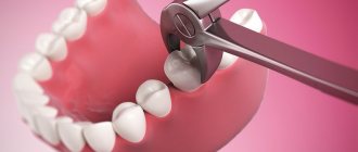
After teeth are removed, the jaw bones significantly decrease in volume over time (atrophy). During the first year, 25% of the bone tissue in the area of the extracted tooth is lost, and in the next 2-3 years the bone tissue decreases by 50% of the original volume. Once the implants are installed, bone loss stops.
During implantation, healthy natural teeth are not prepared.
If you are missing one or more teeth, then it is possible to eliminate the defect in the dentition with the help of a fixed denture. But to install it, the dentist needs to grind the adjacent teeth into crowns and remove the nerves (pulp) from them. The more teeth that are missing, the more teeth that will have to be ground down. For example, if you are missing one tooth, then for a bridge, you need to grind down two adjacent teeth. Unfortunately, the service life of teeth ground for crowns is much shorter than that of healthy ones. Hygienic care of teeth connected by a bridge is much more difficult. The use of implants allows you to keep your natural teeth intact.
The use of implants in case of loss of lateral teeth allows the production of a permanent prosthesis.
When the chewing teeth are removed, it is impossible to restore the defect in the dentition with a bridge, since there are no teeth on which the fixed prosthesis should rest. If you make a fixed prosthesis with one-sided support, then the teeth on which it will rest will be overloaded and will become loose over time. Usually in this situation a removable denture is made. It is inconvenient to use a removable denture; in addition, the bone tissue underneath quickly atrophies. Implantation allows you to install additional supports and make a permanent prosthesis for such a defect.
Implants have a longer lifespan than crowns and bridges.
The results of prosthetics with traditional bridges and crowns are not that good. It has long been scientifically proven that 75% of crowns and/or bridges that rely on “their” teeth successfully overcome the five-year mark. After this period, 25% of dentures need to be replaced. Practice shows that some teeth that served as support for fixed dentures have to be removed.
Statistics on the success of implant-supported dentures are more encouraging. More than 90% of implant-supported prostheses function successfully for 10 years. Service life of the company's implants. And on toothless jaws, the shelf life of implants exceeds 35 years.
Treatment planning and diagnosis are the basis for success!
It is necessary to sign up for a consultation on the possible use of implant treatment with an orthopedic doctor, since the final result: the type of design, the number of required implants is the responsibility of the orthopedic doctor. When an understanding has been reached between the doctor and the patient regarding the scope of the work to be done, an implant surgeon is invited to an extended consultation. During the consultation, X-ray examinations are necessary to evaluate the volume of bone tissue. Most often, orthopantomography (OPTG) is used for diagnosis, which allows one to obtain an image of the upper and lower dentition and adjacent anatomical formations.
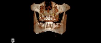
But the image obtained using this method is planar and is not always informative. The most reliable method of x-ray examination is computed tomography (CT), which shows the volume of jaw bone tissue in three projections. With its help, the installation of implants is precisely planned. Only with the help of these preoperative diagnostic procedures can it be established whether the quantity and quality of jaw bone tissue is sufficient for implantation.
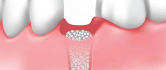
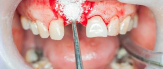
In case of bone tissue deficiency, bone regeneration using your own bone or using artificial bone material is indicated. Preoperative diagnostic measures also include the production of plaster models and surgical templates, which are currently quite widely used.
Indications and contraindications for implantation are established on the basis of a general medical history and examination (blood tests, etc.), assessment of the psycho-emotional state and dental status of the patient.
After determining the indications, the first thing that is necessary is to exclude the presence of contraindications. If you have any serious systemic diseases, a written opinion from your attending physician (cardiologist, endocrinologist, etc.) is required.
Only high-quality planning of the operation allows one to achieve long-term and highly aesthetic results. At the SNK DENTISTRY clinic, we guarantee highly qualified assistance in prosthetics on implants of various systems and choosing the best type of prosthesis for a given clinical case. Your dental health is our responsibility.
What does preoperative examination include?
To make a sinus lift safe and effective, the doctor must ensure that the patient is in normal health. In dental clinics, a number of laboratory and instrumental research methods are widely used for this purpose:
- Inspection. Before deciding to perform bone grafting, the dentist performs an external examination and examination of the oral cavity. An external examination is very informative for diagnosing many dental pathologies; the doctor pays attention to the shape and symmetry of the facial part of the skull, the color of the skin and visible mucous membranes (sclera), the condition of the submandibular, sublingual and cervical lymph nodes. When examining the oral cavity, the condition of the mucous membrane is assessed, using mirrors and a probe, all existing teeth are examined;
- X-ray examination of the upper jaw and maxillary sinus. This simple research method allows us to identify individual features of the structure of the maxillary sinus and the presence of pathological neoplasms in it. X-rays also allow you to assess the condition of the bone tissue of the upper jaw;
- CT scan. The method allows you to obtain accurate information about the condition of the bone tissue of the upper jaw and maxillary sinus. Based on the results of computed tomography, the estimated volume of osteoplastic material that needs to be introduced for implantation is determined;
- Consultation with an otorhinolaryngologist. In addition to assessing the condition of the maxillary sinus using CT or radiography, it is important to obtain the opinion of a doctor specializing in diseases of this anatomical structure. The otorhinolaryngologist must conduct an examination and collect a detailed history of past diseases of the ENT organs, including the maxillary sinus, which may become an obstacle to the intervention;
- Clinical blood test. Allows you to assess the condition of the patient’s entire body, identify a latent inflammatory process (increased levels of leukocytes and ESR), anemia (decreased red blood cells), coagulation problems (assess the level of platelets), as well as a bacterial or viral infection. All these pathologies can be suspected based on the results of a simple analysis, so it is necessarily included in the preoperative examination and sinus lift planning;
- Coagulogram. Since any type of sinus lift involves a violation of the integrity of the tissues in the oral cavity, it is necessary to check the coagulation indicators. If any of the indicators deviates from the norm, then the operation is fraught with the development of bleeding;
- Determination of blood group and Rh factor. Included in the list of mandatory studies before any surgical intervention;
- Blood glucose. The tactics for managing patients with diabetes mellitus are somewhat different, and in some cases, when the disease is decompensated, sinus lifting is contraindicated;
- Screening for hepatitis B and HIV infection. If these diseases are detected, the decision to implant and perform bone grafting should be made together with an infectious disease specialist;
- Wasserman reaction (to detect syphilis);
The list of necessary diagnostic procedures may differ in different clinics. However, it is important to understand that all of them are necessary to assess your health and weigh all the possible risks that accompany the operation.
Diagnosis using computed tomography
After a preliminary assessment of the patient’s condition, the dentist begins a more detailed diagnosis. To do this, a three-dimensional computer image (CT) is taken with a tomograph, on which hidden pathologies can be seen, such as hilar granuloma or cyst. Based on the tomography results, the dentist can estimate the height and width of the bone crest. If the bone volume is small, osteoplasty is necessary, especially if the structure is massive, for example, when installing a bridge supported by implants.
Why is preoperative examination necessary?
Preoperative examination is the most important step in performing any type of sinus lift. A thorough diagnosis of the condition of the oral cavity and the entire body as a whole allows us to promptly identify and, if possible, eliminate contraindications to the upcoming surgical intervention.
Preoperative examination is aimed at identifying or excluding the following relative and absolute contraindications to the procedure:
- Inflammatory gum diseases;
- Caries;
- Neoplasms in the maxillary sinuses;
- Acute or chronic sinusitis in the acute stage;
- Previous operations in the area of the upper jaw and maxillary sinus;
- Anomalies of the anatomical structure of the upper jaw and maxillary sinus;
- Acute rhinitis (including allergic etiology);
- ARVI;
- Pathologies of the blood coagulation system;
- Severe mental disorders;
- Diseases of the heart, lungs, kidneys in the stage of decompensation;
The above pathological conditions can become an obstacle to performing a sinus lift or delay it during treatment (inflammatory gum diseases, acute respiratory viral infections, allergic rhinitis and others).
Stages of 3D planning of dental implantation and preparation for navigational surgery
- Examination and diagnosis
- CT (computed tomography) examination
- Assessment of conditions for implantation (volume, bone density, presence of inflammatory processes)
- Digital dental impressions
- Combining CT scans with digital dental models and creating a dental implantation project
- 3D modeling and selection of the required orthopedic structure (crowns and dentures)
- Selection of a suitable implant system by design based on the volume and density of bone tissue
- Virtual placement of implants
- Control of requirements satisfaction
- Generation of a 3D navigation template with bushings
- 3D printing navigation template
- Creation of a step-by-step protocol for navigational implantation
- Teeth implantation operation using a template