How to pull out a baby tooth with floss
The essence of the method is that a loose tooth is tied with a thread, the other end of which is tied to the door handle.
The door opens and the tooth flies out of the mouth. This technique does not withstand any criticism either from dentists or from psychologists. A morally outdated method that can frighten a child and even cause psychological trauma.
Home manipulations often lead to infection in the socket. Inflammation may develop - flux, pain and fever will appear. The best solution is to have loose baby teeth removed at a dental clinic. It's fast, painless and inexpensive.
Anatomy of permanent teeth
The molar tooth includes three zones: the root, which sits deep in the jaw socket and holds the tooth in place, the neck, located in the periodontal area, and the crown, which extends directly into the oral cavity. After the apex of the tooth appears, a protective film forms on the enamel, the strongest layer of the tooth, which is soon replaced by a salivary layer, formed from the saliva itself. The dental tissues themselves are not just a piece of bone, but a certain heterogeneity, which includes, in addition to enamel, dentin (the main substance of the tooth) and the dental cavity, in which nerves and blood vessels branch. Compared to bones, for example, the phalanges of the fingers, dentin is noticeably stronger - it contains an increased amount of minerals, for example, the same calcium-based compounds. The root zone of dentin is connected to the periodontium using a special layer - dental cement, which communicates with the tissues of the periodontium itself and supplies the dentin with nutrients.
Why do teeth change?
As the body grows and matures, it requires more and more nutrients, which can only be obtained through digestion. To do this, food must enter the stomach well chewed - this ensures its complete breakdown and subsequent absorption of the necessary compounds.
Changing teeth performs three important functions:
- Provides a person with a strong chewing apparatus.
- Allows you to replace teeth damaged by caries with new, healthy ones.
- Increases the number of teeth - the upper and lower jaws of a teenager can accommodate 32 permanent teeth along with 20 milk teeth.
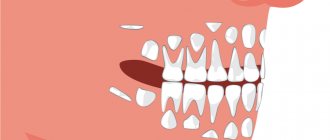
Differences between baby teeth and permanent teeth
The structure of primary and permanent teeth is generally the same, but there are several noticeable differences. Which are better not to neglect:
Baby teeth have whiter enamel, while permanent teeth have a slight yellowish tint. Milk teeth are not as strong as permanent teeth due to the lower percentage of mineral substances. The pulp of baby teeth is wider and thicker, while dentin and enamel are, on the contrary, thinner. This explains the facilitated, spontaneous removal of a shelf tooth - there are often situations when schoolchildren in class independently loosen and remove milk teeth that have served their purpose. In permanent teeth, the length is significantly greater, dominating over their width and cross-sectional area. The root of a baby tooth is thinner and shorter - a person is able to pull out a baby tooth without the help of specialists. Meanwhile, the permanent “embryo” of a real tooth under the shelf one is already ready to grow quickly and be born. The latter is achieved due to the dental gap, which has expanded by the time the non-permanent tooth falls out.
What are caries and pulpitis?
Caries is a disease in which the hard tissues of the tooth are destroyed due to the loss of minerals from the tooth enamel.
How and why caries forms, read our article on the treatment of caries. The carious process, progressing, gradually reaches the pulp chamber, in which there is a neurovascular bundle of fibers that nourishes the tooth - the pulp, and destroys it. Bacteria from the carious cavity penetrate inside, infect the pulp, it becomes inflamed and pulpitis begins.
Both adults and children suffer from caries and pulpitis, but the course of the disease at different ages and its treatment differ.
Read about the treatment of caries and pulpitis in adults and pulpitis in baby teeth in the sections dedicated to these topics. In this article we will tell you about the treatment of caries and, especially, pulpitis of permanent teeth in adolescents, as well as about its features.
Where do teeth come from?
Teeth are formed in the fetus’s body during the mother’s pregnancy. When a mother abused something and undermined her own health, the subsequently born child was guaranteed to have diseased teeth that were no longer permanent. At the 15th week of pregnancy, the mother has hardened dental tissues in the fetus - starting from the crown area and ending with the root zone. The embryos of molar teeth are formed by the 5th month of fetal life. The body of a developing fetus and child is designed in such a way that in the upper jaw the anlage of the permanent teeth is located above the anlage of the milk teeth, and in the lower jaw - vice versa. The formation and development of teeth begins as early as the sixth week of fetal development. The source for them is a special epithelial dental plate. Already by 14 weeks of pregnancy, the unborn baby is actively forming hard dental tissues, initially in the area of the coronal part, and then in the area of the roots of the tooth. When a child is born, primary teeth are the first to grow - by the end of the child's first year of life, they will erupt. However, the dentition contains a group of large molars - they, in turn, do not have milk predecessors and subsequently, when they fall out, grow “on a permanent basis”. Nature has arranged it in such a way that while the child’s jaws are still too small, large molars are not needed there.
In the absence of lateral teeth
With the early removal of several lateral teeth, depending on the adaptation mechanism, the following may form: - deep bite: the height on the sides is lost, and the lower front teeth begin to reach the mucous membrane of the palate. At the same time, the face is visually shortened. - mesial bite: in the absence of lateral teeth, chewing will be transferred to the incisors - they will experience functional overload and wear out. The jaw will move forward, and in this position it will be fixed at the skeletal level. - open bite: in the absence of two or three teeth nearby, muscle balance is disturbed and so-called “bad habits” are formed: tongue protruding, cheeks sucking into the area of missing teeth. In the future, this will interfere with the eruption of permanent replacement teeth, leading to non-occlusion of these teeth. - Crossbite: Removing side teeth on one side will force the child to chew only on the opposite side. Such chewing will lead to a sideways displacement of the lower jaw, the formation of a crossbite and asymmetry of the face and temporomandibular joints.
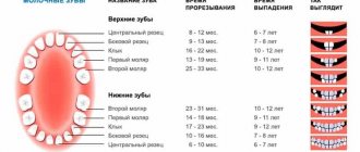
How many primary and molar teeth does a person have?
In children, the size of the jaw is almost half (in terms of the number of teeth that fit on it) smaller than in adults. Initially, the child has up to 20 teeth - 10 on each jaw. That is, one jaw - 4 incisors, 2 canines and 4 molars. Primary molars have not yet been clearly divided into small and large.
After 16 years, a teenager’s jaws reach approximate sizes that are accessible to an adult. A teenager already has approximately 28-30 teeth, and not 20-24, as before. The number of molars is often represented by 2 small molars and 2 large molars - on each side on each jaw. The last 2 or 4 teeth - “wisdom teeth” usually appear by the age of 20-22 - and a person acquires a full set of teeth, numbering 8 incisors, 4 canines, 8 small molars and 12 large molars - on both jaws in total.
Our first baby teeth
A child is born without teeth - physiologically he does not need them, since the baby feeds on breast milk or formula and drinks water. At 4–6 months, the first teeth appear, the baby begins to be introduced to complementary foods in the form of baby cereals and vegetable purees, which involve at least a little chewing. Further, the growth of teeth accelerates and by the age of two there are 20 of them - this is the maximum number that can fit on the baby’s jaws.
The facial skeleton continues to grow, so at the age of 5–6 years, the child erupts 2 more teeth, called “sixes.” At the same time, the processes of resorption of the roots of primary incisors and fangs begin - this is how preparation takes place for the replacement of temporary teeth with permanent ones.
What does the dental formula look like?
The medical record, which the big one keeps in the dental clinic at his place of residence, contains notes about his dental condition. To avoid confusion, doctors number the teeth on each side of each jaw. So, the 1st and 2nd teeth are incisors, the 3rd are canines, the 4th and 5th are small molars (doctors call molars molars), the 6th and 7th are large molars. The 8th - the farthest one - is a “wisdom tooth”; a number of people do not have it, or they do, but not all. Each side of the jaw is also numbered: 1 - top right, 2 - top left, 3 - bottom left, 4 - bottom right. For example, entry 48 does not mean that you are a “Tarkatan” with a combat superset of teeth that does not exist. You simply do not have a “wisdom tooth” on the lower right. Entry 41 - a person lost one of the frontal incisors on the same side on the same jaw. You can write down the formula of the teeth more clearly: for example, “there is no 8th tooth from the bottom right.”
It often turns out that due to lack of space on the jaw, the wisdom tooth develops incorrectly - it can grow crooked, in which case its removal is indicated. For example, it may remain under the gum and, because of this, be affected by caries, which can subsequently develop into pulpitis or a dental cyst. Problematic “wisdom teeth” are quickly and decisively removed, and their absence will not greatly affect the quality of chewing food.
Is the child worried?
Many parents worry that when their children lose their baby teeth, they will develop complexes. Actually this is not true. Of course, loose and falling teeth bring physical discomfort to children, however, they do not worry about the unaesthetic appearance. After all, most of their friends also walk around with lost teeth.
When a child's first tooth falls out, the baby may be bothered by the socket that appears as a result of tooth loss. The baby's curiosity inevitably develops, and he constantly tries to touch the socket and the growing molar with his tongue (or even his finger) in order to explore new sensations.
Parents definitely need to explain to their child that this should not be done: your hands can introduce an infection into the mouth, which can cause teeth and gums to become sore.
There is no need to worry if ichor comes out of the hole after a tooth falls out. In this case, you need to let the baby rinse his mouth with a soda solution. If the wound is actively bleeding, you can let the child lightly bite on a cotton pad or gauze pad.
Teething order
The timing of the appearance of permanent teeth to replace lost milk teeth is generally the same for all children and adolescents. After the child turns 5 years old, the first large molars make themselves felt. Then the central incisors are replaced from below, then the same teeth from above, and the lateral incisors from below. At 8-9 years of age, the lateral incisors on top are replaced. From 9 to 12 years of age, all small molars are replaced. At 13, all fangs are replaced. After 14 years, second large molars appear on all sides, which were not there before. By the age of 20-22, “wisdom teeth” finally appear. There are cases when during the rest of their lives they never erupted.
Oral care
On the first day after removal of the milk jug, you need to brush your teeth without toothpaste, avoiding touching the socket with a brush. If the child cannot carefully clean the oral cavity from food debris without touching the socket, then you can rinse your mouth well with warm water.
Children who are prone to stomatitis on the tongue, gums or lips should rinse their mouths with a soda solution, chamomile decoction or an antiseptic such as Rotokan or a furatsilin solution.
For 4-5 days after the baby tooth has been removed, it is important to observe dietary restrictions - exclude salty, spicy and hard foods.
Author: Zhukov M.A.
How to determine that a child will soon have molars?
According to some of the signs, interdental intervals initially increase. This happens due to the growth of the jaw bones - the teeth begin to not fit tightly together, as happens in adults. Then the temporary teeth gradually become loose - due to the gradual disappearance of the temporary root, which can no longer reliably hold such a tooth, and itself is gradually pushed out of the soft and semi-soft tissues on the jaw. The loss of a temporary tooth is a clear sign that the permanent tooth is already growing in full force, and the top of its crown will soon push apart the gum tissue. The appearance of a new tooth is also accompanied by slight redness and swelling. If a child or teenager has a fever, their health has worsened, and their gums hurt, go to the doctor immediately.
Reasons for delayed teething
At what age teeth fall out and the sequence of tooth loss are important indicators of physiological development. A huge amount of preparatory work goes through a child’s body. The root of the temporary tooth partially resolves, and the rudiment of the permanent tooth pushes the milk tooth upward from the alveolar bed. But sometimes in such a natural process, due to external or internal reasons, failures occur [4].
When children lose their baby teeth too early or late, it is necessary to understand the reasons. After all, this may be a signal of serious internal disorders that require observation or treatment by specialists. The delay occurs due to:
- system violations:
- rickets;
- chronic intoxication;
- metabolic disorders;
- pathologies of hormonal processes, especially hypofunction of the thyroid gland;
- lack of vitamin D.
Violations of local eruption conditions are also important:
- trauma to the tooth germ;
- lack of space in the dentition;
- incorrect position of the tooth germ;
- inflammatory diseases of the jaw bones [2].
No molars
The molars, despite the specific timing specified above, may ultimately not hatch. For example, temporary teeth do not fall out for a long time, and permanent teeth do not grow for a long time after they fall out. At the first stage, the dentist will take an X-ray of the condition of the child or teenager’s jaws. The X-ray machine will clearly project what should grow in place of the supposed teeth, which teeth are ready to hatch, and whether they exist at all. But the problem will immediately become obvious, and the reason for it most likely lies in the physiology of the child, which has undergone some delay. If this is true, be patient: in the end, the teeth will not take long to arrive. But if the picture shows emptiness, you are faced with a complete absence of teeth, the reason for which is a violation of the intrauterine development of the fetus during the mother’s pregnancy. Only prosthetics will help here - artificial insertion of false teeth.
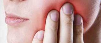
Molar tooth hurts
After the molar has emerged from the gums, the “young” tooth enamel is not fully saturated with all the necessary microelements. During this period, the tooth is especially vulnerable: it is strictly forbidden to subject it to overload. It is not for nothing that children are advised not to chew, for example, candy, large quantities of peanuts, or any solid foods. If you neglect this rule, children may immediately develop caries, and this is the path to problems with specific teeth. Caries gradually turns into pulpitis (damage to the internal cavity of the tooth) and periodontitis (damage to the ligaments surrounding the tooth). The child often experiences toothache and poor general health. Without turning to a professional doctor for help in time, parents can lead their child to various consequences - up to the complete loss of a diseased tooth. If a predisposition to the appearance of carious lesions has been identified, their prevention may involve closing the natural deep pockets surrounding the molars. This is done using high-quality composite materials. In this case, food debris will not accumulate in these places, destructive microflora will not develop, and the period of susceptibility to caries will pass when the teenager grows up.
Useful tips for parents
- Monitor your baby's teeth closely. If there are several children in a family, it is better for each of them to have a sign indicating the lost and emerging teeth.
- Teach your child to take care of their teeth. Teach him to brush his teeth twice a day and rinse his mouth after eating. You can use plain water or herbal decoction of chamomile.
- During the teething period, give your child plenty of calcium. Avoid solid foods and feed your baby soft purees, grated fruits and other foods.
- Discuss the upcoming change of teeth with your child so that it does not come as a surprise to him.
If you observe difficulties with the germination of dental units, this is a reason to immediately consult a dentist. Remember that changing teeth is a stressful process for your child. Support him so that this period passes with minimal psychological losses.
Molars grow crooked
Molars are also subject to crooked growth. They can begin to actively grow before the temporary ones become loose and fall out. Encountering this natural obstacle in the process of their growth, they can grow crookedly than would be intended by nature - they are “led” to the side. If the growth of a molar is detected immediately after a temporary one, this defect leads to a curvature of the bite, which is why the child or teenager will need the help of an orthodontist. In this case, it is necessary to urgently remove the temporary tooth that is interfering with the growth of the permanent one. If time has not yet been lost, it may be possible in this way to remove the predisposition of the same tooth to acquired curvature. Despite the fact that a teenager, having realized the uselessness of a temporary tooth, can independently loosen it and remove it, doctors strongly advise refraining from such a step so that the child avoids, for example, sepsis.
In the absence of front teeth
With early removal of the upper front teeth, we can observe the following changes: - lack of load in the anterior area leads to underdevelopment of this zone, the formation of a lack of space and reverse overlap - the upper lip recedes, the lower lip protrudes forward. The lower teeth tilt too far forward, and the anterior part of the upper jaw does not receive any stimulus for further development. There is a shortage of space. - the process of biting food is disrupted - bad habits arise - tongue protruding, lip sucking. In the future, this will prevent the correct and timely eruption of the front teeth. An open bite may form, the teeth will not be able to reach each other (the tongue will usually lie there) - and will not be able to fully perform their function of biting, inherent by nature. - the absence of more than two front teeth makes it difficult to form speech - mainly hissing and whistling sounds are affected.
Molars fall out
If molars suddenly begin to fall out suddenly, this is a sign that the child’s health is not in the best condition. Tooth loss is preceded by both systemic diseases (immune problems, impaired development of connective tissue) and local ones (caries, pulpitis, periodontal disease, etc.). Tooth from permanent dentition. When lost, it leaves behind a permanent problem. A radical solution could be a complete insertion of an artificial tooth - but... Before it is carried out, the child will have to use a removable and replaceable prosthesis before he grows up.
How is pain relief performed when treating pulpitis in adolescents under 14 years of age?
Pulpitis is often accompanied by severe pain, so high-quality and safe pain relief is especially important for effective treatment and comfortable well-being of the patient.
Treatment of pulpitis in children is carried out completely without pain, for this purpose local conduction anesthesia is used. For severe pain, it is combined with intrapulpal anesthesia.
To prevent a child from having a negative reaction to an injection of an anesthetic drug, in our clinic we use local application gel anesthesia (freezing).
If there is increased anxiety, preliminary sedation is recommended; before visiting the dentist, the child can be given light sedatives - valerian, Novopassit, motherwort tincture, according to the instructions and in the dosage indicated for his age/weight.
Injuries
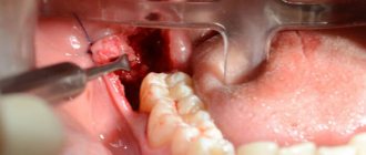
An accident or incident, such as a fight, can cause a tooth injury. And it doesn’t matter whether a small part has broken off, or the tooth has cracked, as they say, “to the point of bleeding” - the help of a doctor is definitely needed. In some cases, lost dental tissue is replenished. If a tooth is broken into pieces, it will most likely need to be completely removed and a prosthesis replaced every year. And the answer is simple - the dental tissues have not yet fully matured, the body is growing. And it is necessary to take full care of your teeth at such an early age. In case of extension, the operation is performed by introducing composite materials that replace enamel and dentin.
Methods for treating pulpitis in adolescents under 14 years of age, and how do they differ from treatment for adults?
Before starting treatment, the dentist’s main task is to determine whether the patient’s permanent tooth has roots, what stage of formation they are in, and also to find out the condition of the tooth, the location of the source of inflammation, the shape and length of the root canals. To do this, the patient must undergo an x-ray.
Treatment will be different for teeth with formed and unformed canals.
There are 3 main methods of treating pulpitis.
- A conservative method of treating pulpitis is without removing the pulp; during the treatment, the canal is washed with an antiseptic, a calcium-containing paste is applied to the pulp chamber and filled.
- Partial pulp removal
This method involves removing the infected coronal part of the pulp, cleaning and rinsing the cavity, which is then filled with a drug. After the inflammation subsides, the tooth is filled.
- Surgical method with complete removal of the pulp (depulpation). In this case, the pulp is removed, the canals are expanded and washed, and then filled.
We do not use arsenic pastes to treat pulpitis - the same popularly known “arsenic” to “kill” the pulp. Modern technologies and materials make it possible to treat pulpitis using more humane methods for patients of all ages.
Read more about these techniques in our articles:
Treatment of pulpitis of baby teeth
Treatment of pulpitis (in adults)
When and what methods of treating pulpitis of a permanent tooth in a child can be used?
- A child’s diseased permanent tooth has unformed roots.
In this case, during treatment ONLY gentle methods of treating pulpitis are used - a conservative method of treatment or partial removal of the pulp while preserving its living root part.
Depulping – i.e. Complete removal of the pulp is not applicable for such teeth, because their pulp connects to the tissues of the growth zone, which will be injured, and this is not allowed. It is necessary to preserve the living growth zone until the formation of the tooth and periodontal tissues is completed.
In the process of treating permanent teeth with developing roots, all manipulations are aimed at relieving inflammation and preserving the growth zone of the tooth. Subsequently, to avoid relapses, it is recommended to regularly monitor the patient using radiography until the tooth root is completely formed, and then carry out high-quality endodontic canal treatment.
- A child’s diseased permanent tooth has already formed roots.
When treating such a tooth, any of the above methods can be used, including complete removal of the pulp (depulpation). The latter is preferable, but the dentist chooses a technique based on the specific situation.
- Conservative treatment is carried out in case of “early” stage of the disease and in case of injuries.
- Partial pulp removal is effective in treating chronic and onset acute forms of pulpitis.
- The indication for depulpation is any form of pulpitis with severe pain and if other methods do not produce results.