The main difference between molars and premolars is their location in the dentition. Premolars are located closer to the smile zone, and molars are removed deeper.
The number of dental units also varies. Premolars are located two on each side on both jaws. There can be a pair of molars on each side, or maybe even three teeth. Third molars are wisdom teeth; they appear later than all other units. Wisdom teeth can grow in at twenty, thirty, or even later. There are cases when wisdom teeth are completely absent.
Structure and functions
The structure of molars includes two parts:
- pulp, that is, soft tissue;
- group of hard tissues - dentin layer, enamel, cement.
Anatomically, the following areas are also distinguished:
- crown part protruding above the gum tissue;
- neck in the form of a border between the crown and root system, clinical and anatomical parts are distinguished;
- root system that provides fixation, located deep in the bone structure.
Eights belong to this group of teeth, but they may not be present in everyone. They do not carry any special functional load; if problems arise during development or eruption, removal of wisdom teeth is recommended.
The article was written with the support of an orthopedic dentist
Dental Center RUTT Sidorov Dmitry Alekseevich
Not all teeth have the same structure. Depending on the shape and their location, permanent dentition teeth, that is, those that grew after the loss of baby teeth, are divided into four types: incisors, canines, premolars and molars. On one half of the upper jaw there are two incisors, one canine, two premolars and three molars, with the third being the wisdom tooth, which grows later than everyone else. The second half of the upper jaw is symmetrical to the first, and the upper jaw in terms of the arrangement of teeth is identical to the lower one.
Types of human teeth. Human teeth. Child's teeth.
Incisors
Incisors are teeth located in the very middle of the dentition, which is why they are also called anterior (frontal) teeth. The main features of incisors are a single root, a slightly flattened crown and a special cutting edge. The front surface of the crown is slightly convex, and the back is slightly concave, with several tubercles at the neck of the tooth. The upper central incisors have the largest crown, and the lower central ones have the smallest. The main function of the incisors is biting food. They are the weakest teeth, so they are not able to withstand heavy chewing and any other load, but their structure allows them to tear off pieces of food most effectively.
Types of teeth
Fangs
Fangs are four teeth, two in each row of teeth, located at the corners of the dental arches. The main function of the fangs is to tear off denser and harder parts of food, when more force is required and the fangs cannot cope with the work. The power of the canine crown is greater than that of the incisors, and there is a well-developed tubercle along the entire cutting edge. The fang has a single root, but it is distinguished by its great length and power. Due to such a strong root and crown, as well as due to the convenient placement of the dental arch, the canine is the most stable tooth of all.
Premolars
This name is given to the smallest molars, located immediately behind the fangs and somewhat similar to them. These teeth have two special cusps designed for grasping and tearing food. Unlike fangs, the surface of these teeth is wider, due to which they can chew food. The crown of the premolars is prismatic, and they have one root. Only the upper first premolar has two roots. There are no premolars in the primary dentition.
Types of teeth
Molars
Molars are the largest teeth in the dentition. They are located behind the premolars in the amount of three on each half of the dental arch. The third molar - the wisdom tooth - may be missing, as it grows in adulthood, and sometimes it does not grow at all. The main function of molars is to grind and grind food, which requires particularly great effort. The crown of molars is very large, with a large chewing surface and 3-5 cusps. The upper molars have three roots, while the lower molars have only two.
The size of the molars gradually decreases from the first to the third, the chewing surface and the length of the roots also decrease. The maxillary and mandibular third molars are often so close to each other that they can fuse together.
In case of dental anomalies or some malocclusions, some teeth may be missing. Most often this happens to molars or premolars. But in normal condition, a person has 28 or 32 teeth, depending on whether his wisdom teeth have already grown.
Source: https://www.32top.ru
Upper molars
For the upper row of the jaw, the molars have the following anatomical surfaces:
- The buccal part is convex, with a prominent groove that is most susceptible to caries. When performing hygienic cleaning, it is recommended to pay special attention to this area.
- The chewing side has some differences and a large number of tubercles, which increases the role of this group in performing the chewing function. One of these features is the triangular trigon, which unites the following types of tubercles - protocone, metacone, paracone.
- The palatal part is distinguished by such a characteristic feature as the Carabelli tubercle, located on the border of the surface from the side of the palate and the medial area. In rare cases it has a separate root.
- The mesial part has a convex or straight line between the cementum and enamel. The root system is complex and can be barrel-shaped, divergent, or cylindrical.
- The distal one resembles the medial one and requires special attention when cleaning (dental floss is recommended to completely remove any remaining food).
What does the diagram of a child’s teeth look like with the numbers of each unit?
The pattern of baby teeth in children differs from the pattern with numbers in adults. Doctors specially designate milk teeth with different numbers so as not to confuse themselves and their patients: after all, a child who was treated for a milk bite in childhood will soon grow up and begin to go to the dentist for treatment of already permanent dental units. And if the same numbering is used in both cases, then there will be incredible confusion in the treatment history - but such a history in the dental record is often extremely important for the further competent and effective elimination of problems of the dental system.
Therefore, the numbering order for baby teeth in children is as follows: the serial number remains the same as in adults (1st, 2nd – incisors, 3rd – canine, etc.), but the segments of the jaws are designated by numbers 50 and 60 for the upper right and left segments and numbers 70 and 80 for the lower left and right, respectively. And when the dentist informs parents that their child has caries on the 82nd tooth, you don’t need to immediately imagine a baby shark with several rows of sharp baby teeth, we are only talking about the second lower right incisor. According to the counting scheme, permanent teeth in children are no different from the scheme of adult molars.
Lower molars
Teeth of this type are located in the distal region of the lower row. They are distinguished by the presence of several anatomical surfaces:
- The buccal side is characterized by a narrowing towards the neck, divided into two or three parts with one and two grooves. The line of the enamel-cementum area is concave or convex, sometimes has an irregular shape. The roots are conical, cylindrical or trumpet-shaped.
- The chewing part has tubercles for chewing. There are also distal, medial-buccal, medial-lingual and other types of tubercles (there are five main ones).
- The lingual part has two convex uniform parts located on the lingual chewing line. There are no enamel streaks, the border is smooth and clear.
- The mesial side has a lingual slope, the contour line is straight or convex. The enamel-cementum border is straight. There is a slight tilt towards the cheek.
- The distal one is distinguished by the presence of a tubercle shifted towards the cheek or along the midline. The root is narrower than the medial one and has a wedge-shaped shape.
Diseases
The following diseases develop more often:
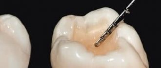
- Caries is a destructive process, as a result of which a cavity appears inside. Most often, the lesion affects the chewing surface. At the initial stage, there is a change in the shade of the enamel and pain. For treatment, it is necessary to remove the affected tissue and perform a filling.
- Pulpitis is a condition in which pathological processes affect the pulp and cause inflammation. Causes: infection, injury, or incorrect previous treatment.
- Cysts form in the root area and are small formations with purulent contents. There are usually no symptoms; the problem can be detected during examination or when the mass increases significantly in volume.
- Periodontitis is a condition in which inflammation develops at the root apex. The reasons are failure to carry out timely treatment, violation of the filling installation technology. Depending on the condition and form, three stages are distinguished - sluggish, chronic, acute.
- Pericornitis is a pathology in which inflammation of the periosteum occurs. Most often it is diagnosed in adults, when the teeth of eights appear.
Molars perform an important function, ensuring the formation of a bite and chewing food. To protect against the development of diseases of these units of the series, regular visits to the dentist for routine examinations are recommended.
Location of premolars
If the fangs are involved in tearing off pieces from the whole product, then the premolars are the rudiments of the chewing group of teeth. They are called small radical elements. They are located behind the fangs in two units, and outwardly they vaguely resemble them. The premolar units have 2 cusps on the chewing surface. This promotes good food grip, and the wide crown successfully chews food. The peculiarity of these rudiments is that they are absent in the primary dentition, but in the permanent dentition there are eight of them. In addition to grinding food, small radical units are capable of crushing it and roughly grinding it.
A characteristic feature of these masticatory organs is the presence of 2 tubercles on top of the crown. They are called buccal and lingual. They have a single root. The first premolar rudiments on both jaws are key stable units, while the second are variable organs that often undergo reductional changes. Some medical scientists view premolars as canines connected to each other. The upper and lower premolar units are structurally different.
Features of the lower premolars
The premolars of the lower jaw have a specific anatomy, a more rounded shape, similar to a barrel. Its crown is significantly inclined towards the tongue. The organ is stable, solid, and has excellent resistance. It is able to withstand high loads and considerable pressure when chewing. Units have one root. Characteristic features of premolar rudiments:
- Weak expression of the lingual tubercle, which is part of the median ridge.
- The furrow between the elevations is weakly expressed.
- The lingual tubercle, which looks like a semicircular belt, is slightly separated.
- Its convexity faces the tongue.
- The sharp tip is located on that tubercle that stands out well.
- The groove divides the lingual tubercle into 2 parts.
The length of the first unit is on average 22 millimeters. Usually she has one channel. It is very rare that two canals that converge are diagnosed. The largest cavity is in the area below the neck. The canal has an oval shape and ends in a narrowing. The root is characterized by a distal deviation. The length of the second rudiments is from 20 to 24 millimeters. In most cases there is only one root. There are organs with two roots. One root can have 2 independent channels. There is one channel inside 1 root. Near the top it can be divided into 2 separate passages.
Features of the upper premolars
The premolars of the upper jaw always have crowns with pronounced angles. Their edges are clearly aligned, and the main elements of the crowns are well defined. The first premolar organ has 2 roots, as well as:
- The average length is 21 millimeters.
- Most often there are 2 roots and two canals.
- There can be one root with one channel.
- Sometimes there are two canals in one root.
- It is rare that a unit has 3 roots and three canals.
One third of the roots have a distal bend. The direction of the organ cavity is buccal-palatal. It is located quite deep, on the same level with the neck of the rudiment. Its covering is a thick layer of dentin. The shape of the canal mouths is funnel-shaped. This allows you to freely enter the canal when opening the cavity. Second units:
- The average length is from 2 to 2.4 centimeters.
- Often such organs have 1 root and one canal.
- There may be 2 roots and two canals.
- Rudiments with 3 roots and three canals are very rare.
Typically, the canals of such teeth have two orifices. The canals are connected and also opened by a single apical foramen. 20 percent of units have 2 holes. The cavity of the chewing organ is located at the level of the neck itself. The shape of the channel is slit-like.