Physics and medicine
Without air, a person cannot live even for a few seconds. This natural mixture, many gases, as well as their absence - vacuum, are widely used in human life. For example, in medicine and some areas of production, aspiration sampling is used. In medicine, this term refers to several concepts:
- a procedure that allows you to collect material using a vacuum;
- a physiological process based on the “suction” effect that occurs due to a decrease in pressure.
In the article we will consider another concept used in medical practice, called aspiration test. As a physical phenomenon, aspiration is the process of sucking air through special instruments or devices, which is then studied and analyzed for the content of substances in the material under study.
The meaning of the test
In medicine, an aspiration test is used for self-monitoring of medical personnel during manipulations, for example, when administering an injection, as well as when removing biomaterial for further study. Used in ecology, sanitary and epidemiological control and in industry, the aspiration method of air sampling allows for analysis of gas or liquid. The aspiration technique is simple and in many cases does not require complex equipment. By drawing the material under study into special equipment using an artificially created vacuum, a specialist can even visually, if provided for by the operating algorithm used, determine the presence or absence of certain inclusions. Air sampling using the aspiration method is a quick and fairly informative way to obtain the necessary information about the composition and quality of the gas. In medicine, aspiration of biological fluid allows competent and high-quality self-monitoring of a medical worker when administering injections. After all, drug delivery must be carried out in a certain way: some drugs must enter directly into the blood, other substances must be delivered intramuscularly. It is the process of correct injection that allows monitoring the aspiration test.
Anesthesia
Self-monitoring of aspiration during procedures has become widespread among medical professionals, because parenteral administration of drugs is used in almost all areas of medicine. Anesthesia is no exception. The drug must act in a certain way depending on the assigned tasks. An anesthesia aspiration test is a method of monitoring before administering a drug by injection to ensure that the syringe needle is positioned correctly to obtain the desired result. Local work of the anesthetic substance is ensured by its introduction into the extravascular space. This is precisely what can be determined by preliminary sampling of the environment from the place where the needle with the active component hit.
It is believed that performing an aspiration test is necessary to prevent the needle from entering a blood vessel and to avoid rapid intravascular injection of a local anesthetic solution, which can cause toxic complications [2]. Aspiration is an important safety factor for local anesthesia, which, according to the American Dental Association (1973) [7], is a desirable element of injection, and according to S. Malamed (2004) [8] - mandatory.
Intraligamentary anesthesia (ILA) has become widespread due to the advent of the non-aspiration pressor syringe. Aspiration with spongy methods of anesthesia cannot be carried out with a pressor syringe, since structurally there is no mechanical connection between the piston rod and the cartridge plug. However, S. Malamed (2002, 2004) [8, 9] believes that with normally performed IPA, intraosseous and intraseptal anesthesia, only a negative aspiration test occurs (!?). A.Zh. Petrikas et al. (1987, 2011) [4, 5, 10], on the contrary, showed that during spongy anesthesia - intraseptal and intraosseous - positive (!) aspiration is almost always observed, which is confirmed by the dissertation works of their colleagues [1, 3, 6].
This is one of the proofs of the vascular mechanism of intraosseous or additional anesthesia. The dentist does not take into account the dangers of vascular injection. We are not aware of studies of aspiration with these anesthetic techniques.
Assuming a vascular mechanism for intraligamentary injection, it is advisable to observe the relationship between positive or negative aspiration and the effectiveness of anesthesia. Of particular interest in this case are the lower lateral teeth - the most difficult for local anesthesia. The use of electrical testing is an important element for laboratory evaluation of pain management [11].
Our objectives were: 1) to investigate the frequency of aspiration test (AT) in IPA; 2) to study, using electrical testing, the depth of the ILA of the pulp of the lower molar with positive and negative aspiration.
Material and methods
A randomized, crossover study was performed on 36 volunteers - 17 men and 19 women - aged 19-24 years, practically healthy (ASA class I). After discussing the nature of the injection with each student, voluntary written consent to participate in the experiment was obtained. The study was approved by the ethics committee of the TGMA with further examination of the protocols.
For ILA, 4% solutions of articaine with adrenaline 1:100,000 and 1:200,000 were used.
ILA was performed on each subject by 1 operator (M.D.V.) using a QuickSleeper computer injector (Dental Hi Tec Cholet, France). Special intraligamentary needles with a length of 9 mm and a diameter of 0.3 mm Intralig-S (Dental Hi Tec) were used. The test object was the mandibular first molar (H6). Teeth with gingivitis, large restorations and endodontic treatment were excluded. The injector provided a constant injection rate of 1.0 ml MA≈102 s. Anesthesia was achieved using 2 classic injections - on the mesial and distal sides of the tooth, and in case of their insufficiency - another 1-3 additional injections [3].
Aspiration was created by reversing the syringe plunger for 5 s. Electroodontometry (EOM) was performed before anesthesia and after each injection. The effectiveness of anesthesia was assessed by the value of the pain sensitivity threshold of the pulp using an IVN-98 Pulpotest-Pro apparatus for electroodontodiagnostics (Kaskad, Russia) on an intact lower first molar. The threshold of pain sensitivity was determined in microamperes (μA) until the sensation of pain appeared. The criterion for the onset of pulpal analgesia was a threshold value of 100 μA, recognized in Russian endodontics as an indicator of pulp death or switching off its sensitivity [2]. Additionally, the second indicator of the depth of anesthesia was taken into account - maximum analgesia (complete anesthesia) - 200 μA.
Statistical analysis of the results was carried out using t and χ2 tests.
Results and discussion
ILA was successful on all 36 mandibular molars. It was achieved by several injections - from 1 to 5 (on average - 3.2), as well as by increasing the administered dose of anesthetic from 0.4 to 1.2 ml (on average - 0.7 ml). In 2 cases, anesthesia was achieved with 1 injection. With each effective anesthesia, 1 of the injections, and sometimes 2, were accompanied by positive aspiration. A positive aspiration was indicated by a thin stream of blood or pink coloration of the anesthetic solution in the cartridge (Fig. 1).
Figure 1. Positive AT for IPA, carried out using a SleepеrOne computer syringe (“Dental Hi Tec”).
As a result, 34 (94.4%) of 36 successful IPAs had positive aspirations. Positive AT was observed in 50 (43.9%) of 114 injections, negative - in 64 (56.1%) injections (see table).
With positive aspiration (50 injections), pulpal analgesia developed in 42 (84%) cases, and in 35 (70%) - with a maximum EOM of 200 μA (complete switching off of sensitivity) and in 7 (14%) - with reduced thresholds - from 102 to 180 µA. When performing 8 (16%) injections, EOM indicators did not reach the level of pulpal analgesia, although they were relatively high - from 60 to 90 μA.
With negative aspiration (64 injections), EOM indicators in 96.9% of cases were below 100 μA. In 2 (3.1%) cases, however, after 3-5 minutes, pulpal analgesia was observed with EOM indicators of 100 and 110 μA.
In 2 cases out of 36 ILA, when pulpal analgesia occurred immediately after a single injection with a positive aspiration test, the EOM indicator was maximum - 200 μA.
Changes in average EOM indicators with the results of statistical processing depending on positive or negative aspiration are presented in Fig. 2.
Figure 2. Average pain threshold values (μA) with confidence limits (p=0.05) for EOM of the lower first molar before and after ILA in cases of positive or negative aspiration with 114 injections of 4% articaine with adrenaline.
The average EOM indicators with 36 ILA after 50 injections with positive aspiration were 171.6±7.9 μA, with negative AT after 64 injections - 38.2±3.4 μA (the differences between all EOM indicators are highly significant; p<0.001). EOM indicators during injections with negative aspiration significantly increased compared to the initial ones (before anesthesia), during injections with positive aspiration - compared to indicators before anesthesia and with indicators with negative aspiration.
In 34 (94.4%) of 36 cases, ILA of the lower first molar was almost always achieved with several injections (114 in total), at least one of which was with positive aspiration. The absolute positive success of the injections was achieved both through several injections and an increased dose of ILA (on average - 0.7 ml). Only in 2 (5.6%) of 36 anesthesia procedures in all injections was aspiration negative. This is contrary to S. Malamed's findings, repeated in several ADA guidelines. Positive aspiration during intraseptal anesthesia was observed by A.Zh. Petrikas in 1987, with intraosseous (Stabident Technic) - L.A. Yakupova in 2006 and with intraseptal - O.E. Efimov in 2011, which is reflected in their dissertation works [1, 4, 6].
Positive aspiration, as a rule, was combined with pulpal analgesia and, conversely, negative aspiration was almost always associated with insufficient pain relief.
The hypothesis according to which positive aspiration during IPA is a guarantee of an analgesic effect is confirmed by convincing data from the mathematical processing of the results of this study (χ2=7.75; p<0.01 and t test, p<0.001 - see Fig. 2).
This hypothesis indicates the presence of a leading vascular component in the mechanism of IPA, although not in all cases. The results of the observation carried out on 36 subjects are reliable; they cover 114 aspiration samples and are supported by electrical testing. These studies indicate a significant, but apparently virtual misconception S. Malamed [8, 9] made in assessing aspiration during intraosseous anesthesia. The consequences arising from this error are beyond the scope of this study and require serious discussion.
In 2 cases, pulpal analgesia developed in the absence of blood in the syringe (AT - negative) only due to injection pressure and diffusion (infiltration) of the anesthetic, i.e. without obvious vascular component. Conversely, in the other 2 cases, complete anesthesia almost under the needle was achieved with positive aspiration with 1 vascular injection. In other cases, there was a mixed mechanism of anesthesia: vascular plus diffuse. Nevertheless, the vascular component prevailed over the diffuse one: 84% versus 3%.
This logical explanation must take into account the anatomy of the anesthetized lower first molar, which consists of 2 roots with different sources of blood supply.
The absence of pulpal analgesia with a positive aspiration test in 8 (16%) of 50 injections in the presence of fairly high pain thresholds when exposed to electric current is probably due to the anesthetic reaching only 1 of the 2 roots of the first molar.
The use of EOM has proven to be a useful manipulation for obtaining reliable, controlled analgesia. In the clinic, when electrically tested anesthesia was insufficient, additional injections always solved the problem [3].
So, in the present study, the phenomenon of aspiration during intraligamentary injection was studied for the first time. Positive aspiration was observed in 94.4% of cases during PLA and is an indicator of effective pain relief; provides clear evidence for the vascular mechanism of IPA in the form of positive aspiration upon injection, which is directly related to its analgesic effect; with ILA performed with negative aspiration, in some rare cases it is possible to develop anesthesia of the dental pulp with some delay (3-5 minutes) of this effect and its decrease.
Dentistry
An aspiration test in dentistry is a technique that allows you to assess the quality of the upcoming anesthesia. In dentistry, there are quite a few types of anesthesia administration; they differ both in the method of delivering the analgesic and in the place of its administration. But in any case, an aspiration test should be a mandatory preliminary procedure. Dental anesthesia should not enter the bloodstream and spread to nearby tissues and organs. Its goal is local work, providing pain relief during the procedure in a small area of the oral cavity. In dental practice, special syringes are used that have not only all the classic details of this instrument, but also special devices on the piston to properly hold it when creating forward and backward movements. At the moment, performing an aspiration test in dentistry using a carpule syringe is considered the most convenient and technologically sophisticated way of both administering an anesthetic and assessing the correctness of the manipulation.
Pain relief methods
The most common method is local anesthesia, designed to reduce the patient's sensitivity to pain in the desired area. Under its influence, a person understands what is happening and can speak and hear calmly. At the same time, he does not feel anything in the place where the dentist is working.
Indications for such anesthesia may be:
- removal of the tooth and its root (not always suitable if we are talking about the “eight”);
- minor surgical interventions;
- removal of tartar (various sprays and gels are used for this);
- treatment of incompletely erupted teeth;
- elimination of periodontal disease;
- treatment of caries, especially advanced ones;
- removal of nerves;
- root canal filling;
- installation of a metal-ceramic crown.
Partial anesthesia is not relevant for major operations, injuries to the bones of the maxillofacial area and allergies to the drugs used.
General anesthesia is used extremely rarely, mainly only when the patient experiences extreme fear and stress. And it can also be recommended for psychosomatic diseases, allergic reactions to local anesthetics and extensive surgical interventions, for example, prosthetics on implants.
General anesthesia in dentistry is not allowed in the following situations:
- if more than six months have passed since the stroke and/or heart attack;
- with cardiopulmonary failure in the stage of decompensation;
- if there are serious heart rhythm disturbances;
- for diseases of the thyroid gland;
- while intoxicated;
- when the patient is under the influence of drugs;
- in case of impaired functioning of the kidneys and liver.
Cosmetology
Aesthetic medicine is developing rapidly. But a procedure such as injection remains the most common way of introducing medications and cosmetics into the subcutaneous and deep layers of the epidermis. This procedure has the potential for tissue embolization. In order to avoid the adverse consequences of the intervention, an aspiration test is performed in cosmetology. What kind of manipulation is this? Its purpose is to control the correct placement of the medical instrument to obtain a high-quality result with minimal complications. When carrying out an intervention, the specialist must take into account many factors, one of which will be the individual scheme of the patient’s vascular system. Therefore, a test to verify the correctness of the procedure is simply necessary. For example, if during the procedure for introducing a gel into the subcutaneous layer, the active substance enters a blood vessel, this will cause a disruption of blood flow, and then ischemia and necrosis. In addition, in some cases, retrograde injection of a special material is indicated, which also involves the use of aspiration.
When “doesn’t hurt” = “safe”
July 26, 2018
It is impossible to imagine a dental appointment without the use of local anesthesia, which is becoming more and more advanced every year. The three pillars of modern pain management are safety, effectiveness and predictability.
However, the use of even the most advanced anesthetics carries risks. Experts note that insufficient awareness of possible complications often creates a false sense of complete security among dentists and prevents them from anticipating situations that threaten the health and lives of patients.
Safety issues should be the highest priority at every stage of patient management. Of course, it is impossible to cover all aspects of local anesthesia within the framework of a journal article - there is specialized literature for this. However, we have tried to summarize the recommendations of leading domestic experts in the field of anesthesia and resuscitation in dentistry and highlight several important rules for preventing a number of local complications when using local anesthetic drugs.
When preparing the material, we used the manual “Safe anesthesia in dentistry,” published by the publishing house “GEOTAR-Media” in 2018. Authors of the manual: S. A. Rabinovich, MD, Professor, Head of the Department of Pain Management in Dentistry, Moscow State Medical University. . A. I. Evdokimova, Honored Doctor of the Russian Federation; E. V. Zoryan, Ph.D., Associate Professor of the Department of Pain Management in Dentistry, Moscow State Medical University named after. A. I. Evdokimova; L. A. Zavodilenko, Ph.D., doctor - anesthesiologist-resuscitator, assistant at the Department of Pain Management in Dentistry of Moscow State Medical University named after. A. I. Evdokimova; Yu. L. Vasiliev, Ph.D., dentist, scientific consultant of the GEOTAR design bureau.
CAREFUL TREATMENT PLANNING
At this stage, it is necessary to collect the patient’s medical history, assess the duration and traumatic nature of the necessary intervention, and decide on local anesthetic drugs and methods of their administration. This will allow you to take into account some risk factors and prevent complications.
The authors of the manual are Fr. Thus, it is indicated that the average half-life of lidocaine in patients without somatic pathology is 1.8 hours, and in patients with severe liver dysfunction it can increase to 6 hours or more. If the patient has concomitant somatic diseases, the authors of the manual recommend that it is mandatory to obtain an extract from the medical history indicating the diagnosis and the basic therapy recommended for use.
When choosing a drug for local anesthesia, you should take into account all the components included in its composition. For example, if you have a history of bronchial asthma, it must be taken into account that about 7% of asthmatics have increased sensitivity to sulfites, which manifests itself in the form of allergic reactions. The manual indicates that sodium bisulfite is often added as a stabilizer to solutions containing epinephrine (adrenaline), which can provoke allergic reactions in patients with an asthmatic component.
If the patient has concomitant somatic diseases, the authors of the manual recommend that it is mandatory to obtain an extract from the medical history indicating the diagnosis and the basic therapy recommended for use. |
When calculating the dose of the selected drug, the authors of the manual suggest paying special attention to the patient’s body weight and age. Thus, for pediatric patients, the minimum toxic doses of all local anesthetics are significantly less than for adults. “To achieve guaranteed complete pain relief and reduce toxic effects in children, it is recommended to choose the most effective and safe drugs based on articaine, mepivacaine or lidocaine,” experts point out.
In elderly and senile patients, the authors of the manual recommend taking into account age-related changes in hormonal balance and metabolism, an increased amount of fat and a decreased amount of muscle tissue. The authors of the manual, citing domestic studies from different years (V.R. Weber, B.T. Moroz, 2003; E.V. Zoryan, S.A. Rabinovich, 2017), recommend reducing the dose of anesthetic: in patients aged 70 years - by one third, at the age of 80 years - by 2 times. “The dentist should strive to use the minimum amount of anesthetic to achieve analgesia,” the guidelines emphasize.
The book also mentions that when planning treatment, it is also necessary to assess the emotional and psychological state of patients. For people with high anxiety, it is recommended to provide additional medication with sedatives.
Having received as much information as possible about the patient, it will be easier for the dentist to make a decision on the possibility of treatment as an outpatient appointment or on referral to a hospital.
RULE FOR ASPIRATION TEST
Before starting treatment, the main parameters of the patient’s physical condition should be determined and recorded in the medical history: blood pressure, heart rate, respiratory rate. The patient must be informed about the possible risks and sign a consent to carry out all planned interventions. |
The most important aspect of the safe administration of local anesthesia is the prevention of intravascular injection of the anesthetic drug. As a reliable way to prevent intravascular injection of an anesthetic, the authors of the manual strongly advise always using an aspiration test: “System complications caused by intravascular injection of an anesthetic can pose a real danger to the patient’s life. That is why an aspiration test should always be performed to prevent unwanted complications. Statistics show that not all dentists perform aspiration tests on a regular basis, but evidence suggests that dental therapists perform aspiration tests 50% of the time, dental surgeons perform the aspiration test 52% of the time, but prosthodontists perform the aspiration test only 25% of the time. cases."
An aspiration test is the collection of a liquid medium into a syringe, which can be performed in different ways. By the absence of blood in the fluid, you can clearly see that the tip of the needle is not in a blood vessel.
The design of disposable general medical syringes is not suitable for performing an aspiration test. But you can still do it - by moving the piston in reverse, holding the syringe with one hand and the piston with the other. This is not very convenient for the doctor, and also creates conditions for the occurrence of microtraumas, the authors of the manual rightly note. Special dental syringes allow this procedure to be carried out with one hand, and self-aspirating dental injectors make this procedure very simple. The authors of the manual emphasize that an aspiration test must be performed before administering the anesthetic, repeat it if the needle has been moved during the injection, and remind that the results of the aspiration test are false negative in approximately 2% of cases.
Even after a single use of the needle, its tip becomes deformed. |
RULE “ONE NEEDLE - ONE INJECTION”
During an anonymous Internet survey conducted by one of the authors of the manual (Yu. L. Vasiliev, 2009), it turned out that out of 3,000 doctors, only 44% of respondents change needles for injections in one patient. The rest do this selectively for different injection options, and a third do not change needles at all.
Meanwhile, the authors of the manual say that even after a single use of the needle, deformation of its tip occurs. When a needle is inserted into soft tissue, a wound channel is formed, which corresponds to the diameter of the needle, and when the needle is removed, it increases due to its deformation and capture of soft tissue. Thus, repeated use of the needle increases the wound channel, and also increases the pain of the procedure for the patient due to dulling of the needle tip. A bent or blunt end of a needle can injure tissue and cause a number of complications, experts warn.
In addition, the breakage of an injection needle is indicated as one of the complications of local anesthesia. To prevent breakages, it is recommended to use thicker needles, and also remember that after immersing the needle into soft tissue, about a third of the length of the needle should remain outside.
| “The dentist should strive to use the minimum amount of anesthetic to achieve pain relief. We recommend using a dose not exceeding 1/2 the volume of the carpule." |
RULE “1/2 MAXIMUM ALLOWABLE DOSE”
In practical work, the authors recommend that dentists use the rule of using 1/2 of the maximum permissible dose of anesthetic.
This is explained by the fact that on the carpule of the finished dosage form of any anesthetic, the manufacturer usually indicates the volume of the anesthetic contained in milliliters, and not the amount of the active substance in grams (milligrams). In addition, the carpul often contains two active substances - a local anesthetic and a vasoconstrictor. Therefore, it can be difficult for a doctor to quickly calculate the maximum allowable dose in each specific case for each of the substances administered.
The authors of the book “Safe Pain Management in Dentistry” introduce doctors to a simplified method for quickly calculating the administered dose, which consists of assuming that each carpule contains 1.8 ml of solution. This increases the volume of the injected anesthetic solution, but at the same time serves as the key to a safer dosage calculation. For example, if 2.5 carpules were injected, then the injected volume can be taken as 4.5 ml (2.5 times 1.8 ml). The list of references provides a link to a detailed description of this method for those who want to familiarize themselves with it in more detail: Becker DE, Reed KL Local anesthetics: review of pharmacological considerations // Anesth. Prog. - 2012. - Vol. 59. - No. 2. - P. 90–101.
In cases where the volume of dental intervention requires the use of local anesthetics in a dosage exceeding 50% of the maximum allowable dose, the manual indicates the need to ensure the ability to provide the patient with anesthesia, including access for intravenous injections, assisted or artificial ventilation.
PREVENTION OF LOCAL COMPLICATIONS
Local complications include:
- pain and burning during injection;
- paresthesia;
- facial nerve paresis;
- needle breakage;
- tissue damage and necrosis;
- lockjaw;
- formation of hematomas;
- local and systemic infectious complications (abscesses, phlegmon, bacterial endocarditis, etc.).
The manual also describes such types of complications as anesthesiophagy, erroneous administration of solutions other than local anesthetics, and others.
Pain and burning during injection, according to the authors of the book, are often associated with errors in anesthesia technique, and can also be observed when a drug with a low pH value is administered too quickly. In this case, they advise that the injection rate should not exceed 1 ml/min. “The anesthetic will then spread slowly, and if the needle enters a blood vessel, this will help avoid the toxic effects of high concentrations of the anesthetic solution,” the manual says. To reduce the likelihood of local complications when injecting into dense connective tissues, experts recommend further reducing the rate of drug administration to 0.5 ml/min. It is also possible to administer the anesthetic fractionally, with pauses of 15–30 seconds, and when introducing large portions of the anesthetic, the injection interval should be increased to reduce accumulation.
The manual discusses in detail situations in which, due to non-compliance with the anesthesia technique and individual structural features of the maxillofacial region, the target nerves to be anesthetized or adjacent ones lying near the target are injured. For example, it is indicated that with injury to the branches of the trigeminal nerve, temporary paresthesia and neuralgia are possible for a period of several days to several months. If the inferior alveolar nerve is damaged, paresthesia and neuralgia may occur in the area of the entire half of the lower jaw and especially the lower lip and soft tissues of the chin on the corresponding side. To prevent this type of complications, it is recommended to clarify the anatomical and topographical features and carefully follow the appropriate anesthesia technique. In addition, the authors also write about the important role of the condition of the needle: a curved or blunt needle tip can also injure tissue and cause a number of complications.
The development of trismus is mentioned as one of the local complications, which can be caused either by injury to the blood vessels or muscles in the infratemporal fossa during local anesthesia, or by contamination (for example, alcohol or hypertonic solution) of the injected local anesthetic solution. The authors say that trismus develops in the first days after dental intervention and most often goes away after 2-3 days, however they clarify that “the development of trismus can be associated not only with anesthesia, but also with overstrain of the muscles and ligamentous apparatus of the temporomandibular joint during long-term interventions requiring maximum mouth opening.”
Speaking about tissue necrosis, experts mention that during anesthesia this phenomenon is rarely observed. Nevertheless, it is important to be aware of this complication, since “cases of sterile abscesses have been described when local anesthetic solutions containing the vasoconstrictor norepinephrine (norepinephrine) or high concentrations of epinephrine (adrenaline) are administered, especially when the drug is injected under high pressure in the area of the hard palate. Patients suffering from hypertension and diabetes need special monitoring and precautions when choosing local anesthetic drugs due to the high risk of developing local avascular necrosis.”
The main components of modern anesthetic drugs for injection methods of pain relief
procaine (novocaine, lidocaine, prilocaine, mepivacaine, articaine, bupivacaine, ropivacaine
In Russia - epinephrine (adrenaline). In some countries, local anesthetic medications may include: norepinephrine (noradrenaline), phenylephrine (mesaton), octapressin (felypressin)
sodium and potassium sulfites
parahydroxybenzoates (added only to local anesthetic solutions sold in ampoules), EDTA (ethylenediaminetetraacetic acid) |
The manual notes that the transition to carpule technology has significantly reduced complications associated with the erroneous administration of solutions other than local anesthetics. Factory production of anesthetics eliminates the human factor and ensures sterility and high precision in manufacturing technology. “The introduction of carpule technology made it possible to transfer responsibility for the quality of drugs administered from the carpule to manufacturing companies. By choosing a well-known manufacturer, the doctor largely saves himself from trouble,” experts say. It is also noted that the use of carpules and disposable needles has led to the fact that infection during local anesthesia is extremely rare. Sometimes the authors associate post-injection infections with the spread of the process when an anesthetic is administered under pressure into infected tissues. To prevent anesthesiophagy, it is recommended to select drugs with a duration of action corresponding to the planned intervention. Patients should be warned to refrain from eating or chewing gum until the anesthesia wears off.
Unfortunately, the format of a journal article does not provide the opportunity to consider another group of complications that arise during local anesthesia—somatic ones. Somatic complications include arterial hypertension or hypotension syndrome, acute coronary syndrome, acute cerebrovascular accident syndrome, acute ocular hypertension syndrome and the development of a systemic toxic reaction to local anesthetics. Detailed information on the prevention of these complications, as well as on resuscitation measures using modern equipment, can be found in the above manual.
Tags: Anesthetics
More on the topic
When “no pain” = “safe”
It is impossible to imagine a dental appointment without the use of local anesthesia, which is becoming more and more advanced every year. The three pillars of modern pain management are safety, effectiveness and predictability. Return to section
Collection of biomaterial
Carrying out medical manipulations may also include the removal of a cytological sample for examination. An aspiration test also helps with this. The need for a biopsy, for example, is determined by the doctor. Often this procedure is carried out simultaneously with other diagnostic measures, for example, bronchoscopy, colonoscopy, and so on. The collection of tissues or liquids necessary for analysis is carried out either with a syringe or with an aspiration gun - a more powerful design intended only for these purposes. The cytological sample is then delivered to the laboratory for examination, so the sampling procedure is the most significant stage of the biopsy.
Fine needle aspiration biopsy in the diagnosis of cancer
Find out the cost of PTAB Thyroid gland
Sign up through your personal account
Biopsy (puncture) is a diagnostic method in which cells or tissues are taken intravitally from the human body and then examined microscopically.
In the structure of mortality in Russia, cancer is in second place after cardiovascular diseases. Cancer statistics in Russia show that approximately 1,500 patients with cancer are registered in our country every day. In total, at least 2.5 million patients with various forms of cancer are registered in oncology clinics in Russia.
The main reason for increased mortality from cancer in Russia is late diagnosis. An important reason for the late detection of malignant neoplasms is that people do not consult a doctor on time due to lack of knowledge in this area or insufficient funds. This leads to the fact that in Russia every third cancer patient dies within a year from the moment of diagnosis. For example, in the United States, more than 80% of patients pass the five-year survival mark.
In the majority of cancer patients, the pathology preceding thyroid and breast cancer is usually benign neoplasms. Nodules in these groups of patients can be detected by ultrasound. The widespread use of high-resolution ultrasound devices makes it easy to diagnose nodular formations, including small ones (up to 1 cm in diameter).
The main goal of a diagnostic search for nodal pathology is to identify patients with neoplastic changes in the mammary and thyroid glands, since it is this category of patients that is primarily subject to surgical treatment and, if necessary, radiation therapy, etc.
It is no secret that it is almost impossible to reliably determine the type of tumor (benign or malignant) using ultrasound; moreover, ultrasound is a rather subjective method in which the human factor (ultrasound diagnostic operator) is present. Significant variability in ultrasound diagnostic indicators depends on two main factors - the level of qualification of the diagnostician and the technical parameters of the ultrasound scanner used.
The insufficient information content of the used methods for studying diseases of the thyroid and mammary glands, combined with the need to clarify the diagnosis, forced clinicians to actively use invasive manipulations with morphological verification of the process.
Ultrasound-guided fine-needle aspiration biopsy (FNAB) is the most accurate, highly informative, safe method used in many diagnostic algorithms for focal diseases of the thyroid, breast and other superficially located structures.
The study of punctate allows not only to recognize the nature of the process (benign or malignant), to clarify the histogenetic affiliation of the neoplasm, but also to identify cancer in the early stages of development, when clinical manifestations of the disease are not yet present.
In patients operated on for thyroid cancer, if tumors are detected in the surgical site, a puncture biopsy provides a clear differential diagnosis between tumor recurrence, hyperplasia of the remaining gland tissue, or postoperative changes such as granuloma.
Main indications for TAPB:
- Single or multiple nodes in an organ, when their benign nature is in doubt.
- The presence of altered lymph nodes, when it is necessary to determine the source of metastasis.
- Focal formations of the mammary glands with characteristic signs of a tumor process, various cysts (atypical, simple). When a cyst is punctured, a therapeutic function is also performed, since its contents are removed as much as possible.
- Neoplasms of the salivary glands, soft tissues of the neck, in some cases, median or lateral cysts of the neck.
- Recurrence of the primary tumor
According to the American Association of Clinical Endocrinologists (AACE), fine-needle aspiration biopsy of nodules measuring 10 mm or less is not indicated unless ultrasound findings are suspicious for thyroid cancer and there is no high risk of cancer according to medical history. However, all these recommendations are not absolute and in certain situations leave the clinician the right to decide on the need for fine-needle aspiration biopsy, even in cases where nodular formations do not meet the above criteria.
Main contraindications to TAPB:
- blood diseases (blood clotting disorders can lead to bleeding, which may require urgent hospital treatment);
- the patient has mental illness, since TAPP can provoke an exacerbation of the underlying disease.
Methodology for performing FNAB of nodal changes in the thyroid and mammary glands
The fine-needle aspiration biopsy method is simple, as is drawing blood from the patient's arm. It has virtually no contraindications and can be performed on an outpatient basis without anesthesia. The introduction of anesthetics causes a deterioration in ultrasound visualization of the area under study, a change in the cellular pattern, and the anesthesia itself is as painful as the puncture. The patient is in a supine position. The skin surface is treated with a solution of 70% ethyl alcohol. An injection (puncture) is made through the skin into the tumor-like formation of the organ and the contents of the formation are aspirated. As a result of puncturing the formation under study with a needle, tissue fragments are collected. The movement of the needle and all manipulations are visually controlled on the screen of the ultrasound machine. After the tip of the needle is inside the formation, the doctor performs aspiration (sampling) of the contents of the node several times with a syringe. To obtain a sufficient amount of biological material, 2 - 3 injections are performed in different areas of the formation. The needle is then removed and the resulting material is applied to glass slides. The slides are sent for examination to the cytology laboratory. Next, these slides are specially stained, and a cytologist examines their contents under a microscope. By carefully examining the cellular material taken through a biopsy, the doctor will be able not only to determine the malignancy or benignity of the pathology, but also to identify the degree of damage to the organ. When planning surgery, the need for a biopsy increases. After the study, a written conclusion is issued. The duration of a puncture biopsy depends on the number of lesions being punctured. No special preparation is required to perform a fine-needle aspiration biopsy. The TAPB procedure is well tolerated by patients. A hypoallergenic sterile patch is glued to the injection site, which can be removed after 20 minutes. 5-10 minutes after the biopsy, the patient can lead a normal life.
Side effects and possible complications of TAPP
The occurrence of moderate pain at the site of TAPE is associated both directly with tissue damage in the manipulation area, and sometimes with the possible development of delimited hematomas, which resolve safely. Discomfort, pain in the neck, and headaches can be triggered by throwing back the head during TPA if the patient has cervical osteochondrosis.
Fine-needle aspiration biopsy is today the most valuable and informative method for preoperative differential diagnosis of various nodular formations. This is the only method that allows one to reliably confirm or refute the malignant nature of the process. Fine needle aspiration biopsy is critical in determining treatment strategy.
Using fine-needle aspiration puncture biopsy, you can obtain reliable and accurate information about changes in the tissues of the mammary and thyroid glands in order to identify the disease at an early stage and begin effective treatment.
FAQ
Is a fine needle aspiration biopsy really appropriate for me?
Fine needle aspiration biopsy is the only non-surgical method for determining whether a nodule in the organ being examined is benign or malignant.
The large tumor must be removed. In this case, am I also indicated for a fine-needle aspiration biopsy?
Yes, it is indicated to determine the extent of surgical intervention. Sometimes a node may have tumors of different histological structures. Knowing this in advance, the surgeon plans and carries out more competent treatment accordingly.
Should fine needle aspiration biopsy be ultrasound guided?
Yes, definitely! Tumors are often located close to blood vessels, neurovascular bundles and other organs. In order to avoid their traumatization during biopsy, the latter should be carried out under ultrasound control.
Can a node “degenerate” into a malignant one after a biopsy?
NO!!! Fine-needle aspiration biopsy does not cause malignancy (degeneration into a malignant neoplasm)!
Can a biopsy cause the tumor to spread?
There is no data in world practice to prove tumor spread after fine-needle aspiration biopsies.
The life of each of us depends only on ourselves. How to get maximum pleasure and minimum suffering from it? - try to maintain your health, strengthen your immune system, carry out the necessary prevention and treatment in a timely manner, and, accordingly, diagnosis.
Make an appointment for ultrasound diagnostics and an appointment with an oncologist-mammologist at the Clinic in Severny by calling +7.
Author of the article: ultrasound doctor Irina Yurievna Novikova.
Microbiology
Particular importance is attached to such procedures as the aspiration method of air sampling, microbiology. Science that studies the inhabitants of the microworld helps many areas of knowledge and human activity to develop, taking into account and using the characteristics of the life of microorganisms. Aspiration samples of air intake are widely used in sanitary and epidemiological control. Moreover, the collection of material is the most critical stage, the correct execution of which determines the quality of the analysis performed. An aspiration test in this case is a forced method of deposition of microorganisms from the environment onto a nutrient material or a special collecting liquid. This way, experts can monitor their growth and conduct research.
Indications
Aspiration biopsy is prescribed in the following cases:
- diagnosis of endometrial cancer
- bleeding when using hormonal drugs
- bleeding during menopause
- suspected endometriosis
- diagnosis of endometrial hyperplasia
- endometrial polyps
- suspicion of the development of oncological processes in the uterine cavity
- menstrual irregularities
- diagnosis of uterine fibroids
- inflammation of the uterine mucosa
- examination of the uterus for infertility
- endometrial examination after hormone therapy
- obtaining a tissue sample for bacteriological examination
Sample collection techniques in medicine
In medicine, the same syringe used for injection is used as an aspiration device. The basic principle of operation of instruments and devices for carrying out aspiration manipulation is vacuum suction of the medium. For example, the reverse movement of the syringe piston, which creates negative pressure.
The technique for monitoring the correctness of the injection recommends following the following algorithm:
- The plunger of the syringe, already in the working position, must be pulled back gently, without applying excessive force.
- The piston stroke can be only 1-2 millimeters to obtain sufficient aspiration vacuum.
- It is unacceptable to change the position of the syringe both during the aspiration test and during the injection. To do this, it is fixed using a stop with the ring finger and little finger of the medical worker performing the procedure.
- If the needle is thin, it may take a few seconds for the test results to appear. If the test is positive (which depends on the expected result), then the injection is placed in place without displacing the needle. If the test result is negative, then the needle is either moved a few millimeters without removing it from the tissue, or the injection site itself is changed.
When injecting into a highly vascularized area, multiple aspirations may be required to determine the optimal location for the procedure.
Improving the technique of aspiration test when performing local anesthesia in dentistry
S. V. Tarasenko Doctor of Medical Sciences, Professor, Head of the Department of Faculty Surgical Dentistry of the First Moscow State Medical University named after. I. M. Sechenova
E. A. Belyaeva , dental surgeon, MAU "Balashikha Dental Clinic"
A. V. Kuzin Assistant of the Department of Faculty Surgical Dentistry of the First Moscow State Medical University named after. I. M. Sechenova
A. A. Kurtyshov student of the First Moscow State Medical University named after. I. M. Sechenova
Systemic complications caused by intravascular injection of anesthetic can be life-threatening for the patient. Some dentists perform the aspiration test on an ongoing basis, but evidence suggests that therapists perform it in 50% of cases, surgeons in 52% of cases, but orthopedists only in 25% of cases (S. A. Rabinovich, O. N. Moskovets, M. V. Lukyanov, E. V. Zoryan). When blocking the inferior alveolar nerve in children and adolescents, the needle more often ends up inside the vessel than in adults.
Ophthalmological disorders after intraoral anesthesia are a common complication. Symptoms include blurred vision and blindness (temporary blindness). In addition, motor neuron anesthesia can lead to mydriasis (dilation of the pupil), ptosis (dropping of the eyelids), and diplopia (double vision). Symptoms of similar manifestations involving ptosis, enophthalmos (collapse of the eyeball in the orbit) and miosis (papillary restrictions) have also been described [6].
According to many authors, intravascular injection is a common phenomenon in general anesthesia. To prevent intravascular injection of anesthetic, it is imperative to carry out an aspiration test.
Aspiration - the reverse absorption of the medium from the area of injection of the local anesthetic - is used to ensure that the tip of the needle is not inside a blood vessel by the absence of blood in the solution. This is necessary in order to prevent the introduction into the bloodstream of highly concentrated substances used in modern local anesthesia technology.
The purpose of our study was to improve the safety of local anesthesia in dentistry using an aspiration test. To achieve this goal, we compared the effectiveness of performing an aspiration test with carpule syringes of different designs, studied the frequency of positive aspiration tests during anesthesia of the inferior alveolus nerve with various conduction methods of pain relief, and also developed an algorithm for the prevention of intravascular injection of anesthetic during conduction anesthesia of the LLN (inferior alveolar nerve) .
Aspiration is used to ensure that the tip of the needle is not inside a blood vessel by the absence of blood in the solution. This prevents the introduction into the bloodstream of highly concentrated substances used in modern local anesthesia technology.
In our study, we used anesthetic carpules with rubber pistons of different structures: with a retention rim around its perimeter (Ubistesin 3MESPE), without retention (Septanest). Also carpule syringes, which differ in the structure of the end part of the piston (Fig. 1 a, b): a syringe with an “anchor” type piston, which has three hooks in its end part that hold the rubber piston; a syringe with a “corkscrew” type piston, having at its end part one corkscrew-shaped hook that is screwed into the rubber piston of the carpule; a syringe with a “hook” type piston, having at its end part two mutually oppositely directed hooks holding the rubber piston of the carpule; a syringe with an “arrow-shaped” piston, which has an arrow-shaped hook at its end that holds the rubber piston of the carpule.
The practical part of the study was carried out as follows. The needle of the assembled carpule injector was positioned into the dye-tinted water. An aspiration test was carried out with each syringe by capturing the solution into the cartridge with the reverse movement of the piston. Data on the results of the aspiration test were photographically documented and entered into a table.
The clinical part of the study was carried out on the basis of LHO No. 2 of the Balashikha Dental Clinic. The study involved 270 patients aged 25 to 75 years with a diagnosis of chronic periodontitis of the lower jaw teeth.
As a technique for anesthetizing the inferior alveolar nerve, we used the five most popular methods in domestic dentistry:
- mandibular anesthesia by palpation;
- torusal anesthesia;
- anesthesia using the Gow-Gates method;
- anesthesia using the Vizirani-Akinozi method;
- anesthesia using the Ibragimov method.
Pain management was carried out under careful assessment of systemic hemodynamic parameters. When using the ArmedYX 300 pulse oximeter, a monitored increase in heart rate of 30 units immediately after local anesthesia indicated intravascular administration of the anesthetic. Additionally, the doctor conducted a survey about the patient’s well-being for the presence of palpitations, fear, abdominal pain, nausea, which also indicated the cardiotoxic effect of epinephrine in the local anesthetic solution.
The results of our study were the following data.
When comparing various systems for performing aspiration testing, the best performance was found in combinations of syringes with “anchor” and “corkscrew” type pistons and carpules with a retention notch. More informative data on the results of the practical part of the study are given below (Table No. 1).
Table No. 1. Possible combinations of injection instruments used in dentistry and the effectiveness of aspiration
| Rate of successful dye aspiration per 100 samples | ||||
| Carpule type | Syringe with an “anchor” type piston | Syringe with a “corkscrew” type piston | Syringe with a hook-type piston | Syringe with arrow-shaped piston |
| With retention rim | 100 | 89 | 55 | 5 |
| Without retention rim | 0 | 38 | 37 | 15 |
Based on our results, we believe that the use of carpules with a retention notch facilitates the aspiration test. We can consider them to be “intended” for performing an aspiration test. The retention notch allows the use of most carpule syringes, with best results when used with syringes with an "anchor" and "corkscrew" plunger style. The “anchor” grip of the piston is the most reliable; it fixes the rubber piston of the carpule so strongly that it is possible to remove it entirely by applying significant force to the injector ring, not to mention carrying out an aspiration test.
It is worth mentioning that the type of piston of the carpule injector involves the use of only carpules with a retention recess. Hook-type syringes can be used with any type of carpule, but require a lot of time and force to assemble the syringe. We do not recommend the use of syringes with a “arrow-shaped” piston, since they are not suitable for performing an aspiration test, which is confirmed by the data of our experimental study.
In the clinical part of the study, when conducting an aspiration test in patients undergoing outpatient surgical treatment, the following data were obtained. The lowest frequency of intravascular injection was observed during anesthesia using the Gow-Gates technique, and the highest frequency of positive aspirations was obtained in the group where torus anesthesia was performed. Data on performing aspiration tests during anesthesia using other methods were also obtained and documented (Table No. 2).
Table No. 2. Frequency of positive aspiration tests during blockade of the inferior alveolar nerve using various methods of anesthesia used in domestic dentistry.
| Anesthesia method | «-» | «+» | % | Symptoms |
| Mandibular | 103 | 8 | 7,6 % | 0 |
| Torusal | 51 | 9 | 17,6 % | 1 |
| Go - Gates | 67 | 3 | 4,91 % | 0 |
| Akinozi | 21 | 1 | 4,76 % | 0 |
| Ibragimov | 28 | 2 | 7,14 % | 0 |
From the above data it is clear that approximately every eighth patient who undergoes torus anesthesia may be at risk for complications of local anesthesia. Perhaps such a high frequency of positive aspiration tests when using this method is due to the peculiarities of the anatomical structure of the maxillary artery (a.maxillaris). Further research is required to confirm this hypothesis.
Of interest are the data obtained on the frequency of positive aspirations when performing other methods. A certain pattern can also be observed. Mandibular anesthesia by palpation and the Ibragimov technique are carried out at approximately the same level and have a similar value in terms of the frequency of intravascular administration of anesthetic (7.6 and 7.14%, respectively). The Akinozi and Gou-Gates methods are also carried out at approximately the same level in relation to the jaw branch (they are also called high) - they do not differ from each other in the frequency of intravascular administration. This fact once again indicates the presence of a pattern in the structure of the maxillary artery in relation to the branch of the mandible. It is possible that the main branch of the mandibular artery is located at the level of the lower jaw - the zone of anesthesia of the same name.
The aspiration test must be performed multiple times. In the case of positive aspiration when blood appears in the carpule, it is recommended not to redo the anesthesia, but to change the position of the needle.
When performing the aspiration test technique, we assessed the general condition (pulse, oxygen saturation) of the patient at all stages of dental treatment. Of 270 patients, positive aspiration was detected in 23 patients; after preventive measures: changing the position of the needle, repeated aspiration, symptoms of intravascular injection were detected in only one patient. The patient presented “typical” complaints for this complication: sudden palpitations, fear, nausea. Objectively, on the face in the area of the infraorbital foramen, the area of facial ischemia from the side of the anesthesia was determined. The pulse oximeter reading changed, heart rate increased from 128 to 165 (20-30 units).
This case clearly demonstrates the clinical picture of such a systemic complication of anesthesia as intravascular administration. Prevention of intravascular injection using an aspiration test, carried out by us, allows us to completely eliminate this complication of local anesthesia in dentistry, reducing the risk from 8.52 to 0.37%.
Practical recommendations
Based on our experience, we have developed the following practical recommendations (algorithm) for performing an aspiration test to prevent intravascular injection of local anesthetic.
We recommend using carpuled forms of local anesthetic with a retention rim. Carpule syringes with a “corkscrew” type piston and an “anchor” type piston allow you to tightly grip the rubber carpule piston; these gripping systems do not fly off the retention rim.
The aspiration test must be performed multiple times. In the case of positive aspiration when blood appears in the carpule, we recommend not to redo the anesthesia, but to change the position of the needle, performing aspiration repeatedly.
Conclusion
Prevention of intravascular injection of local anesthetic is a critical aspect of local anesthesia in dentistry. The success of its implementation depends on the type of carpule syringes and carpules used. Carpules with a retention rim and injectors with an “anchor” and “corkscrew” type piston allow an aspiration test to be performed in 89-100% of cases. It can be assumed that this combination of injection equipment used will significantly increase the safety of local anesthesia. Based on the results of our clinical study, we can conclude that the aspiration test is a highly effective method of preventing intravascular injection of anesthetic. Among all methods of anesthetizing the inferior alveolar nerve, torus anesthesia results in a higher rate of intravascular (anesthetic) administration. We do not recommend using this method of pain relief without following the aspiration test technique.
- B. Webber; H. Orlansky; C. Lipton; M. Stevens. Complications of an intra-arterial injection from an inferior alveolar nerve block. JADA, Vol. 132, December 2001.
- Brodsky CD, Dower JS. Middle ear problems after a Gow-Gates injection. J Am Dent Assoc 2001;132:1420–4.
- Blanton PL, RodaRS.The anatomy of local anesthesia. J Calif Dent Assoc 1995;23:55-65.
- Gary J. Wilkie Temporary uniocular blindness and ophthalmoplegia associated with a mandibular block injection. A case report; Australian Dental Journal 2000;45:(2):131-133.
- Heasman PA, Reid G. An unusual sequela to an inferior dental block injection. Dr Dent J 1995;179:97-98
- Ngeow WC, Shim CK, Chai WL. Transient loss of power of accommodation in one eye following inferior alveolar nerve block: report of two cases. J Can Dent Assoc 2006;72:927–31.
- Penarrocha-Diago M, Sanchis-Bielsa JM. Opthalmologic complications after intraoral local anesthesia with articaine. Oral Surg Oral Med Oral Pathol Oral RadiolEndod 2000;90:21–4.
- Pickering T, Howden R. Gray's anatomy, descriptive and surgical. 15th edition. London: Chancellor Press; 1985. p. 703–9.
- Pretterklieber ML, Skopakoff C, Mayr R. The human maxillary artery reinvestigated: I. Topographical relations in the infratemporal fossa. ActaAnat (Basel) 1991;142:281–7.
Analysis Tools
Carrying out manipulations to collect material and monitor the expected results requires the use of special tools or devices. In medicine, such a tool is a syringe with a needle. For example, aspiration test with a carpule syringe is widely used in dentistry. This instrument can have several design options, which depend on both the carpule itself (a two-chamber flask containing an anesthetic and a medicinal solution) and the design of the piston. The prerogative of such instruments is the aspiration test in dentistry, although the design features, which make it possible to fix both the instrument itself and the movement of the piston (both direct and reverse), are used in many branches of medicine where injections are necessary. Also in practice, a suction gun is used - a more powerful device designed for collecting biomaterials. In medicine, aspiration is not only a purely medical procedure. For example, many parents know what an aspirator is - a special bulb with a nozzle that allows you to remove the contents of the nasal cavity of an infant who cannot yet independently get rid of accumulated mucus.
A wider range of tools is used in other areas of human activity where it is necessary to collect the environment. Air sampling using the aspiration method in sanitary epidemiology is carried out using the following instruments:
- Seitz apparatus;
- Krotov apparatus;
- Rechmensky bacteria trap;
- Andersen devices;
- Dyakonov device;
- air sampling device (POV-1);
- Kiktenko device;
- aerosol bacteriological sampler (PAB-1);
- bacterial-viral electroprecipitator (BVEP-1).
Such an abundance of devices for aspiration in sanitary epidemiology and other fields of science suggests that each of them has its own shortcomings or is not capable of performing a set of functions necessary in each specific case.
An aspirating air sampling device is an instrument or device used in industry or in public health inspections. Depending on the conditions, devices are used to draw in air, filtering its contents for subsequent study or removal.
Devices for aspiration and extraction of solid elements from the air are widely used in workshops, both industrial and amateur. For example, during wood processing, air is taken from under the tool along with chips, which are separated and removed from the workplace.
If you don’t pay too much attention to some features, then any exhaust device that operates on a forced negative pressure can be called aspiration.
“Carpule” anesthesia in therapeutic dentistry: choice of drug, features of implementation
Painlessness of all diagnostic and therapeutic procedures is the most important condition for the effective treatment of dental diseases. However, unfortunately, many patients still associate a visit to the dentist primarily with pain. The prevalence of fear associated with possible painful dental treatment, according to various authors, is 61–92%. And 5-14% of the population do not go to the dentist at all because of fear! In addition, the very atmosphere of the dental office, even in an emotionally neutral situation, contributes to the appearance of noticeable negative vegetative reactions in patients. Their typical manifestation is a sharp increase in blood pressure (BP) and heart rate (HR). The frequency of complications and systemic adverse reactions during injection anesthesia at an outpatient dental appointment remains high (about 10% of cases).
Attempts to solve the problem of increasing the effectiveness of local anesthesia in dentistry while simultaneously reducing the impact of psychotraumatic factors on the patient and the risk of complications do not stop to this day.
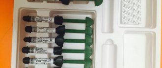
To a large extent, this problem can be solved by “carpule” anesthesia (Fig. 1). However, it should be recognized that quite often, carpuled anesthetics are not used optimally by practicing dentists, without taking into account the peculiarities of their composition, pharmacological characteristics and possible undesirable side effects. Often, the dentist does not even know what anesthetic is in the carpule, what medicinal additives it contains, what possible side effects and complications are...
Rice. 1. A set of instruments and medications for “carpule anesthesia”:
a – carpules with anesthetic
b – needles
c – carpule syringe
Injection anesthesia has found wide application in therapeutic dentistry. Under local anesthesia, treatment of caries and other lesions of hard dental tissues, endodontic procedures, professional teeth cleaning and periodontal interventions, and sometimes even teeth whitening, are currently carried out.
In addition to the “classical” requirements for all local anesthetics (minimal toxicity and allergenicity, absence of damaging effects on body tissue and nerve structures, low likelihood of complications and side effects, rapid biotransformation in the body, etc.), to the drugs used for local anesthesia in therapeutic dentistry, requirements are imposed that reflect the specifics of therapeutic and prophylactic manipulations carried out by dental therapists.
The most significant additional requirements for anesthetics used in a therapeutic dentistry clinic, in our opinion, can be formulated as follows:
− provide deep, complete anesthesia of hard tissues and dental pulp, periapical tissues and oral mucosa in the area of intervention, including being effective in infiltration anesthesia of molars and premolars of the lower jaw;
− have the fastest possible onset of anesthesia (1-5 minutes);
− the duration of effective anesthesia should ensure the painlessness of all treatment and preventive measures, incl. and at the final stages of treatment (removing the matrix and interdental wedges, grinding and polishing restorations in the subgingival area, filling root canals with hot gutta-percha, suturing after periodontal interventions, etc.). Thus, given the duration of the most common therapeutic dental procedures, the duration of effective action of the local anesthetic should be 60-75 minutes.
− ensure rapid and complete cessation of anesthesia of teeth and surrounding soft tissues after completion of dental procedures (optimally – 2.5-2 hours after anesthesia);
− maintain chemical stability and pharmacological properties throughout the entire recommended storage period (up to 2-3 years) at room temperature, including possible fluctuations.
The widest range of carpuled anesthetics on the Russian dental market is represented by (France):
− Septanest 4% 1:100.000;
− Septanest 4% 1:200.000;
− Scandonest 2% Special;
− Scandonest 3%.
Septanest 4% 1:100,000 (Fig. 2), in our opinion, should be considered as the main anesthetic for use in therapeutic dentistry. This drug has a deep analgesic effect. The rapid onset of anesthesia – 1-3 minutes – saves the doctor’s working time. The duration of anesthesia is 60-75 minutes.
Rice. 2. Septanest 4% 1:100.000 (Septodont).
Septanest 4% 1:100,000 penetrates well into bone tissue, allowing most manipulations to be performed under infiltration anesthesia, including the treatment of molars and premolars of the lower jaw. When using this drug, we resort to conduction anesthesia on the lower jaw (torsal, mandibular) only when it is necessary to anesthetize a large area of the dental system (for example, with the simultaneous depulpation of 3-4 chewing teeth, surgical intervention on the periodontium of 1-2 segments of the dentition, etc.). d.).
As our clinical experience shows, the use of Septanest 4% 1:100,000 is effective for conduction and infiltration anesthesia for any therapeutic dental procedures: treatment of caries and other lesions of hard dental tissues, long-term aesthetic restorations of teeth with composite materials, endodontic manipulations, professional teeth cleaning, periodontal interventions etc.
Septanest 4% 1:100,000 is a 4% solution of articaine with increased adrenaline content. Due to the high concentration of the vasoconstrictor, the duration and depth of anesthesia increases due to local tissue ischemia and slower leaching of the anesthetic from the injection area [Petrikas A.Zh., 2009]. Slowing down the general absorption of the anesthetic due to local constriction of blood vessels also reduces the severity of possible “general” side effects from the anesthetic. In addition, local tissue ischemia in the anesthesia zone reduces bleeding, facilitating therapeutic procedures.
An important feature of Septanest 4% 1:100,000 compared to analogues is the presence of sodium salt EDTA in its composition. This substance in combination with sodium bisulfite significantly increases the stability of adrenaline. It is thanks to the presence of these two additives that the concentration of adrenaline in the carpul remains constant throughout the entire recommended shelf life (2 years). Concerns expressed in some publications that the presence of EDTA in the anesthetic may cause headaches and nausea in the patient are rather theoretical. We have not encountered these phenomena throughout the entire period of using this anesthetic in our clinical practice.
Due to this composition, Septanest 4% 1:100,000 is the only “non-American” anesthetic based on articaine approved for use in the USA (it is supplied to the American market under the brand name “Septocaine”). Only Septodont exports carpuled anesthetics based on articaine to almost all countries of the world, including the most difficult to register drugs: the USA, Japan, Australia, England, Scandinavian countries.
Septanest 4% 1:200,000 (Fig. 3) has a “normal” adrenaline content. Due to this, the strength of its analgesic effect is slightly lower than that of Septanest 4% 1:100,000 [Petrikas A.Zh., 2009], and the duration of effective anesthesia is 30-45 minutes. However, this drug has an analgesic effect that is quite sufficient for most therapeutic dental interventions. We use Septanest 4% 1:200,000 for conduction and infiltration anesthesia for short-term manipulations associated with the preparation and restoration of teeth with composites, during professional teeth cleaning, non-traumatic and short-term surgical interventions. In addition, the use of Septanest 4% 1:200,000 is indicated for anesthesia in children, pregnant women and patients at risk. We consider Septanest 4% 1:200,000 as the anesthetic of choice for therapeutic dentistry and the primary anesthetic for use in dental surgery.
Rice. 3. Septanest 4% 1:200.000 (Septodont).
Scandonest 2% Special (Fig. 4) is a carpuled anesthetic based on 2% mepivacaine with a high content of adrenaline (1:100000). It has no analogues on the Russian dental market. A special feature of this drug is its pronounced and long-lasting vasoconstrictor effect. This provides an increased duration and depth of anesthesia, reduced bleeding of the surgical field due to local tissue ischemia and slower leaching of the anesthetic from the injection area.
Rice. 4. Scandonest 2% Special (Septodont).
The onset of anesthesia is 2-3 minutes. The duration of anesthesia on the upper jaw is 60-120 minutes, on the lower jaw – 120-240 minutes. As noted above, Scandonest 2% Special provides a long-lasting and deep analgesic effect, sufficient for most dental procedures. We recommend using this drug for conduction and infiltration anesthesia for any dental procedures, incl. long and complex. It may also be used by dentists as a “primary” anesthetic.
Adrenaline-containing anesthetics are undoubtedly the basis of local anesthesia in modern dentistry, however, it should be remembered that in the following clinical situations their use is contraindicated:
− the patient has cardiovascular diseases (arterial hypertension, paroxysmal tachycardia and other types of tachysystole, coronary and cerebral circulation disorders, heart disease, etc.);
− recent myocardial infarction (within the last 6 months);
− angle-closure glaucoma;
− treatment with tricyclic antidepressants (amitriptyline, melipramine, etc.), monoamine oxidase inhibitors (MAO), non-selective beta-blockers, antipsychotics, cardiac glycosides;
− severe forms of diabetes mellitus, especially in the stage of decompensation;
− severe thyrotoxicosis, taking thyroid hormones (thyroidome);
− sharply increased level of anxiety;
− the patient’s upcoming doping test.
In the clinical situations listed above, the use of anesthetics that do not contain adrenaline and its stabilizers (sodium bisulfite and EDTA), for example, Scandonest 3% (Septodont) is indicated (Fig. 5).
Rice. 5. Scandonest 3% (Septodont).
Scandonest 3% is a carpuled anesthetic based on a 3% mepivacaine solution without the addition of adrenaline and other vasoconstrictors. The onset of anesthesia takes 1-3 minutes. The duration of anesthesia is 10–20 minutes. This drug provides a mild, shallow analgesic effect, sufficient for only short-term and non-traumatic therapeutic procedures.
We consider Scandonest 3% as a “reserve” anesthetic intended for “at-risk” patients and recommend its use in the following situations:
− conduction and infiltration anesthesia for short-term and non-traumatic therapeutic and diagnostic procedures;
− providing anesthesia to patients for whom the use of anesthetics with vasoconstrictors is contraindicated (hypertension, diabetes mellitus, coronary insufficiency, etc.);
− carrying out anesthesia for patients with a burdened allergic history: with bronchial asthma, allergic dermatoses, the risk of an allergic reaction to sulfites and EDTA.
Speaking about the clinical use of carpulized anesthetics, several points should be emphasized.
1. The problem of reducing injection pain.
Carrying out injection anesthesia in dentistry is usually accompanied by pain when inserting a needle, moving it into tissues and introducing an anesthetic drug. This causes the patient discomfort and fear not only of dental procedures, but also of anesthesia itself, for example, with subsequent injections in children. To avoid this and minimize the listed negative phenomena, we recommend using three-stage anesthesia:
The first stage is the application of a local anesthetic to the site of future needle injection for 1-1.5 minutes (Fig. 6, 7, 8), for example, “Yilonor gel” (Septodont) (Fig. 9). It is an anesthetic gel based on 5% lidocaine and the antiseptic cetrimide, which allows for simultaneous anesthesia and antiseptic treatment of the mucous membrane in the area of the needle insertion site before injection anesthesia. Before injection, the gel from the surface of the mucous membrane must be removed (Fig. 10).
Rice. 6. Three-stage anesthesia: a local anesthetic drug is applied to a paper block.
Rice. 7. Three-stage anesthesia: a local anesthetic drug is applied to a cotton swab.
Rice. 8. Three-stage anesthesia: application of a local anesthetic drug to the site of future needle insertion (exposure - 1-1.5 minutes).
Rice. 9. "Xilonor gel" (Septodont).
Rice. 10. Three-stage anesthesia: removal of local anesthetic gel from the surface of the mucous membrane with a dry cotton swab.
The second stage is submucosal (submucosal) administration of 0.2–0.3 ml of anesthetic solution (Fig. 11).
Rice. 11. Three-stage anesthesia: submucosal injection of anesthetic.
The third stage - after 1-2 minutes - subperiosteal or intraligamentary injection of a solution of 1.0-1.5 ml of anesthetic (Fig. 12). The optimal injection rate is 1 ml/min.
Rice. 12. Three-stage anesthesia: subperiosteal injection of anesthetic.
This technique, although it takes a little longer than the traditional one, in our opinion, is more preferable, since it allows you to reduce pain to a minimum.
2. Amount and speed of anesthetic administration.
Due to the high efficiency of carpuled anesthetics, a small amount of them is sufficient to achieve effective anesthesia (Table 1).
Table 1. Recommended volumes of local anesthetics for injection anesthesia during dental procedures in adults
In some cases, one injection is not enough and the doctor has to “add” anesthesia. In this case, the maximum permissible dose of the anesthetic should not be exceeded. A one-time administration of an anesthetic in an amount not exceeding half the maximum dose is considered the safest (Table 2).
Table 2. Maximum and safe single doses of carpuled anesthetics
Note: 1 carpule = 1.7 ml.
The introduction of a local anesthetic drug into the lumen of a blood vessel can lead to complications associated with the general toxic effect of the anesthetic and vasoconstrictor (toxic reaction). Therefore, when performing injection anesthesia, in order to avoid this phenomenon, it is imperative to carry out an aspiration test.
It is also recommended to maintain a safe rate of anesthetic administration. To ensure that articaine, when accidentally directly introduced into the bloodstream, does not have a general toxic effect, the contents of the carpule (1.7 ml) should be introduced into the tissues no faster than within 20-25 s (corresponding to the rate of inactivation of articaine by enzymatic blood systems). The optimal rate of administration is considered to be 0.5 ml in 15 s, which corresponds to 1 min for a carpule.
Drugs containing mepivacaine, which is metabolized in the liver, should be administered even more slowly. The rate of their administration should not exceed 1 ml per minute.
3. The problem of reusing carpools.
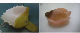
Often, a fairly large amount of anesthetic drug remains in the carpule after anesthesia (Fig. 13). Some dentists keep these carpules to use again.
This is absolutely unacceptable! It is prohibited to reuse the carpule with the remaining anesthetic solution for another patient, even changing needles!
Rice. 13. Such carpools cannot be reused!
Even if no blood is visible in the carpule, the risk of cross-transmission of infection (AIDS, viral hepatitis, etc.) in case of repeated use of the carpule is almost 100%. This is due to the fact that, due to the elasticity of the rubber piston plug, after the initial administration of the anesthetic and the cessation of pressure, microscopic particles of blood and tissue invisible to the eye are aspirated into the carpula. However, this amount is quite sufficient for the transmission of infection, primarily viral hepatitis, from one patient to another.
Thus, only an integrated approach to “carpule” anesthesia, which involves taking into account the properties and composition of local anesthetics, the general and local dental status of the patient, compliance with anesthesia technology and, most importantly, treating it as an effective, but potentially dangerous medical procedure, allows the dental practitioner to provide effective pain relief in combination with a minimal risk of complications and unwanted side effects.
Authors: Nikolaev A.I., Nikolaev D.A.
Smolensk State Medical Academy, Department of Therapeutic Dentistry
Activity factors
An aspiration test is an effective way to monitor the medical procedure being performed, as well as to collect material for further study. Any medical procedure must first and foremost be safe. To assess the risk and effectiveness of aspiration manipulation, the specialist must take into account several factors:
- rheology, that is, fluidity, of the drug and the biomaterial being taken;
- geometric parameters of the needle: its diameter and length;
- the dimensions of the syringe itself, its dimensions and, therefore, the physical capabilities of creating back pressure;
- aspiration time;
- back pressure value;
- patient's blood pressure.
Result
A positive aspiration test depends on what result is needed to obtain when performing it. When performing an intravenous injection, a test is considered positive if the result is the presence of a small amount of blood in the syringe. This means that the needle is intravascular. However, such testing is not always completely adequate, therefore, when carrying out certain medical procedures, for example, when “pumping” silicone or other liquids into the layers of the skin, care must be taken so that the procedure does not result in tissue embolization, which can lead to serious complications, even necrosis.