184
Dysplasia is a disorder in the formation of tissues, organs, or individual parts of the body. Bone dysplasia is a pathological replacement of bone tissue with fibrous tissue with or without elements of dysplastically transformed bones and calcifications.
Most often, the pathology is congenital, but its development in the postnatal period and even in adults is possible.
What is cementoma
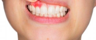
This is a type of benign tumor that forms in the area of the tooth root. It consists of fibrous cement-like tissue and at first does not manifest itself in any way - only a specialist can diagnose the problem. The development of the pathological process is accompanied by the gradual destruction of bone and epithelial tissue in the oral cavity. When the tumor reaches a large size, deformation of the jaw occurs. In some cases, the disease can lead to a fracture of the jaw bone. Women, as well as persons under the age of 20, are more susceptible to pathology.
Only a specialist can diagnose the problem
Most often, cementoma forms in the area of the central teeth of the lower jaw. In other cases, the tumor may be localized in the upper jaw, but this situation is especially dangerous, since here the pathological process can affect the maxillary sinuses. According to ICD-10, cementoma belongs to category D16 - benign neoplasm of bones and articular cartilage, subtype D16.4 - bones of the skull and face.
Massage and prevention
Treatment of the disease at an early age ends positively. In some cases, there is no need to wear stirrups - just massage is enough. But since the treatment is complex, massage is prescribed with the intake of vitamins and medications, supported by physiotherapeutic measures.
The massage does not involve anything complicated in its execution - you just have to watch how a professional does it, so that later you can carry it out yourself to prevent the disease. With the help of massage, spinal dysplasia can be completely cured.
Reasons for the development of pathology
Experts in the field of dentistry still cannot identify specific reasons that are guaranteed to lead to the formation of a benign tumor on the jaw bone. Today, experts identify several factors that can provoke the development of pathology:
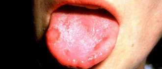
- chronic course of inflammatory processes in the oral cavity - osteomyelitis, actinomycosis, fibrous periodontitis and others,
- severe injury to the jaw bone due to a blow, bruise,
- systematic mechanical damage to hard or soft tissues of the oral cavity - incorrectly installed fillings, dentures, orthodontic apparatus (brackets).
The photo shows osteomyelitis.
Genetic predisposition can also be added to possible predispositions. Negative external influences, such as smoking or radiation therapy, can also contribute to the development of pathology.
Characteristic symptoms
One of the most dangerous characteristics of cementoma is its asymptomatic course. In the first stages of development of the pathology, the patient does not experience any discomfort or pain. Therefore, most often the tumor can only be detected at a dentist’s appointment.
“Dangerous disease! I noticed it already at the last stage, when I went to the dentist to treat a tooth, I just wanted to put a filling and remove one. As a result, they discovered cementoma... And indeed, there was no pain at all, I didn’t feel anything. I’m sure that I’m still very lucky that everything coincided like that...”
Nataly, from correspondence on the forum www.32top.ru
The tumor can be identified by x-ray.
As the size of the tumor increases, the thickness of the cortical plate decreases, which creates unnecessary pressure on the periosteum. At this stage of pathology development, the patient may experience the following symptoms:
- discomfort and moderate pain when eating or talking,
- unpleasant sensations at the moment of mechanical impact on the causative area,
- slight deformation of the enamel structure, change in its color.
When the tumor reaches a large size, it can lead to rupture of the mucous membrane and soft tissues in the oral cavity. In the most advanced cases, there is a risk of deformation of facial features and even fracture of the jaw bone.
Types of dental cementoma - classification
Experts identify several forms of cementoma. Each variety is characterized by its own characteristics and symptoms. Let's look at each type in a little more detail.
Benign cementoblastoma
We are talking about a true cementoma, which is understood as a neoplasm of coarse fibrous tissue with varying degrees of mineralization. The tumor is covered with a kind of film - this capsule separates it from healthy cells. This form is characterized by a rather slow course, but the tumor itself can grow to unlimited sizes. The initial stages of this process are not accompanied by any symptoms, but over time there is pronounced resorption of the cortical plate. As a result, the patient experiences discomfort and unpleasant sensations during mechanical action on the causative area, for example, when brushing teeth or eating.
The tumor is covered by a peculiar capsule
An advanced stage can lead to deformation of the jaw bone and the eruption of a tumor, which is fraught with infection and the development of inflammatory processes. Typically, the tumor occurs on the lower jaw in the premolar area.
Cement-forming fibroma
The tumor grows slowly, and the process of its development is usually accompanied by mild symptoms. A lesion with weak mineralization of the neoplasm is formed in the bone tissue, which is due to its fibroblastic structure. In this case, cement-like tissue is formed already at an advanced stage of the pathology, when tumor growth stops. This form of cementoma is especially dangerous when localized in the upper jaw, where its spread is fraught with damage to the maxillary sinus1.
Periapical cement dysplasia
The pathological process occurs in dental cement and bone tissue, which leads to thinning of the walls of the root system of molars. This situation is dangerous due to root fracture. Cement dysplasia usually has many pathological foci, but all of them, as a rule, do not exceed 1 cm in diameter. Tumor formation is also asymptomatic. In this case, there is no deformation of the jaw. Most often, the neoplasm is localized in the anterior parts of the lower dentition.
Periapical cemental dysplasia is often localized in the mandible
Gigantoform cementoma
This form of cementoma is characterized by intensive development, namely the rapid transformation of connective tissue into cementum. The x-ray image clearly shows oval-shaped pathological foci located in close proximity to the root system of the tooth. In this case, the localization can be very different, but usually tumors arise in places that are symmetrical relative to each other. Many experts agree that giganoform cementoma has genetic background, so often a similar diagnosis is given to several members of the same family.
This variety has oval-shaped lesions
How is diagnostics carried out?
Diagnosis of cementoma is possible only in a dental clinic. It is often possible to identify it after the patient contacts him, but for a different purpose, for example, for the treatment of caries or standard prevention. If, during a visual examination, a specialist develops appropriate suspicions or characteristic symptoms occur, the patient will be sent for an x-ray.
Histology helps to conduct a thorough study of the structure and structure of deformed tissues. The sample is placed on a microslide (slide for the object being studied), after which it is carefully examined in a multiply enlarged format, that is, under a microscope. This allows you to accurately determine the specific form of the tumor and make a final diagnosis. Histological examination also makes it possible to differentiate cementoma from other types of tumors, including malignant ones.
Histological examination is also performed for diagnosis
Disease in adults
In the adult population, dysplasia is not diagnosed so often, and its detection is associated exclusively with birth defects. If dysplasia was not detected at an early age, then gradual disturbances in the lumbocartilaginous structure of the pelvic joints occur throughout life. Such a problem, identified in childhood, can subsequently negatively affect the development of other areas of the spine, as well as even the elbow joint. Disease of the left pelvic region is most often diagnosed in men. The development of the problem begins as a result of presentation during intrauterine development.
This diagnosis has unfavorable heredity, which is important to understand at the time of birth of children. Wide and tight swaddling should occur as a preventive measure for subsequent generations. Activity, the absence of bad habits, a balanced diet, and massage will be especially necessary for female representatives who were overtaken by this disease in childhood while carrying a baby.
It is almost impossible to get rid of spinal dysplasia in adults, since the skeleton is completely formed
If dysplasia was not detected in females, do not forget that it develops in more cases in boys. It is during pregnancy and especially after birth that it is worth paying attention to the lumbosacral spine, the joints of the knees, pelvis and elbows.
When pelvic joint dysplasia is detected in an adult, according to medical terminology, such a case is interpreted as untreated childhood dysplasia.
Therapy for existing joint disorders in an adult is quite difficult and in most cases does not lead to any results. This disease can be cured in childhood or adolescence, when the growth period has not yet stopped.
What treatment is required
The choice of direction of therapy directly depends on the form of the pathology. Thus, benign cementoblastoma and cementing fibroma are treated by surgical removal, including partial excision of the affected area of bone tissue. In this case, the causative tooth has to be removed.
In the case of periapical cement dysplasia, the growth of the tumor does not occur as intensively. The surgical method is practically not used, since such an intervention can create a threat of tissue necrosis. If there are no symptoms, therapy is based on regular observation by a specialist, enhanced oral hygiene, and careful monitoring of the periapical lesion area.
In the presence of pronounced symptoms and the development of the inflammatory process, rejection of the sclerotic masses occurs. This process may take an impressive period of time, but upon completion, complete recovery will be achieved. To speed it up, a specialist can create a small depression in the area of the affected bone.
Conservative methods of therapy are also used to treat gigantoform cementoma. The doctor will definitely prescribe anti-inflammatory drugs and put the patient on regular monitoring.
Diagnostic measures
Due to the many forms, stages and localizations of the disease, as well as the similarity of its manifestations with other diseases of the dentofacial apparatus, diagnosing the pathology is considered difficult and requires a highly qualified dentist.
The following diagnostic methods are used:
- Questioning and inspection. The patient's complaints of pain, discomfort and dysfunction are listened to. Facial symmetry is assessed. The oral cavity is examined and palpated for the presence of bulging of the alveolar process, exposure of sclerotic masses and other manifestations of dysplasia.
- Radiography.
- Tomography (MRI or CT).
- Histological examination of altered tissues.
Laboratory tests are not informative, since metabolic processes in dysplasia are usually not impaired.
The gold standard for diagnosing the fibrotic form is radiography of the affected area + biopsy (for histological studies).
On an x-ray, the lesion appears to be of reduced density (ground glass type) relative to adjacent healthy tissue, without clear boundaries. Pathological tissue smoothly transitions into healthy tissue.
It is possible to alternate between areas of high and low density. The cortical plate is often thinned. The dental roots are not resorbed.
Factors indicating fibrous dysplasia are the presence of other FD lesions, as well as skin pigment spots or Albright's syndrome.
Differential diagnosis of MFD consists of distinguishing it from odontogenic cysts, adamantine cysts and osteoblastoclastomas. In the case of MFD, when the lesion is opened, overgrown osteogenic tissue is discovered, rather than tumor fluid.
MRI or CT are very informative and the final point in diagnosis for fibrous dysplasia.
The type of radiographs for CDD depends on its stage. The first shows the resolution, the second shows a mixed picture consisting of resolution and compaction, and the third shows the compacted area.
Let me introduce the similarities between radiographs of periapical dysplasia and destructive periodontitis (periapical granulomas, radicular cysts), the former is often mistaken for periodontitis. EDI of teeth helps to isolate these diseases. Unlike periapical granulomas and cysts, periradicular dysplasia is not inflammatory in nature.
An open biopsy may be needed to confirm the diagnosis. The altered tissue located under the cortical plate manifests itself as bleeding and a grayish tint.
Possible complications
Complications most often arise when cementoma is localized in the anterior part of the upper jaw. As the tumor grows, there is a serious risk of its spreading to the nasal sinuses, which can subsequently lead to the development of nasopharyngeal pathologies.
Another possible complication of cementoma may be a breakthrough of the oral mucosa with the tumor coming to the surface. Perforation leads to the appearance of a hole in the bone tissue and mucous membrane. As a result, the risk of infection of the affected tissues, the development of inflammation and the spread of pathological processes to adjacent areas of the jaw increases.
Preventive measures
To minimize the risk of developing cementoma, it is important to ensure proper and complete oral hygiene, and also, if possible, eliminate the risks of accidental injury to the teeth and jawbone. Experts provide a number of specific recommendations in this regard:
- brush your teeth twice a day, rinse your mouth after every meal, use floss and preventative rinses, visit a dental hygienist twice a year to remove plaque and dental deposits,
- promptly seek dental care for injuries to the teeth and mucous membranes,
- do not postpone a visit to the doctor if there is a traumatic factor in the oral cavity, for example, an incorrectly installed filling, crown or prosthesis.
It is worth having professional teeth cleaning twice a year.
Late detection of cementoma can lead to disruption of the integrity of bone tissue, spread of the tumor to healthy parts of the jaw, and the appearance of perforation in the jaw bone and mucous membrane. To avoid all these complications, it is necessary to carefully monitor the condition of your teeth and oral cavity, as well as regularly visit the dentist’s office for preventive maintenance - at least twice a year.
- Trigolos N.N., Makedonova Yu.A., Firsova I.V., Starikova I.V., Piterskaya N.V., Marymova E.B., Poroyskaya A.V. Cement-bone dysplasia of the jaws, 2015.
Reviews
Diseases of the jaw bones (dysplasia, osteitis, odontogenic tumors, etc.), if not life-threatening, can significantly complicate it if not treated in a timely manner.
If you or your child have been diagnosed with jaw dysplasia, tell us what form it was and how the treatment ended.
A live, direct story from a participant in an event is much more valuable for ordinary users than dry information from a doctor. You can leave a comment at the bottom of this page.
If you find an error, please select a piece of text and press Ctrl+Enter.
Tags jaw
Did you like the article? stay tuned
Previous article
The unique qualities of hyaluronic acid and its use in dentistry
Next article
Types of incomplete amelogenesis and ways to eliminate it