If the tooth is not severely damaged, but there is a problem with its roots or in the bone surrounding them, it is not at all necessary to remove the tooth completely; it is always possible to save it. However, such “tooth-preserving” operations can only be performed by highly qualified surgeons. Such surgeons work in our clinic under the guidance of a leading specialist in this area, Doctor of Medical Sciences, Professor Leon Andronikovich Grigoryants, who himself is the author of many methods of tooth-preserving operations.
Instead of completely removing the tooth, an incision is made in the gum and the problematic tooth root or part of it is removed. Such operations are called:
Tooth root amputation is the cutting off and removal of the entire tooth root at the point where it departs from the bifurcation (trifurcation) without violating the integrity of the coronal part of the tooth.
Hemisection of a tooth root is the cutting off and removal of one of its roots from a tooth along with the part of the tooth adjacent to it.
Corono-radicular separation is a dissection of the tooth crown into two parts in the area of its bifurcation, followed by curettage.
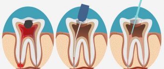
Tooth replantation (primary, delayed). This operation involves tooth extraction, endodontic treatment, curettage of periapical tissues and return of the tooth to its alveolus.
Resection of the apex of the tooth root . Resection of the root apex of a tooth is understood as cutting off and removing the apex of the root of the “causal” tooth and the pathological tissues surrounding it.
The defect formed at the site of the removed tooth root or cyst is filled with bone material and the wound is sutured. Such operations are performed in one day, they are painless, do not require special preparation of the patient and long-term rehabilitation. Below are typical clinical cases.
What is bifurcation
The bifurcation of a tooth is the area on the tooth itself where the branching of its roots begins (you can see what it looks like in the photo below). The term "bifurcation" means "branching into two", i.e. for two roots. But some teeth have not two, but three roots - in this situation the term “trifurcation” would be more correct, but doctors often still use the word “bifurcation” as a more common concept. Moreover, bifurcation does not occur on any tooth, but on multi-rooted ones - molars have two or more roots (“sixes”, “sevens” and “eights”).
Is it necessary to treat bifurcation?
In fact, when a dentist talks about tooth bifurcation, he means not only the area of root branching itself, but also the pathological processes occurring here. But there should be a clarification here. For example, perforation in the bifurcation area - i.e. the formation of a hole that violates the integrity of the tooth. This pathology not only causes complications (pulpitis, inflammation of the bone socket, etc.), but often calls into question the very existence of the tooth. Therefore, treatment of perforation or other pathologies in the area of root branching should begin as early as possible.
On a note! If there is perforation in the bifurcation area, the tooth is quite difficult to treat and will not last more than 3-5 years, after which it will have to be removed. Such a tooth is not suitable for a prosthesis, since it can no longer withstand increased chewing loads.
Removing a broken instrument from the root apex area
| After poorly performed endodontic treatment, it is necessary to remove the broken instrument from the area of the apex of the root canal of tooth 15. The tooth hurts when biting. Chronic periodontitis develops, which can lead to serious complications. | A root apex resection operation was performed. A control photograph was taken after 3 months. The inflammatory process has been stopped. The tooth is saved! |
In case of removal of the root apex, it is necessary to perform retrograde canal filling of the remaining part of the root. Such operations make it possible not to involve the crown part of the tooth; in this case, we can save not only the tooth, but also the expensive fixed prosthesis installed on it, for example, a metal-ceramic bridge.
Why problems arise
Involvement of the furcation area in the pathological process occurs in two cases:
- infection penetrates from the outside (through the gum or bone): normally, the furcation area is in close contact with the periodontium and the bone walls of the tooth socket. But if the bone begins to sag (which happens with periodontitis, periodontal disease), then the furcation area becomes accessible to microbes from the external environment. Also, inflammation of the bone (for example, when an infection enters it through the bloodstream) leads to the “melting” of dental tissue. Over time, the root cement is destroyed, a hole appears in the tooth, and microbes penetrate into the pulp,
- from inside the tooth: for example, with pulpitis or perforation of the bottom of the pulp chamber (during drilling or passage of root canals), which is located just above the furcation area. In this case, microbes from the oral cavity penetrate into the bone socket, causing its inflammation.
Retrograde root canal filling
Indications for retrograde filling:
1. Poorly sealed root canal with phosphate cement, resorcinol-formalin paste or other materials that make re-treatment impossible.
2. Poorly sealed root canal and the presence of a metal object in it: a) stump insert; b) anchor pin; c) fragments of an endodontic instrument.
3. Obliteration of the root canal.
4. Extremely long and curved root canal when conventional endodontic treatment is impossible or unsuccessful.
5. Canals with apical deltas or funnel-shaped expansion.
6. The presence of metal-ceramic or other crowns or fixed structures in combination with the above conditions.
| A root apex resection operation was performed. A control photograph was taken after 3 months. The inflammatory process has been stopped. The tooth is saved! Leaky sealed canals of 12, 11, 21 and 22 teeth. Radicular cyst, core inlay of tooth 12, metal-ceramic crowns. | 2 years after retrograde filling with Trioxident cement. The cavity is filled with newly formed bone tissue. Teeth saved! In most cases, doctors would recommend such teeth to be removed and then made with a removable denture. |
What symptoms should you pay attention to?
In many people, pathology in the area of branching of dental roots is asymptomatic. But some patients note the following signs:
- pain when biting and pressing,
- darkening of the enamel,
- bad breath,
- unsteadiness of an artificial crown or bridge,
- discharge of blood or ichor from under the gums.
Read on the topic: why a tooth hurts “without a nerve” - and how to remove the pain, advice from a dentist.
How is diagnostics carried out?
Diagnosis of the pathological process in the area of bifurcation of tooth roots includes several studies. An X-ray examination is required - computed tomography, which shows the smallest changes, is more informative here. The dentist also probes the gum pocket (normally, the gums should not move away from the tooth by more than 3 mm).
It is important to know! If necessary, the artificial crown is removed or the old filling is removed and the dental cavity is examined for the presence of perforations. It is optimal if the doctor uses a microscope during such an examination - after all, with multiple magnification, you can see the smallest defects.
If pathology in the bifurcation area occurs during treatment, the dentist should pay attention to the appearance of blood in the cavity. In this case, we can speak with 99% certainty about perforation, which needs urgent restoration.
Delete or restore?
Tweet This is a question that not only patients but also doctors argue over. But there are clear indications for which a tooth can only be removed. We will talk about one of these testimonies today.
The divergence (destruction) of the roots in the bifurcation area is subject to mandatory tooth extraction, and there can be no talk of any crowns, inlays, pins or fillings. Imagine that your tooth has collapsed, the filling has fallen out, the wall has broken off and you have delayed visiting the doctor to such an extent that the doctor gives you a verdict - removal... the tooth, unfortunately, cannot be saved. But you heard somewhere that extraction is the last step, that you can always remove a tooth, that you need to take all measures to resuscitate it! And believing that the doctor, as is typical in principle for everyone, made a mistake, or does not want to take on a difficult tooth, or even doubts his manual skills to save it, you decide to turn to another specialist. Having interviewed all your friends (friends) in search of the phone number of a miracle specialist who should save your tooth, you get an appointment with a doctor who tells you that he knows how to do this, that he has had many such cases in his practice and the results just super!, and you can do everything right now, in one hour (it’s like a whole slogan)).
This should immediately alert you! There are no miracles. Unfortunately…
But the tooth will be restored. And it will be like new, and will not bother or hurt. And in your joy, you won’t even pay attention to how much you pay for treatment - because it’s not that important at the moment, they don’t save on health!! and rush to share your joy with your loved ones and friends, along the way idolizing the doctor who did the impossible.
But the most important question you should ask your doctor is how long his work last ? What are your guarantees ? If they answer you - a standard year, then think before you sit down in a chair whether you are ready to pay about 10,000 rubles for the restoration of a tooth in order to have it removed anyway in a year.
Could it be worse?... unfortunately it can...
Such doctors are driven not by a feeling of compassion for their patients, not by a sense of help and medical duty... but by one feeling... of profit and greed...
A year later you will come to him, and with the question of what happened, that everything fell apart again, you will be sentenced to a new account.
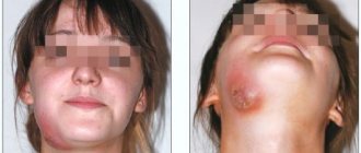
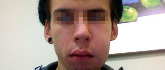
Let's look at a specific example. Patient L., a year and a half ago, was diagnosed with chronic periodontitis of 24 teeth and treatment was prescribed - removal, without explaining the reasons. But not believing the doctor, he decided to turn to another specialist, who treated the canals, installed a stump inlay and covered it with a crown worth 20,000 rubles. A year and a half later, sharp pain began in the area of this tooth and soft tissue swelling developed. After an examination by Dr. V.S. Fedotov, they took a 3D photo and this is what they saw:
Periostitis
The absence of bone tissue in the selected area only indicates that the tooth is based on honest word, if of course it is honest) and is not far from other complications, such as perforation of the bottom of the bay of the maxillary sinus and loss of the adjacent 25th tooth due to the lack of bone tissue, despite the fact that the tooth was cured correctly.
This is what we observe when removing the crown and removing the core tab from the disturbing tooth:
Root bifurcation
The roots of the tooth have already separated, are mobile and must be removed.
We take an impression to make temporary metal-ceramic crowns so that our patient does not go without teeth; after all, the 24th and 25th teeth are an aesthetic zone.
impression for temporary crowns
We remove the roots, curettage the hole, fill it with bone material with collagen and apply sutures.
After 2 days (time to make the crowns) (and these couple of days were also needed for the swelling to subside after root removal and bone grafting) we get the finished work:
temporary metal ceramics
And we fix the finished work in the oral cavity:
metal ceramics
Next, after the integration of bone tissue and restoration of the socket of the extracted tooth, a two-stage operation is planned to implant the 24th tooth using the same temporary metal-ceramic crowns during the integration of the implant into the bone.
to be continued...
Modern methods of treatment
When pathology is detected in the area of root bifurcation, a logical question arises: is it worth saving the tooth? Only a dentist can give an accurate answer after a thorough diagnosis, because... There are many determining factors here. It is important to correctly predict the “functionality” of the tooth if treatment is chosen - so that severe inflammation of the gums and bones does not begin (which, by the way, can affect the roots of neighboring teeth).
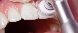
Therapeutic treatment
Conservative treatment of the bifurcation area is possible if the hole size does not exceed 1 mm, and is most often carried out using the following technologies:
- closing the perforation with special filling materials: these can be glass ionomer cements that absorb moisture - since it is not possible to properly dry the work area. Suitable brands for root canal filling are ProRoot MTA, MTA-Angelus,
- closing the perforation using calcium preparations: they restore the structure of damaged tissues (which mainly consist of calcium),
- reinforcement with metal threads and filling of the hole in the bifurcation area.
In all of the above cases, after restoration of the hole in the bifurcation area, the tooth cavity is closed with a temporary filling, and after 5-7 days the patient comes for a control x-ray to assess the quality of the perforation closure. There may be several such visits. After achieving a positive result (the perforation is completely closed, there is no inflammation near the roots), a permanent filling is installed.
On a note! If the pathology began from the outside, then treating the tooth alone is not enough - you also need to cure all the tissues that surround it (this is called “periodontal”). Treatment of periodontitis is carried out comprehensively - i.e. conservatively and surgically. First, tartar and granulation tissue are removed using curettage, and then regeneration drugs are introduced.
Surgery
If the perforation cannot be closed, or the hole is 1-1.5 mm or more in size, then surgical intervention is indicated1:
- cystectomy: cutting off the root of a tooth is called
- hemisection: in this case, the root is removed along with the adjacent part of the crown,
- complete removal followed by prosthetics.
“During the treatment of pulpitis in the clinic, they “punched” a hole to the bone with a drill. Of course, I didn’t feel it, but the doctor’s face even changed a little. I thought that I would have to remove it, but the filling seemed to fit normally. Although the dentist immediately said that it would not last long, and in 3 years it would have to be removed. But I don’t feel any particular problems yet.”
Andrey, 34 years old, Kirov, review from forum.stom.ru
When the cause of the pathology is inflammation of the bone tissue or periosteum, removal is often required (even along with a section of the bone). The bone volume is replenished with a special graft, and after some time a prosthesis is installed.
Symptoms
In most patients, pathologies in the bifurcation zone are asymptomatic. However, some note discomfort, manifested in the following signs:
– painful sensations during palpation, chewing;
– darkening of the tooth surface;
– unpleasant odor from the mouth;
– loosening of crowns, bridges;
– appearance of ichor from under the gums.
Diagnostics
To identify pathological processes in the bifurcation zone, radiography, computed tomography, and probing of the gum pocket are performed.
To examine the cavity for the presence of perforation, if necessary, remove the artificial crown and old cement.
If pathology occurs during treatment, the dentist pays attention to the appearance of blood in the cavity. It indicates perforation, requiring immediate treatment.
Treatment
When choosing a treatment method, the dentist prevents the development of inflammation of the soft and hard tissues, which can affect neighboring units.
1.Therapeutic method. Conservative treatment in the bifurcation zone is possible if the perforation size is no more than 1 mm. Pathology is treated in the following ways:
– close the perforation using a filling material (glass ionomer cement that absorbs moisture);
– eliminate perforation with calcium-containing preparations that restore tissue structure;
– reinforced with metal threads, cemented holes.
With the above methods, the dental cavity is then filled with temporary material. After 5–7 days, the patient undergoes a control radiography to assess the quality of perforation removal. There may be several such visits.
When a positive result is achieved, permanent cement is installed.
In case of external causes of pathology, the tissues surrounding the problem unit are treated - tartar and granulation tissue are removed using curettage, and regenerating drugs are introduced.
- Operative method. If therapeutic treatment is ineffective or there is a large perforation hole (over 1–1.5 mm), surgical methods are used:
– cystectomy (tooth root is cut off);
– hemisection (the root is removed along with the adjacent tissues of the crown);
– extraction (the tooth is completely removed).
If the cause of the pathology is identified in the periosteum or bone tissue, the lesion is eliminated along with the bone area. The lost volume is replaced with a graft, subsequently installing a prosthesis.