Benign tumors of the oral cavity are neoplasms of various nature located in the mouth. Tumors of this localization do not tend to develop into malignant ones; as a rule, they have limited growth. The following neoplasms fall into this category:
- Papilloma;
- Myxoma;
- Cyst;
- Fibroma;
- Fibromatosis;
- Hemangiomas;
- Lymphangiomas.
The most common tumors are of epithelial origin, that is, they originate in glandular or squamous epithelium. In rare cases, tumors appear from muscle fibers or adipose tissue. A benign neoplasm can be localized on the tongue, on the inner surface of the cheeks, on the palate (hard and soft), in the area under the tongue, on the lips and on the gums.
The causes of benign tumors are currently not fully understood. Among the dominant risk factors are bad habits, various injuries, one-time (as, for example, during tooth extraction) or permanent (microtrauma caused by the edge of a broken tooth).
Anatomical structure
The oral cavity is the initial section of the digestive tract, in which food is chewed and saliva is produced to digest food. It is involved in the process of breathing, swallowing, articulation and speech.
The composition of the oral cavity includes:
- vestibule (lips, front side of teeth, inner surface of cheeks);
- gums;
- the bottom on which the tongue lies;
- two thirds of the tongue;
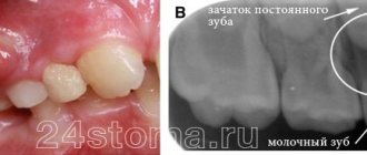
- teeth;
- retromolar triangle - the space on the lower jaw behind the third molar;
- hard and soft palate.
Let's sum it up
Mucous fibroids on the inside of the cheeks, gums, lips, and tongue grow very slowly and do not cause severe discomfort. They do not pose a danger to the patient, but the problem must be eliminated as early as possible. The fact is that a benign seal in the oral cavity with regular trauma can degenerate into a malignant one, and the oncological process requires serious therapy and is extremely dangerous to life and health.
It is impossible to get rid of the disease using traditional methods. It is important to undergo high-quality dental diagnostics and consult with specialists. In most cases, surgical excision is indicated, but with small growths, the absence of severe symptoms and the elimination of provoking factors, there is a possibility of rapid tissue restoration without external influence.
There is no need to worry if the doctor has decided to undergo surgery. Modern clinics practice laser or radio wave removal on an outpatient basis. These are the most gentle and highly effective methods. An extremely important point in a positive prognosis for the treatment of dental fibroids is an early visit to a doctor and proper excision by an experienced physician.
Classification
Oral cancer is divided into three types:
- papillary. The nodule in the mucous membrane increases in size and hangs into the oral cavity. The neoplasm progresses slowly;
- infiltrative. The seal on the pinkish mucosa is distinguished by a whitish color, clear contours and shape, and thinning of the membrane around it. On palpation from the side of the cheek, a dense infiltrate is felt. The tumor tends to grow rapidly. The patient complains of unbearable pain;
- ulcerative The most common form of the disease. Ulcers on the mucous membrane do not heal, they grow, and the border around them turns red. The outline is torn and its edges are bleeding.
Tumor metastases appear quickly. Malignant cells grow into the mental, submandibular, and deep jugular lymph nodes. This process is influenced by the thickness and depth of the tumor. Thus, when the tumor deepens by 4-5 mm, metastases occur in 98% of cases. At the T1 stage of oncology, metastasis is detected in half of the cases, and when the T4 stage is reached, distant spread of cancer cells is observed in 85% of cases.
Epithelial tumors of the oral cavity
Papillomas
These formations are characterized by rapid growth and an increase in the size of the oral mucosa. The neoplasms are characterized by a smooth surface with a pinkish tint, a soft structure and a stalk with clear boundaries. Papillomas are single and not grouped together. In more than 50% of cases, patients are diagnosed with strains of human papillomavirus (HPV) 11 and 6. They provoke the disease.
Nevi
Pigmented nevi, or birthmarks, are rare. Most often they appear on the face. Patients may not pay attention to the formation until it begins to cause noticeable discomfort. Among the unpleasant symptoms of nevi are bleeding, changes in color and increase in size.
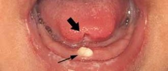
Glands of Serra
These formations are diagnosed before the child is 1 year old. They are located in the mucous tissues of the lower jaw and alveolar processes. These tumors look like small round formations that are located on a flat base and are yellow in color. Serre's glands can be single or multiple, occurring in a simultaneous group of 3 to 5 pieces.
Causes
The prevalence of oral cancer is growing and is currently diagnosed in 2% of patients among the total number of cases. Since 2009, the incidence has increased by 25%, with mostly squamous cell carcinoma being detected and only in isolated cases adenocarcinoma.
Most foci of oncology are observed in the tongue. Slightly less malignant formations on the floor of the mouth. Cancer of the soft and hard palate, gums and cheeks is detected in 20% of cases. Much less frequently diagnosed is damage to the alveoli of the lower jaw - 4%, the arches of the palate, retromolar region and vestibule - 3%.
Based on practice, men are more susceptible to oral cancer than women. This is due to bad habits, for example, the abuse of cigarettes or chewing tonic mixtures increases the production of saliva, which washes away beneficial elements from the mucous membrane. The risk group includes patients with HPV, elderly people, workers in hazardous industries, patients with lichen planus, people whose oral mucosa is systematically injured by fillings, prostheses, and metal objects.
Symptoms
A malignant ulcer from ordinary stomatitis in the mouth can be identified by swelling and swelling of the cheeks, pain and constant discomfort even at rest. You should be wary of prolonged non-healing of the wound and its bleeding.
As the disease progresses, the symptoms intensify:
- swelling increases and spreads to the neck;
- the red or white spot on the oral mucosa intensifies;
- discomfort when chewing and swallowing;
- difficulty speaking due to friction of the mucous membrane on the teeth when moving the jaw;
- the appearance of bad breath;
- feeling of a foreign object in the throat;
- anemia of the mouth.
In the late stages of cancer, teeth fall out and body weight rapidly decreases.
Vascular tumors
Hemangiomas
Such tumors arise due to the growth of the walls of blood vessels. There are three types of neoplasms depending on which vessel wall has enlarged: arterial, capillary and venous.
The tumor can be located on the lips, cheeks and tongue. It has the appearance of a blastoma with a soft structure, a purple tint and rough protrusions. Hemangioma increases rapidly, especially during puberty of patients. If injured, the tumor may begin to bleed. It can easily become infected, which can lead to tissue necrosis and thrombosis.
Lymphangiomas
This tumor appears due to the proliferation of lymphatic vessels on the lower or upper lip. The new growths have an uneven surface with blisters that are colored red or light yellow. They are easy to damage. Lymphangiomas are prone to purulent inflammation. They can grow quickly, which causes pain. The tumor is removed using multiple coagulation.
Diagnostics
At the initial consultation, the doctor examines the oral cavity, examines ulcers, erosions, damage to the mucous membrane, and then takes a smear for examination. To confirm the inflammatory process, the patient is sent for a general and biochemical blood test.
The diagnosis is confirmed by the results of the examination:
- MRI and ultrasound of soft tissues of the neck. The images reveal the localization of the pathology, the depth of germination and the structure of the tumor, compaction from blood and lymph, decomposition of the cortical layer of the bone;
- if metastases are suspected, a fine needle aspiration biopsy of the lymph nodes under the chin, under the jaw and in the upper third of the neck is performed;
- positron emission tomography. Shows the depth of the tumor, as well as early metastases;
- osteoscintigraphy. Skeletal bones are examined to look for displaced cancer cells;
- CT scan of facial bones with contrast. The images show the tumor growing into the neck vessels, jaw or base of the skull.
Connective tumors
Fibroids
This tumor is formed from fibrous connective tissue and is detected at the age of 6–15 years. Such formations are covered with a mucous membrane, have a smooth pinkish surface and are located on a base or stalk. Fibroids are located in hard-to-reach places and may not grow for a long time.
Fibromatosis of the gums
This is a disease of genetic predisposition. It is characterized by significant gum growth, without an inflammatory process. At the time of diagnosing fibromatosis, doctors identify hypertrophy of the interdental papillae, gingival margin and gums, which become pinkish and dense in consistency.
Myomas
They are formed from muscle tissue and are located on the tongue. They look like nodular formations with a smooth surface. Myoma is actively growing, which causes problems with diction and chewing food. Often provokes aching pain.
Myxomas
Such tumors most often form in the upper jaw in patients aged 10–35 years. The formations grow rapidly due to an increase in mucoid substance and have a tuberous, papillary or rounded surface.
Pyogenic granuloma
A granuloma looks like a small nodule on the skin. It consists of a bundle of blood vessels and can therefore bleed when exposed. Such neoplasms do not degenerate into malignant ones. Their removal is carried out by cauterization with electric current.
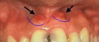
Epulis
These formations with a diameter of 0.5 to 7 cm are most often located on the alveolar process. They arise due to rubbing of the mucous membrane with poorly selected dentures. When injured, the epulis may bleed.
Neuromas
Neoplasms with a diameter of about 1 cm are formed against the background of an active increase in the cells of the Schwann sheath of nerve fibers. Patients usually do not report any discomfort when they are present. Only upon palpation may pain appear.
Treatment
The choice of treatment tactics depends on the stage and extent of the tumor. When the tumor grows rapidly, treatment methods are combined.
Operation
The doctor determines the principle of surgical intervention after determining the stage of the tumor and its spread. If cancer cells have penetrated the periosteum and surrounding tissues, a wedge-shaped, planar or sagittal resection of the jaw is performed. If the examination reveals the growth of cancer cells directly into the bone or the defect is noticed during surgery, segmental resection of the lower jaw is performed. The doctor assesses the lesion on site and determines the thickness of the excised layer.
The next stage of the operation is partial or complete excision of the cervical lymph nodes to prevent metastases if the thickness of the tumor is more than 4 mm or the location of the tumor in the floor of the mouth or on the tongue. If the tumor is located in the midline, then the cervical lymph nodes are excised on both sides. The operation ends with the immediate replacement of damaged tissue.
After removal, the tumor is sent for histological examination. Its size, thickness, depth, edges are assessed. Further treatment is affected by cell growth beyond the boundaries of the capsule of the removed lymph node, and the spread of cancer cells to neighboring organs.
Radiation therapy
Radiation after surgery is prescribed when diagnosing T3, T4, N2, T3 stages of the disease no later than six weeks after tumor removal. The need for radiation therapy increases with perineural invasion of the lymphatic vessels. The total focal dose for all sessions is 60 g, and the single focal dose for one session is 2 g. When metastases are detected on the neck, the SOD increases to 66 g, and if there is no risk of metastasis, the SOD decreases to 50 g.
As the main treatment, radiation therapy is used in a total focal dose of 60-70 g. The procedure is performed five days a week and is combined with chemotherapy. Every three weeks, 100 mg of cisplatin is administered.
Chemotherapy
Anticancer drugs are prescribed before surgery or along with radiation therapy to reduce the size of the tumor. Sometimes therapy is prescribed simultaneously with surgery.
Treatment involves the use of a 5-fluoroacyl regimen together with cisplatin or other agents - carboplatin, methotrexate, bleomycin. They cause a number of side effects, for example, vomiting or nausea, hair loss, decreased appetite, and increased bleeding. Symptoms disappear after treatment, but permanent hearing loss is sometimes observed after taking cisplatin.
The prognosis of oral cancer depends on the stage at which the disease is detected. If treatment is started at stage zero, the disease will stop. It is worth noting that smoking provokes relapse or degeneration of the tumor, so repeated surgery or radiation may be required. Surgery at the first stage increases survival rate to 80-85%, and the combination of radiation therapy with surgery at the second stage by 60-80%. Already at subsequent stages of cancer development, the survival rate is no more than 50%, and all three treatment methods are used simultaneously.
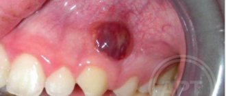
Hematoma
Description
If there is a swollen lump on the gum filled with blood, most likely it is a hematoma. This tumor occurs as a result of an incorrectly removed tooth or injury. Such a lump on the gum above the tooth, as a rule, does not hurt.
Treatment
Dissolves on its own. But if the lump hurts, consult your dentist for advice. The doctor will prescribe painkillers.
Dispensary observation
Since the tumor can recur and metastasize, after completing the course of treatment the patient is registered with the oncology clinic. The first year you should visit a doctor every month, the second year a preventive examination is carried out every 4-6 months, and then once a year or in case of any ailments. The examination involves an examination - ultrasound and contrast MRI of the soft tissues of the neck, PET, osteoscintigraphy. Consultation with an otolaryngologist, dentist and oncologist is required. The doctor may shorten the period of medical examination if there is a high risk of relapse.
List of references on the topic:
- Gantsev Sh.H. Oncology – M, 2012 – P.204-205.
- Golovin D.I. Errors and difficulties in diagnosing tumors, D.: Medicine. Leningr. department, 2015 305 pp.
- Selected lectures on clinical oncology/Ed. IN AND. Chissova, S.L. Daryalova. – M., 2010
- Matyakin E.G., Alferov V.S. Chemotherapy of head and neck tumors // Mat. 2nd Ros. oncol. conf. “Current trends in the development of drug therapy for tumors” December 8–10, 2016 – M., 256 p.
- Tumors of the head and neck: hands/ A.I. Paches. - 5th ed., add. And revised - M.: Practical Medicine, 2013. -478 p.
- Shine A.A. Oncology. M – 2014 365 pp.
- Encyclopedia of Clinical Oncology/Ed. M.I. Davydova. – M., 2014 –P.140-179.
- Bityutsky P.G., Kitsmanyuk Z.D., Trofimov E.I. Diagnosis and treatment of cancer of the oral mucosa // Medical consultations. - 2014. - No. 1. - P. 23-27.
- Byakhov M. Yu. Options for combined and complex treatment of locally advanced cancer of the oral mucosa and oropharynx: Dis. Dr. med. Sci. - M., 2013.