From this article you will learn:
- structural components of enamel,
- the structure of its organic and inorganic matrix,
- electron microscopy images.
Tooth enamel (enamelum) is the outer shell of the crown of the tooth, representing the hardest tissue in the human body. This hardness is explained by the fact that 95-97% of tooth enamel consists of mineral components (mainly calcium phosphate in the form of hydroxyapatite crystals). Organic matter accounts for only 1-2%, plus about 2-3% water. The strongest are the surface layers of enamel - especially on the occlusal surfaces of the teeth, and towards the enamel-dentin border, as well as when approaching the neck of the tooth - its hardness decreases.
The hardness of enamel is 397.6 kg/mm², which is comparable to quartz. Such hardness allows it to withstand extreme mechanical loads, but on the other hand, it makes it very fragile. The enamel of human teeth does not crack or chip only due to the presence under the enamel layer - a layer of dentin, which has a moderate elasticity coefficient. However, despite its hardness, enamel has good permeability to calcium and fluoride ions contained in saliva (or toothpastes and mouthwashes), as well as to pigments contained in food and drinks.
Structure and development of tooth enamel –
Tooth enamel can have different shades - from yellow to various shades of gray and white, which depends on its transparency coefficient. The more transparent the enamel, the more the underlying layer of dentin, which is physiologically yellow, will be visible through it. In addition, the enamel may have a blue tint - at the cutting edges of the incisors (where there is no underlying dentin layer), as well as at baby teeth. The transparency of enamel depends on the degree of its mineralization and homogeneity, which is associated with the ratio of its organic and inorganic matrices. Transparency also depends on the thickness of the enamel layer.
The thickness of the enamel will vary on different surfaces of the tooth crown. For example, in permanent teeth, the thickness of the enamel ranges from 1.7 to 2.5 mm in the area of the chewing cusps of the molars, and to only 0.01 mm in the cervical part of the tooth (where the enamel borders the root cement). Thus, the closer to the neck of the tooth, the less its thickness will decrease. In baby teeth, the thickness of the enamel will be two times less than in permanent teeth, not exceeding 0.8-1.0 mm.
Composition of tooth enamel
Dental enamel is the most durable, wear-resistant and hard component in the human body. It represents the outer or surface layer of the teeth, completely covers its coronal and partially cervical part, and also performs protective functions. The main characteristics of enamel include:
- The color of tooth enamel varies from white to yellowish, and it can change shade (color) depending on a person’s dietary habits or bad habits.
- The thickest areas of enamel cover the chewing molars in the area of their anatomical tubercles; their thickness varies between 2.3-3.5 mm;
- The thinnest areas of tooth enamel are localized in paroxysmal areas (places of contact in their lateral projection), here the thickness of the protective layer reaches approximately 1.3 mm;
- The enamel that covers all the teeth in the human oral cavity is not capable of regeneration, because there are no living cellular structures in the tissues of this protective layer;
- Depending on the characteristics of each person’s body, up to 95% of the chemical composition of tooth enamel is presented in the form of mineral compounds. The remaining percentage is divided between water and organic matter in approximately a 2:1 ratio, respectively. In addition, depending on the percentage of mineral content of tooth enamel, it can be more or less transparent (the higher the percentage of minerals, the more transparent the enamel becomes).
Teething
This stage is characterized by the active movement of temporary teeth from their places of origin inside the jaw until the crown completely exits into the oral cavity. This process is accompanied by active changes in the surrounding tissues, increased development of the root part, restructuring of the alveolar bone structure, as well as the formation and strengthening of the periodontium.
The first temporary teeth, which are usually the lower central incisors, erupt at the age of 5-6 months. Behind them appear the upper antagonists, other incisors and first primary molars. The last to erupt are the lower and upper canines. Typically the process lasts from 4-6 months to 2-2.5 years. With late eruption, the timing of the appearance of temporary teeth shifts from 8-10 months. up to 3.5 years.
Other components in the enamel
In addition to the already mentioned main components of the enamel layer of the tooth, its chemical composition also contains a set of other components:
- Neonatal line - present exclusively on baby teeth, it looks like a dark-colored stripe (almost black). This line is located in the area of contact between two types of enamel, the first of which was formed before the baby was born, and the second after.
- Bundles and plates of dental enamel are special enamel formations containing prisms of a hypomineralized type, between which the interprismatic substance consists of the same material. It is noteworthy that the molecular structure of this material involves a large number of protein compounds. Many dentists are of the opinion that through the mentioned bundles and plates, various microorganisms penetrate into the enamel from the oral cavity, making their way to deeper dental tissues, causing caries, etc.
- Gunter-Schräger stripes are lines that stand out on the tooth enamel in a darker or lighter shade, the width of which does not exceed 100 microns. They are located perpendicular to the surface of the enamel layer and are formed as a result of opening its prisms.
- Retzius lines - in shape they resemble arches offset from the central one, located symmetrically in relation to each other. When cut across a tooth , these formations resemble rings inside a tree trunk. The formation of Retzius lines corresponds to different periods of mineralization of the enamel layer.
Stabilization
This period is characterized by a complete stop of all processes of tissue formation and development. The crown and root reach the required shape, size and level of strength that allows them to perform their basic functions.
The period of stabilization of primary teeth lasts on average 2.5-3 years. At this time, it is important to ensure optimal chewing load on the bite, which will ensure the normal development of facial and other muscles, as well as periodontal tissues and jaw bones. If a child has caries or other diseases, it is during this period that treatment of temporary teeth will be most effective in terms of preserving them, preventing the spread of infection and ensuring normal conditions for changing the bite.
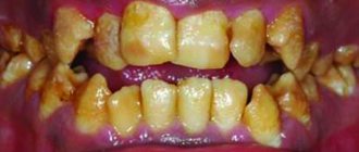
Features of the structure of the enamel of baby teeth
The main distinguishing feature of the enamel layer of children's teeth is that it is less durable and also much thinner than the enamel of permanent teeth. This is explained by the lower content of mineral compounds in teeth in relation to water and organic substances. Considering these features, if you examine baby teeth and their enamel layer under a microscope, you will notice the following differences:
- Due to the fact that the service life, as well as periods of mineralization and the tendency towards this process are shorter, the Retzius lines are much less pronounced in the structure of primary dental units.
- If in permanent teeth enamel prisms are located apically, then in milk teeth their direction is completely different, they are located horizontally.
- In children's primary teeth, the final enamel layer is much less pronounced; prisms are clearly visible on its surface, while its structure is much more porous, with microscopic cracks present.
Under the influence of each of these features, tooth enamel in children is more susceptible to wear and damage. For this reason, children develop caries much more often and it progresses faster, which is why it is important to visit the dentist regularly and have their teeth treated in a timely manner.
The recipe is ready
That's all, in short, what is needed to create teeth. Thus, the recipe for creating a tooth looks something like this:
- Stem cells - assorted
- Scaffold is a natural product
- Growth factors - to taste
Regenerative medicine technologies are progressing incredibly quickly. And now, probably, the most basic provisions for dental tissue engineering have already been developed. They all stem from our knowledge of the cellular and molecular basis of tooth development. We understand that the best result in tooth bioengineering can be achieved only in the presence of two types of cells, and not one: these are both epithelial cells and mesenchymal cells (where would we be without them?) [28]. However, you cannot build a tooth on cells alone. Thus, the role of growth factors and extracellular matrix cannot be excluded here. Fortunately, science does not stand still, and new provisions are being actively developed. Perhaps, in the near future, the treasury of knowledge called “dental tissue engineering” will be replenished with another equally valuable “coin”.
But, despite all the promising potential of tissue engineering in dentistry, there are still problems to be solved related to the conduct of clinical trials, the innervation and blood supply of the bioengineered tooth, its ligamentous apparatus, the timing of its eruption, as well as the choice of a pool of stem cells and technology for working with them , and a number of other equally pressing tasks [10], [29].
As for the most basic thing, namely stem cells: in the experiments performed (it is worth noting that almost all of them were carried out on mice), they mainly used embryonic stem cells. But in the clinic their use is sharply limited, including by law. Therefore, only postnatal stem cells remain (not counting iPSCs, where things are also not calm), and here we face the following snag: unlike mice, humans lack a niche of dental stem cells, which is why our teeth do not have the ability to constantly grow. Those MSCs that are suitable for use cannot be obtained without damaging the tooth, or even more so if the tooth was previously treated endodontically, that is, with pulp removal. Those to which access is open do not have odontogenic competence, for example, gingival MSCs. This is just one of the dilemmas that remain to be resolved (Fig. 9).
Figure 9. The fight for healthy teeth for humanity.
website accuratedentistry.net
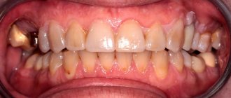
Types of damage to tooth enamel
Over the course of life, even if you provide your teeth with proper care and follow the rules of oral hygiene prescribed by dentists, the enamel layer gradually wears out and is destroyed. This contributes to the occurrence of various diseases of the oral cavity; it is influenced by the food a person eats, etc.
Among the main causes of damage and destruction of tooth enamel, dentists identify:
- Erosion is damage to the enamel layer, and then dentin, which is not associated with carious lesions of the teeth . The essence of this pathological process lies in disorders of mineral metabolism. As a result, disturbances occur in the crystalline structure of the enamel, which is manifested by its focal thinning and destruction. Externally, erosions look like local darkening on a round or oval tooth. The occurrence of erosions is provoked by the consumption of foods with high acidity levels, pathologies of the gastrointestinal tract, the use of certain medications, and the use of aggressive tooth powder or paste.
- Excessive sensitivity of tooth enamel - this disorder is especially pronounced in the form of painful sensations when the teeth cold or hot food, drinks, and even as a result of contact with cool air. The sensitivity of tooth enamel develops due to its thinning under the influence of the factors already described above. A thinned enamel layer puts teeth at increased risk of caries and other dental pathologies.
- Necrosis - this term characterizes multiple lesions of the hard tissues of the tooth, especially the enamel layer and dentin. The pathological process is initially expressed in the appearance of small light spots on the surface of the tooth , which subsequently darken and deepen. The progression of pathology threatens tooth destruction and is accompanied by a number of other oral diseases. The main reasons for the development of necrosis include gastrointestinal diseases, hormonal imbalances, metabolic disorders in the body, and work in hazardous industries.
- Caries is a carious lesion that threatens teeth , primarily affecting the enamel layer of the structure , gradually destroying it and spreading to deeper tissues. There are many reasons for the development of caries, from non-compliance with the rules of oral hygiene and irregular tooth brushing, to pathologies of the structures of the oral cavity, diseases of the gastrointestinal tract and metabolic disorders. If you start caries treatment at the stage when the lesions have affected only the enamel layer, you can only get by installing a filling or even restoring the enamel. But progressive caries is dangerous for teeth due to destruction, which may lead to the need for its complete removal.
- Mechanical damage - due to the fact that the main function of the enamel layer is to provide protection to the teeth , it primarily suffers from the effects of external adverse factors. Mechanical damage to the enamel includes cracks and other violations of its integrity due to blows, bruises, eating too hard food, etc. If the enamel of at least one tooth has been subjected to aggressive mechanical action, you should consult a doctor for an examination and, if necessary, subsequent treatment.
- Wedge-shaped defect – this term characterizes the pathological process in which the area of the dental neck is exposed. In such cases, the thinnest and most vulnerable areas of the enamel layer, located at the base of the teeth, are negatively affected. In addition to visible receding gums, damage to the enamel is indicated by a change in its color, as well as an acute reaction to hot and cold.
Strengthening tooth enamel
Today in dental practice there are many effective ways to strengthen the enamel, which allows you to maintain its integrity and prevent destruction and diseases of the dentition. At the same time, methods of strengthening and protecting the enamel layer are divided into two groups, the first are intended for adults, the second for children.
Resorption and preparation for shift
At the age of 5-6 years, the process of completely replacing the temporary bite begins. This period begins immediately after the resorption of milky roots and the beginning of the growth of permanent rudiments. Temporary teeth begin to undergo resorption and ejection from the alveoli. Odontoclasts, which carry out demineralization and intracellular destruction, as well as pulp tissues that secrete osteoclast-like cells, which are responsible for the destruction of dentin and predentin from the internal side, actively participate in the process. Removal of the crown of temporary teeth, as a rule, occurs under the influence of chewing pressure. The root remaining in the hole undergoes a natural process of destruction and resorption. The location of the crown is quickly epithelialized due to granulation tissue.
The process of loss of temporary teeth occurs symmetrically on both jaws and ends with the appearance of permanent teeth. The individual nature of its course is determined genetically.
Strengthening baby teeth enamel
As was said earlier in relation to baby teeth, their enamel is more vulnerable. To protect it, saving the child from premature loss of dental units and problems in the future, doctors perform the following actions to provide temporary protection:
- Fluoridation involves treating teeth with special fluoride-based compounds; it is recommended to repeat this procedure 2-3 times a year.
- Fissure sealing - the dentist performs the procedure of filling the recesses and grooves of chewing teeth with temporary filling material, protecting the dental structures from the negative effects of harmful microorganisms and other unfavorable factors.
- Application gels and preventive mouth guards for teeth - the method is based on enriching the enamel layer with useful components (fluorine, calcium, vitamins) through the use of special products.
Strengthening the enamel of molars
There are more methods to preserve molars and maintain the condition of their enamel layer. Firstly, this is due to fewer contraindications for adults. Secondly, molars require long-term strengthening.
The main methods of strengthening the enamel of permanent teeth include:
- Drug therapy is based on the use of vitamin complexes containing vitamins of groups B6, B12, D. In addition, the patient is selected drugs that promote better absorption of calcium and fluoride by the body.
- Special gels and oral hygiene products – this technique uses specialized toothpastes and gels containing components necessary for teeth to strengthen and maintain the condition of the enamel layer. Also, teeth are subjected to unimaginative cleaning in a dental office.
- Mineralization and preventive cleaning – mineralization is performed using special means to increase the strength of the enamel and reduce its susceptibility to a number of negative factors. As for cleaning, such procedures are performed by dentists in the clinic using special equipment. During cleaning, plaque and tartar are eliminated , pathogenic bacteria and microorganisms that can harm the enamel layer are removed.
- Home prevention - to maintain healthy teeth and enamel, patients are advised to perform a light massage of the gums, enrich the diet with fresh vegetables and fruits rich in vitamins.
Author: Zhukov M.A.