28.10.2019
Between the ages of 6 months and 3 years, a child grows 20 baby teeth, after which the period of permanent teeth begins to appear. Despite the fact that the growth of molars is almost painless, the process requires control and attention from adults. What do parents need to know about changing teeth in children?
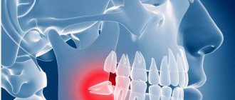
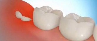
Should wisdom teeth be preserved?
We think yes.
Although wisdom teeth are not involved in the process of chewing food, they can still benefit a person. There must be clear medical indications for their removal. And now we will provide you with another proof of the benefits of your own teeth-eights. Our patient Kim is about 20 years old, she is from Korea and lives in Moscow, as her parents work at the Korean embassy. I contacted the German Implantology Center due to inflammation of tooth 4.6 – the lower right six. The tooth was destroyed
, an abscess developed. There was a temporary filling on the tooth (placed in another clinic):
Where a temporary filling was placed, she was told to remove the tooth. That's why Kim came to us with one simple goal - to atraumatically remove the six, which began to greatly bother and hurt her. After doing a CT scan and looking at the tooth, we offered her an option, which, upon hearing, Kim at first did not believe her ears. Our proposal was to remove the six and immediately in its place... transplant its lower right wisdom tooth! This is called autotransplantation surgery.
Why do you need to build bone tissue?
Increasing bone volume (osteoplasty) is necessary for the primary stabilization of the titanium root. If the height and width of the alveolar ridge are insufficient, the implant simply will not stay in the bone. The method of osteoplastic intervention depends on the clinical picture.
The following methods are used to build bone in the lower jaw:
- Guided bone regeneration.
Increasing the height and width of the alveolar process by replanting osteoplastic material. The surgeon peels off the gum, fills the jaw bone with osteoplastic material, closes it with a barrier membrane and applies sutures. - Splitting of the alveolar process.
Increases bone width. The doctor makes a cut in the center of the ridge, alternately screws spreaders of different diameters into it (from smaller to larger), and fills the resulting space with bone granules. - Bone block transplantation.
An autogenous (taken from the patient) bone block is screwed to the bone, covered with a collagen membrane and sutured. The 6th tooth implant is installed after the block has healed. - Increasing the volume of the maxillary bone.
Because the maxillary structures are located close to the root system of the first molars, a sinus lift is required in 90% of cases. It can be closed or open. The essence of the operation is that the doctor carefully lifts the lower part of the maxillary sinus, fills the resulting space with osteoplastic material and installs a six implant.
Wisdom tooth transplant as a way to replace a defect
Of course, Kim was very surprised, but we showed her photographs of successful operations (and we have all of them successful), apparently our persuasiveness and the patient’s desire played a positive role in making the decision to transplant the eighth tooth. She was happy and agreed to the operation.
Moreover, in this case we did not even have to make a stereolithographic model of the tooth, since all surgical procedures (extraction and transplantation) were performed on the very first day of her visit to our clinic.
Dental transplant surgeries typically require several days of preparation, as we describe in the following clinical case study. But in this case, taking into account the current clinical situation and relying on our many years of successful experience in tooth transplantation, we were able to transplant tooth 8 into the place of tooth 6 as quickly as possible. We had all the indications for current dental transplantation.
The donor was an impacted (completely hidden under the gum) wisdom tooth 48. In fact, as we joked, it was a neighbor tooth to the problematic six. That is, the tooth grew and grew for 20 years, waited in the wings, and that hour struck! Now this donor tooth is for another 30 years
at least he will live in a new place.
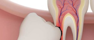
If you look at the sixth tooth from the side, you can see bluish, literally purple gums
:
This color of the gums is due to the developed inflammatory process. The patient developed destruction of bone tissue against the background of advanced periodontitis. There was a crack in the root of the sixth tooth.
The role of primary teeth in child development
The baby needs milk teeth to chew food, they help him to experience the taste of the world around him, and most importantly, they participate in the development of the entire maxillofacial apparatus. It is the baby teeth that pave the way for the permanent row. The main functions of a time series also include:
- Development of the baby's speech skills;
- Bite formation;
- Participation in the development of facial muscles;
- Development of facial bones.
The formation of baby teeth occurs at 6-7 weeks of fetal life, in the womb. The baby's first tooth begins to erupt at 6-8 months, accompanied by an increase in temperature and very unpleasant sensations.
When is removal appropriate?
Pediatric dentists prescribe baby tooth extraction for children only in advanced cases when the organ can no longer be saved. As a rule, the procedure is prescribed if:
- Most of the crown is damaged by caries;
- The tooth becomes loose and causes discomfort to the baby, but does not fall out;
- The tooth is broken, the sharp part of the crown damages the mucous membrane;
- The permanent tooth cannot erupt in the area of the baby tooth, which causes the baby severe pain and discomfort;
- A fistula forms in the gum;
- Pulpitis and periodontitis develop;
- A cyst, phlegmon and other inflammations form in the gums, requiring surgical treatment;
- The root of the tooth or the surrounding areas become inflamed.
Consequences of early removal of baby teeth
As we have already said, baby teeth play an important role in the development of the maxillofacial apparatus. Early removal of baby teeth can cause consequences such as:
- Violation of aesthetics. Children are not too concerned about the aesthetic problem of missing teeth. However, the older the child gets, the more embarrassed he becomes about his smile. Remember the fairy tale about sold laughter? Like Tim Thaler, the baby will become withdrawn, uncommunicative and aggressive. The issue of premature absence of teeth can disrupt the process of social adaptation of a child, imposing on him a stereotype of aggressive behavior with people.
- Displacement of the maxillofacial apparatus. As Aristotle argued, nature abhors a vacuum. Part of a row of teeth may shift to the place of a prematurely removed baby tooth. When changing, the permanent teeth may move slightly to the side, which will greatly spoil the child’s appearance. Even natural facial features can change.
- Speech development disorder. The front teeth are involved in the formation of sounds. If baby teeth are removed prematurely, the baby begins to “get creative” by uttering certain sounds. Subsequently, such a habit is difficult to change.
Milk teeth, like permanent teeth, have roots. Let's consider the consequences of premature removal of specific baby teeth:
- Loss of primary canines. Unexpected elimination of the upper primary canines can cause malocclusion and speech distortion. Removal of the lower fangs causes the child to chew food inadequately and develop gastrointestinal diseases. Losing canine teeth can also lead to a shift in cosmetic focus.
- Removing primary chewing teeth also reduces the baby's chewing activity. As a result, the load on the front teeth increases, and the milk rows begin to wear off. Consequences - the child, worried, refuses solid food. As a result, due to lack of stimulation, the development of the temporomandibular joint may be impaired. Permanent teeth have less space, the rows may develop crowded, the teeth “climb” on top of each other or grow into a second row.
- Removal of primary incisors can cause the canines to shift towards the socket. Sometimes babies suffer from the rotation and tilting of the remaining teeth.
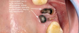
The difference between transplanting your own tooth and implantation with a dental implant
By the way, for your transplanted tooth, the presence of an inflammatory process in the soft and bone tissues does not matter, because all the tissues there are their own and, naturally, the body will protect its transplanted tissues. That is, the body does not treat the transplanted tooth as “friend or foe,” which always happens in the case of tooth implantation with a titanium or zirconium implant.
If there is an inflammatory process, an artificial implant cannot be placed in this place. And you can put your own tooth, because it has the so-called. the ligament of the tooth that has all the cells that will fight.
Naturally, after the tooth extraction, we processed everything as much as possible, carefully cleaned and rinsed it.
In this photo we have begun to treat the wound. It can be seen that there was an inflammatory process in the gums, the bone tissue was partially but severely destroyed. Signs of bone tissue destruction are clearly visible on computed tomography:
By the way, even if the bone tissue is destroyed, when we transplant our own teeth, we do not need bone grafting; there is no need to replant additional bone. And that's why. The tooth itself nourishes and regenerates. Even if there were no front wall of the tooth, if there were no vestibular buccal wall, if you transplant such a tooth, then it is restored from its ligament, even the bone grows.
Therefore, if there is a choice between transplanting your own tooth and implanting a dental (titanium, zirconium) implant, it is better to opt for transplanting your own tooth.
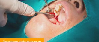
Let's begin atraumatic wisdom tooth removal
In 40 seconds
We made an incision in the area of the donor tooth, which we planned to remove and replant in place of the previously removed six. We carefully removed the wisdom tooth, placing it in a saline solution for a short time:
Many people wonder: do wisdom teeth have roots? Yes, of course there is – this is a molar. We draw your special attention to the fact that this wisdom tooth 48 has not yet finished growing its own roots, its maturation is not yet complete. The growth zone of the root of the lower wisdom tooth is preserved and this tooth subsequently, after 2 weeks, does not require nerve removal! That is, it not only grows in, but also completely becomes accustomed to its nerve.
a powerful ligamentous apparatus he has.
, which provides high survival potential. Indeed, this tooth has taken root, and besides, it has a living nerve.
Transplantation of a wisdom tooth into place 4.6
The donor tooth was clearly placed in the place of the problematic sixth tooth. It was as if he had been there all his life: ideal external dimensions, the wisdom tooth was transplanted to the place of the removed one without any external grinding. Ideal autotransplantation operation.
The implanted tooth is tightly sutured
to ensure maximum tissue contact with each other and a normal healing process.
Why does a person's teeth change?
The structure and development of the dentition in different animals differs significantly from each other. In vertebrates and some species of reptiles, for example, teeth change several times in their lives, and over time, a new one grows in place of the lost tooth; in rodents, permanent teeth immediately grow. This is due to the characteristics of the body, residence and nutrition of representatives of the animal world.
There are several species of mammals whose teeth change once in their lifetime during childhood. This is necessary not only for the growth of the body, but also for survival - after the baby stops feeding on mother’s milk, its life largely depends on the capabilities of the dental apparatus. Humans can also be classified as mammals in this category, so changing teeth is an integral process of growing up for them.
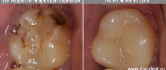
After autotransplantation 2 weeks
Patient Kim came to us after 2 weeks
for medical examination and suture removal:
Nothing bothered her anymore. The tooth had acceptable physiological mobility. The gums have become lighter. Of course, all the purple color is not gone yet, but she has already become more pink.
Pay attention to the white area - this is not pus, but fibrin plaque, which we will talk about in more detail below.
The picture shows that the donor tooth has clearly positioned itself in the position of the extracted tooth 46:
Our Kim has a new six molar. Both the patient and the medical staff of the Research Center are very pleased with the outcome of the tooth transplantation operation.
Indications and contraindications for molar tooth extraction
Reasons for removing a molar tooth may be:
- purulent periostitis;
- abscess;
- phlegmon;
- large tumor in the tooth area;
- purulent periodontal disease;
- the presence of a cyst in the area of the diseased tooth;
- exposed pulp;
- longitudinal tooth fracture;
- critical tooth decay;
- critical tooth curvature;
- disease of the dental bone tissue;
- sinusitis.
The following reasons are contraindications to the removal of molars:
- acute heart diseases;
- acute viral diseases;
- flu;
- angina;
- renal failure;
- pancreatitis;
- hepatitis;
- acute diseases of the nervous system;
- oncological diseases
- initial and final stages of pregnancy;
- stomatitis;
- gingivitis
- general degeneration of the body;
- alcohol poisoning.
About fibrin and fibrin plaque
Fibrin
- a high-molecular, non-globular protein formed from fibrinogen, synthesized in the liver, in the blood plasma under the action of the enzyme thrombin; has the form of smooth or cross-striated fibers, the clots of which form the basis of a blood clot during blood clotting. Thus, fibrin promotes rapid wound healing by forming fibrin plaque.
Fibrin plaque
- a natural phenomenon that occurs in the oral cavity in the area of surgical intervention (at the site where a tooth was removed, stitches were applied, etc.). It occurs on the 3-4th day after surgery in the oral cavity. And this is one of the normal and inevitable stages of healing of a hole covered with a blood clot. This is followed by tissue restructuring, covering the wound with protective epithelium and restoration of normal mucosa.
Patients often confuse fibrin plaque with pus, causing them to worry and call their dentists.
Why does fibrin plaque occur?
Healing of a wound in the oral cavity occurs in a moist environment. If you have a cut on your skin, once the bleeding stops, the wound will be covered with a scab. We all know from childhood that this crust cannot be torn off. A blood clot covered with fibrin plaque is essentially the same “crust”, only in the mouth. And you can't touch her either! Otherwise, the healing process will take longer and the result will be less favorable.
There is no need to rinse out fibrin plaque!
After tooth extraction, tooth transplantation or dental implantation, intensive mouth rinsing is contraindicated, otherwise you may rinse out blood clots and fibrin plaque. As a result, painful sensations will appear, and the wound healing process will be worse.
Therefore there is no need to rinse
, and especially antiseptic and refreshing rinses, which often contain alcohol. However, the mouth can be washed. Make a kind of bath to wash away food residues. For this, a solution of chamomile or even simple black tea is suitable, which you just need to take into your mouth, hold for a while on the injured area and carefully spit out.
Molar removal technology
Before proceeding with the removal of a molar, the doctor must make sure that there is no curvature of the roots. For this, an x-ray is taken. If the roots are not bent, the patient is given an anesthetic injection. After 10 minutes, the tooth can be removed.
First, the dentist carefully separates the gum from the body of the tooth. Then he grabs the tooth with pliers and begins to swing it and rotate it at the same time. After several such manipulations, the doctor pulls the tooth out of the alveoli.
In this sequence, both the upper and lower root teeth are removed. However, it is somewhat more difficult to grasp the upper molars with forceps.
Sometimes, when removing a molar, the doctor must first cut it into two or three parts and then pull out each part separately. This usually happens when the roots are bent.
In case of severe curvature of the roots, when they intertwine with the roots of neighboring teeth, removal must be done in fragments and under general anesthesia. In this case, the doctor completely exposes the gum around the diseased tooth.
If there is a cyst in the area of a diseased molar, the doctor is required to be highly qualified. This removal is done under both local and general anesthesia.
Two days after the removal of a molar, the patient must visit the dentist again to take an x-ray. This is necessary to ensure that all tooth roots have been removed.
Impaired eruption of permanent teeth
Parents should start to worry if there are no signs of eruption of a permanent tooth after the loss of a primary tooth for 5 or more weeks. There are several reasons why permanent teeth are “late” to appear:
- Adverse effects on the fetus and newborn baby. What matters are the damaging factors that affected the developing organism during pregnancy and in the first year of life. Illnesses of the mother and child, past infections, taking certain medications, and the consequences of birth trauma can prolong the eruption of permanent teeth.
- Absence or damage of a permanent tooth germ. The formation of teeth occurs in the 2nd trimester of pregnancy. As a rule, under the influence of unfavorable factors, the formation of an entire group or groups of teeth is disrupted. In this case, after the baby teeth fall out, there is no need to wait for permanent teeth to appear. Damage to the tooth germ is in most cases the result of careless dental treatment.
- Incorrect position of the tooth germ in the jaw bone. If the rudiment is located too deep in the tissues, oriented horizontally and not vertically, then tooth eruption will be difficult, if not impossible.
There are no external symptoms by which one could judge the cause of the disorder. You can only conduct an examination, based on the data of which a treatment plan is drawn up. For this, the child must be shown to the dentist. Perhaps, already at this stage, he will offer a consultation with a pediatric orthodontist, who, in turn, will prescribe treatment - orthodontic plates or a more modern and convenient method - aligners.