Symptoms
In patients with a violation of the continuity of the series, certain groups of masticatory organs are overloaded, chewing function is impaired, as well as diction, and the temporomandibular joint unit does not function properly. If there is no therapy, secondary malocclusion develops, in which the masticatory muscles are seriously affected. If the absence of frontal units negatively affects the appearance, then the terminal defect of the dentition leads to disturbances in the functioning of the gastrointestinal tract. The following groups of units are gradually formed:
- Chewing organs with preserved functions;
- Incisors, canines, premolars, molars with lost ability to act;
An uneven load causes displacement of the arch as a whole or the incisors, and deformation of the occlusal zones also occurs. Sometimes apraxia of speech is diagnosed, and incomplete atrophy of bone tissue is possible. Subsequently, areas with high loads shift more strongly from the normal axis. With the loss of units, the interalveolar distance often decreases, and the lower part of the face is shortened, wrinkles appear, soft tissues sink, and folds form on the skin. Aesthetic defects and incorrect speech lead to the patient’s reluctance to communicate with others.
Classification of dentition defects:
According to Kennedy, they are divided into four groups:
- the first is the jaw line with bilateral defective endings;
- the second is the presence of a unilateral distal effect (the distal support is the outer teeth in the row);
- the third is a unilateral defect that occurs in the presence of support;
- fourth - defects of the anterior section.
According to Gavrilov, there are also 4 groups of defects:
- the first - dental arches with end defects (both on one and both sides);
- the second is the presence of included lateral and anterior defects (also on one or both sides);
- the third is a combined defect;
- the fourth involves singly preserved units.
According to Betelman, there are two classes:
Class 1 represents rows with end defects, they are divided into:
- one-sided;
- bilateral.
Class 2 - included defects:
- one/several defects extending up to 3 teeth;
- one/several defects with at least one of them extending over 3 teeth.
Diagnostics
To successfully treat patients, a thorough examination must be performed. When initial signs appear, patients should consult a dentist. He will examine the oral cavity, assess the condition of the gums and chewing organs. If necessary, the patient will be referred for additional studies. According to indications, instrumental diagnostics are prescribed. If consultation with an endocrinologist, therapist, or gastroenterologist is needed, the patient is referred to these specialists. Consulting an orthodontist is important for drawing up a correction and prosthetic plan.
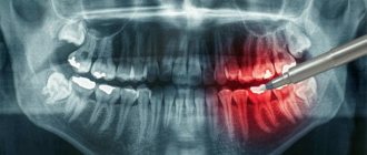
As a rule, the pathology has a specific severity, so it is diagnosed during a visual initial examination. Establishing provoking factors most often requires additional diagnostic procedures. In order to make a final diagnosis, as well as determine therapeutic methods, doctors may refer the patient for radiography. It will allow you to obtain complete information and create the correct treatment plan.
Causes of formation of defects of the lower jaw
Defects of the lower jaw are more often acquired than congenital. Violations of the integrity of the bone tissue of the lower jaw are observed for the following reasons:
- Large loss of bone tissue can prevent the regeneration of the alveolar process (bone edge); fusion of the edges does not occur due to the large gap, forming bone defects.
- In the case of a small loss of bone tissue (up to 10 mm), fusion of fragments of the lower jaw does not occur due to atrophy of soft tissues or the presence of foreign bodies, and a false joint is formed.
Treatment
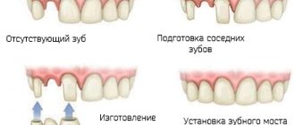
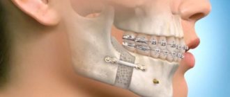
Preliminary treatment of the patient before dental implantation is performed includes sanitation of the oral cavity and special therapeutic measures. The pathology can be corrected with a bridge, plate or arch prosthesis. Depending on the size and nature of the anomalies, different types of structures are used:
- Non-removable bridges cover small flaws. Also, in such a situation, inlays and veneers are used.
- When large areas are damaged, correction is performed using bridge-shaped implants. Crowns can be metal-ceramic, metal, or other materials.
- Extensive lesions and edentia are treated with removable prosthetics. Implants are often made from acrylic.
- If there is no large group of units, it is practiced to install partially removable structures.
- For patients with an excessively deep bite and increased enamel abrasion, experts recommend clasp prosthetics.
Most clients note that modern designs and systems are convenient, practical, durable, and durable. Orthodontists are involved in prosthetics. High-quality materials allow you to achieve high aesthetic results. After selecting the structures, sanitation and removal of severely affected crowns and roots are performed. Professional cleaning of the oral cavity, removal of plaque and stone, and caries therapy are required.
The supporting units are prepared, after which a jaw cast is made. Using it, artificial crowns are created in the laboratory and their color is selected. The final prosthesis is made after fitting. It is fixed with cement. Removable structures are made by injection molding. After this, they undergo polymerization. When a whole group of units is missing and the patient does not agree to preparation, partially removable systems are used. Nylon structures solve minor pathologies, significant anomalies, and the problem of edentia. Ceramics is a popular material for prosthetics of the anterior group of teeth.
Preventative measures are very important. You should constantly observe the rules of oral hygiene and undergo systematic examinations by the dentist in order to promptly eliminate any resulting pathologies. It is easier to eliminate dental defects at the initial stage. This will help prevent serious complications.
Diagnosis of anomalies of the dentofacial apparatus
In modern orthodontics, various diagnostic methods are used, allowing specialists to make accurate diagnoses and select an effective treatment protocol. One of the diagnostic stages for malocclusion pathologies is the calculation of TRG.
TRG - teleroentgenogram involves taking an x-ray in the lateral and direct view, which clearly displays the soft tissues and bone structures of the skull.
How to calculate TRG orthodontics
: The interpretation of the TRG image is carried out by a diagnostician. The conclusion indicates the exact dimensions of the jaws, the angle of inclination of the teeth, the type of growth of the facial skeleton and other important details, on which the correctness of the diagnosis and the choice of treatment method largely depend.
In modern dental centers, TRG diagnostics are carried out using digital equipment. The images are interpreted using special computer programs that show the most accurate research results.
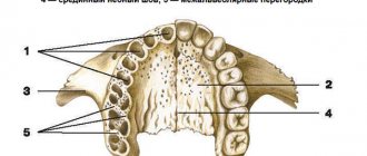
Systematization according to Eichner
A simpler option for dividing abnormal dental development than the previous one. Eichner hypothesized that in the human dental arch there are four so-called protective zones, called molars and premolars for the right and left sides. They are responsible for maintaining the interalveolar height.
In the case of dental defects, they can be divided into three groups:
- first group A - includes rows that have antagonist teeth in all four premolar and molar zones;
- second group B – in some protective zones there are no teeth, this can affect either one zone or several at once;
- third group C – antagonist teeth are lost in all 3 zones.
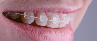
Clasp dentures
Clasps will correct both bilateral and unilateral terminal defects of the teeth.
Their main difference is the arc (from German “bugel”), which can be plastic or metal. In addition to the arch, artificial teeth and imitation gums in the form of a base are built into the clasp prosthesis. The entire system is secured in the mouth using hooks, telescopic crowns or mini-locks. However, the main indication for clasp prosthetics is the impossibility of implantation. A prerequisite is the presence of supporting teeth to fix the puller.
Anomalies of individual teeth
Anomalies in tooth size
Giant teeth are teeth with disproportionately large crowns. Giant teeth are more common in permanent dentition and less common in primary dentition. Typically, the incisors of the upper or lower jaws are giant, but other teeth can also be giant. The etiology of this anomaly in the shape of the teeth is known; disturbances in the developmental process are assumed, leading to the fusion of the rudiments of the teeth, as well as disruption of the activity of the endocrine system. Giant teeth can cause abnormalities in the position of other teeth, interfere with the eruption of neighboring teeth, and cause crowding of teeth. Sometimes they are located outside the dentition. The main disadvantage of giant teeth is their unusual appearance, which attracts the attention of others.
Small teeth are teeth with disproportionately small crowns that have a regular shape. Small teeth are found in permanent dentition. More often than other teeth, the incisors are small, especially the upper lateral ones. The etiology of this anomaly is unknown; it is assumed that the reasons for this discrepancy in the size of teeth and jaws may be hereditary, i.e. a combination of small teeth of one parent and large jaws of the other. Small teeth are usually spaced at large intervals and disrupt the harmony of the face.
Anomalies of teeth position
Vestibular deviation is the displacement of teeth outward from the dentition. One or more teeth of the upper or lower jaw may be in the vestibular position. Most often the incisors are displaced. The reasons may be: delay in the replacement of milk teeth, lack of space in the dentition, incorrect position of the tooth germ, the presence of supernumerary teeth, bad habits, and impaired nasal breathing.
High or low position of teeth - displacement of teeth in the vertical direction. In the upper jaw, supraocclusion is a high position of the tooth that does not reach the closure plane of the dentition, and infraocclusion is an advanced, low position of the tooth relative to the occlusal plane, and infraocclusion is a low position of the tooth. Sometimes there is supra- and infraocclusion of a group of teeth. The cause may be underdevelopment of the alveolar process or some mechanical obstacle.
Diastema is a gap between the central incisors and is much more common in the upper jaw. The reasons may be: low attachment of the powerful frenulum of the upper lip, the presence of a wide, dense bone septum between the central incisors, adentia, anomalies in the shape and size of the teeth, the presence of supernumerary teeth, incorrect placement of the frontal teeth, early loss of one of them.
Mesiodistal displacement of teeth is the location of teeth in front or behind the normal place in the dental arch. Both frontal and lateral teeth can shift. The cause is early loss of baby teeth, early loss of permanent teeth adjacent to a displaced tooth, incorrect position of the tooth germ, edentulism, and bad habits.
Oral inclination is the displacement of teeth towards the inside of the dentition, towards the tongue or palate. When tilted, the root of the tooth is located in the alveolar process, and only its coronal part is tilted orally; when displaced, the tooth is located body-wise outside the dental arch. One or more teeth may be in this position. The reasons are: delay in the replacement of milk teeth, early removal of milk teeth, narrowing of the dentition, incorrect position of the buds of permanent teeth, the presence of supernumerary teeth, shortened frenulum of the tongue, bad habits.
Rotation of the tooth around the longitudinal axis - most often the incisors of the upper and lower jaw are rotated along the axis. This type of anomaly causes aesthetic and functional disturbances. Sometimes axially rotated teeth injure the teeth of the opposite jaw and loosen them. The reasons may be a lack of space in the dentition due to its narrowing or underdevelopment of the alveolar process, delayed loss of a baby tooth, the presence of supernumerary or impacted teeth.
Crowded arrangement of teeth is a close position of the teeth, in which they stand with rotations along the axis and overlap each other due to lack of space in the dentition. The reason is underdevelopment of the alveolar process or the basal part of the jaw or the relatively large size of the teeth, as a result of which the teeth cannot be placed in the correct position.
Transposition of teeth is a positional anomaly in which the teeth change places. The reason is the incorrect formation of the rudiment of teeth.
Tremas are spaces between teeth. There are three types: physiological and pathological. Physiological problems refer to the characteristics of the primary occlusion in its second period; they arise as a consequence of jaw growth. Pathological threes are observed after the replacement of primary teeth with permanent ones in distal and mesial occlusions with protrusion of the upper or lower frontal teeth, with edentia, anomalies in the shape and size of the teeth, anomalies in the location of the teeth, and displacement of the teeth.
Anomalies in tooth shape
Spiny teeth are teeth whose crowns are shaped like a spike. The central and lateral incisors, as well as the lateral teeth of the lower and upper jaws, can have a tenon-like shape. The etiology is not clear; a violation of the development of tooth germs is assumed.
Ugly teeth are teeth of various irregular shapes, which are most often observed on the upper jaw, in its frontal area. The etiology is not clear; a violation of the development of the jaws and tooth buds is suspected.
Anomalies in the number of teeth
Adentia is a congenital absence of teeth and their rudiments. A distinction is made between partial and complete adentia. A violation of the development of the ectodermal germ layer, from which the tooth germ is formed, disturbances of the endocrine system are assumed, and heredity plays a certain role.
Supernumerary teeth are teeth that are excessive in number. They are most often located in the area of the front teeth. Supernumerary teeth are often tenon-shaped, but may have the shape of adjacent teeth. It is assumed that the cause is disturbances in the development of the epithelial dental lamina.
Examples of supernumerary teeth
Read more about the number of teeth people have in the article “How many teeth does a person have? How many teeth do you have?
Prevention
Modern dentistry treats wedge-shaped dental defects at any stage. The following recommendations will help minimize the risk of pathology:
- Use toothbrushes with soft bristles of the same length;
- do not chew seeds, nuts, grilled meats;
- brush your teeth with sweeping movements, from the gums to the bottom edge of the teeth, without pressing on the enamel;
- limit consumption of soda and natural juices;
- after fruits and wine, rinse your mouth and do not immediately use a toothbrush;
- keep chronic diseases under control;
- Have it professionally cleaned twice a year.
The prognosis is favorable, especially if you visit the dentist in a timely manner, when there is no damage to the deep layers of dentin. In advanced cases, it is almost impossible to do without prosthetics.
Types and stages
- Initial
- at the initial stage, the pathology does not manifest itself in anything; it looks like
microcracks
or
superficial abrasions
on the enamel that cannot be seen with the naked eye. - Medium
- as the destructive process spreads, these damages begin to deepen, grooves 0.2-03 mm deep are formed, taking the shape of a wedge.
There is increased sensitivity
to cold, hot, sour, sweet. - Developed
- the depth of the damaged area
is more than 0.5 mm
. The tooth reacts painfully to cold, hot food, and a toothbrush. - Severe
- destructive changes affect the deep layer of dentin, the defect is clearly visible, its depth and length
exceed 5 mm
. With intense load, there is a high risk of breaking off the coronal part.
Pathology differs from the carious process in terms of development, course, and clinical manifestations. In contrast to carious cavities with pigmented dentin, a non-carious cervical defect has shiny walls, smooth borders, and a hard bottom.
Complications
Since the cervical wedge-shaped defect does not cause pain for a long time and does not pose a threat as a source of pathogenic microflora, some patients have the impression that the disease does not require prompt medical intervention. This misconception is costly, since the pathology not only looks unsightly or makes hygiene difficult. Destruction of dentin in the cervical area contributes to the development of the following complications:
- Pulpitis, pulp dystrophy;
- gum recession, exposure of the neck of the tooth;
- crown fracture;
- periodontitis;
- loosening, tooth loss.
The most obvious complication is a violation of the aesthetics of the dentition. In the area of the non-carious cervical defect, the enamel is pigmented. The affected unit takes on an irregular shape - the tooth becomes like a tree trunk, half chopped down with an ax.
The dentist talks about the wedge-shaped defect - common questions:
Treatment methods
Treatment of a wedge-shaped defect can be general or local. General therapy involves the prescription of vitamins and microelements to strengthen the structure of the teeth. Local methods depend on the stage and extent of the destructive process. Treatment begins with identifying and eliminating the cause of the disease. In the case of a wedge-shaped lesion, a number of negative factors are important, so therapy is complex.
- Most often, a non-carious lesion occurs due to a violation of the occlusion, so the first stage is an orthodontic consultation
. The doctor will conduct a diagnosis and select an effective treatment regimen (selective grinding, braces, plates, mouth guards). If the bite is disturbed due to the loss of a dental unit, an implant or bridge is installed. - In case of a mild form of pathology (enamel damage 0.2 mm deep), in order to prevent the risk of deepening the defect is remineralized
, fluoridated, and applications of strengthening drugs are prescribed. - For moderate and developed
damage,
filling
. Restoration of a wedge-shaped defect is carried out with light-curing composites; the elasticity of the material compensates for the increased chewing load and ensures an accurate marginal fit of the filling. To improve aesthetics and prevent relapse of the disease, filled units can be additionally covered with veneers and lumineers. The type of microprosthesis depends on the thickness of the enamel layer. - Deep damage (0.4-0.5 mm), in which there is a risk of breaking off the coronal part, is eliminated by installing prostheses (metal-ceramic, ceramic, zirconium crowns). Loose, non-viable units are removed with further prosthetics
(implantation, bridge).
Hygiene products and dental cleaning techniques are selected individually. Use brushes with soft or medium-hard bristles and non-abrasive pastes. The cleaning technique eliminates horizontal brush movements on the front and back surfaces of the dentition. Use vertical, sweeping, circular movements.
Complex therapy involves the elimination of all provoking factors, including changes in diet, timely treatment of concomitant endocrine and gastrointestinal pathologies.
Features of the Zhulev system
Zhulev E.N. is a representative of the Nizhny Novgorod Medical Academy, a professor who in 1989 put forward his own version of the systematization of dentofacial abnormalities. His system consists of five classes:
- Class 1 – cavities, various types of depressions and pits that appear in the fissures;
- Class 2 – cavities that have appeared in the fissures, as well as the pits of the front teeth;
- Class 3 – cavities present on the surface of any front or lateral tooth;
- Class 4 – cavities in the cervical or gingival region;
- Class 5 – combined cases, including mixed variants from the previous specified problems.
There are many other, less well-known classifications by class, but they are used quite rarely, since the above systems of Kennedy, Gavrilov and some other doctors cope quite well with the task at hand - choosing the most suitable prosthesis for typical, frequently occurring dental defects.
Using such systems, dentists make life much easier for themselves and the patient and can choose the most optimal version of the prosthesis to be installed, which will take into account the anatomical characteristics of a particular person to the maximum.
Of course, each case is unique and it is not easy to predict all possible original combinations of deviations, but in most cases, these classification systems solve the tasks assigned to dentists quite well.
Causes
Any solid body, no matter how strong it may seem, is subject to abrasion under the influence of systematic mechanical loads. Tooth enamel is no exception. The tooth tries to compensate for the high load that occurs during chewing by microbending. That is, it bends, and the greatest tension occurs in the cervical region. Since the enamel here is the thinnest, it cracks with further peeling, forming a wedge-shaped defect. This is the mechanism for the development of pathology.
The main causes of a wedge-shaped defect:
- Violation of occlusion
- inharmonious closure of the dentition (abnormal bite) involves improper redistribution of the chewing load. The units that account for the main force during chewing are erased faster than their neighbors. The enamel cannot withstand it, it cracks and chips. - Improper cleaning
- the use of abrasive pastes, hard brushes, and improper cleaning techniques lead to excessive sanding of hard tissues. - Erosion of enamel
- an imbalance of diet in favor of acid-containing products (soda, juices, fruits, wine, fizzy candies, etc.) contributes to the demineralization of enamel and its rapid destruction. - Poor oral hygiene
- plaque and tartar weaken the protective properties of the enamel, which is a prerequisite for the development of a wedge-shaped defect.
Also, negative factors contributing to the development of the disease include gingivitis, periodontitis, gastrointestinal pathologies, and endocrine disorders.