Central lower incisor
Average age of eruption: 6-7 years
Average age of root formation: 9 years
Average length: 20.7 mm
Narrow and flat in the labial-lingual direction, the lower central incisor is the smallest adult tooth. Radiologically it is visible in only one projection and thus appears more accessible than it actually is. The crown, narrow in lingual projection, has a limited area for access. For gentle access formation, a fissure bur and a spherical bur No. 2 are used. The access cavity should be oval and performed on the lingual side.
The lower central incisor often has two canals. One study reported that 41.4% of mandibular central incisors studied had two separate canals, of which only 1.3% had two separate apical foramina [1]. After completing the access formation, the doctor must examine the tooth cavity to identify an additional canal. Endodontic treatment failures in the lower incisor area are most often associated with an unidentified canal, usually in the lingual location. To create conditions for the straight entry of endodontic instruments into the additional canal, access can be expanded in the incisal direction.
There is a great danger of perforation of the vestibular wall, but it can be avoided if the doctor remembers that it is almost impossible to perforate in the lingual direction, since the bur axis is in contact with the incisal edge. The slit-like lumen of the canal is so common that it can be considered a normal variant, and this requires special attention when cleaning and shaping.
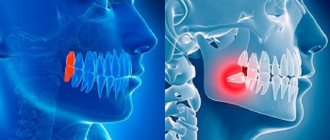
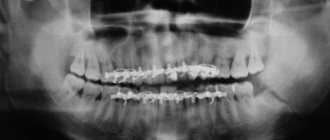
Lateral perforations and pulp anatomy will be discussed in an illustration of the lower second molar.
Diagnostics
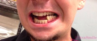
Most people aged 50 years and older experience the physiological type of abrasion. The doctor needs to determine in time when the natural process turns into a pathological one. One of the main methods remains measuring the actual parameters of crowns. In addition, it is important to carefully study the shape of the dental units and determine areas of contact with antagonists. The height of the coronal part is compared with age standards established scientifically.
Diagnostic measures include assessment of fissures and enamel condition. The doctor pays attention to the patient’s facial expressions and appearance. It is important to talk in detail about your complaints and describe your feelings.
To assess how correctly the temporomandibular joint functions, electromyography is indicated. X-ray examination, electroodontodiagnosis and other procedures are carried out.
Lateral lower incisor
Average age of teething: 7-8 years
Average age of root formation: 10 years
Average length: 21.1 mm
Very similar to the lower central incisor, so access cavity preparation follows the same principles.
Their similarity can cause rare but serious errors. Hasty installation of a rubber dam, identical fillings and carelessness can lead to preparation of the access cavity on the wrong tooth. This error can be prevented by marking the vestibular surface of the tooth with a felt-tip pen before applying the rubber dam.
Trauma, periodontal disease, carious lesions and malocclusion can lead to obliteration of the canal. When moving apically to identify the orifice, great care must be taken to prevent unnecessary destruction of the crown and root. Labial perforations were discussed in Illustration IX. If the bur is not oriented along the long axis of the tooth, there is a risk of lateral perforation. The situation becomes more complicated with the traumatic loss of an anatomical crown. Without anatomical landmarks, a lateral perforation can be easily made when moving in a coronal direction. To prevent this, access is carried out without a rubber dam so that the root can be palpated.
Lateral perforation with endodontic files and Gates-Glidden burs is facilitated by the presence of slit-shaped canals with a narrow, hourglass-shaped cross-section. To avoid vertical fracture along the approximal root wall, minimal expansion and preparation of the space for the abutment pin is indicated.
Apical curves and accessory canals are common in the lower incisors.
Complications
What to do if teeth are worn out depends on the provoking factors. As discussed above, the lack of timely therapy leads to cosmetic defects and disruption of the chewing process. In addition, there are other consequences that affect the quality of life of patients:
- inflammation of the gums in the interdental spaces;
- gingivitis;
- diseases of the temporomandibular joint;
- loss of teeth;
- damage to nerve fibers and muscle tissue;
- uneven load distribution leading to traumatic articulation;
- malocclusion.
Mandibular canine
Average age of teething: 9-10 years
Average age of root formation: 13 years
Average length: 25.6 mm
The canine of the lower jaw is more powerful and significantly wider than the incisors in the mesial-distal direction. It rarely causes treatment problems. The atypical form with two roots can be problematic, but is rare.
The access cavity is oval and can be expanded in the incisal direction to facilitate vestibular-lingual access. In the cervical region the canal is oval, in the middle third it is rounded. To completely clean its walls, directed instrumental action is necessary.
If there are two roots, one of them will always be easier to instrument. The other canal must also be opened and funnel-shaped in accordance with the first to prevent dentine filings from entering it and impairing access. Pre-bending the instruments during the initial approach will allow the clinician to walk along the walls of the buccal or lingual root until the tip of the instrument enters the orifice. Once a difficult-to-reach canal has been identified, every effort must be made to shape and create a funnel-shaped orifice to keep access open.
Let's sum it up
What to do if the lower front teeth or upper teeth are worn out, pain and other unpleasant symptoms appear? First of all, you need to go to the dentist to conduct a thorough diagnosis and identify the exact causes of the pathology. The longer the patient ignores the presenting signs, the more severe the consequences for him. In order not to encounter this phenomenon, it is necessary to take preventive measures, especially for those at risk.
Sometimes the enamel wears away due to age-related changes. In this case we are talking about a natural process. Which group should the problem be classified into, and whether treatment should be prescribed, should be decided solely by the doctor.
First lower premolar
Average age of teething: 10-12 years
Average age of root formation: 12-13 years
Average length: 21.6 mm
Often considered a mystery to endodontists, the mandibular first premolar, with its two canals separating at different levels of the root, can be very difficult to machine.
The crown consists of a well-developed buccal cusp and a small or almost non-existent lingual enamel protrusion. The approach is performed buccally from the central sulcus and directed along the long axis of the root to the central cervical region. The oval-shaped pulp chamber is opened using fissure burs with a cutting apex and elongated spherical burs No. 4 or 6. In teeth with one canal, the pulp cavity in the neck area has an almost circular cross-section, and in teeth with two canals it is oval.
One study reported that “at least 23% of first mandibular premolars have a second or third canal”[17]. The canals can split almost anywhere in the root. Due to the lack of direct access, cleaning, shaping and filling these teeth can be extremely difficult.
In a recent study, Vertucci [13] showed that the first lower premolar has one canal at the apex in 74.0% of cases, two canals in 25.5% and three canals in the remaining 0.5% of cases.
Classification and frequency (%) of canal types in first and second lower premolars
By Vertucci, F. J. Am. Dent. Assoc. 97:47, 1978.
Second lower premolar
Average age of teething: 11-12 years
Average age of root formation: 13-14 years
Average length: 22.3 mm
The second lower premolar, which is very similar in crown shape to the first premolar, has a less complex root.
Its crown has a well-developed buccal cusp and a much better formed lingual cusp than on the first premolar. The access is made slightly oval, wider in the mesial-distal direction. They begin to form access in the central sulcus with a fissure bur with a cutting apex, and then expand and form the contour of the burr hole with spherical burs No. 4 and 6.
Researchers reported that only 12% of mandibular second premolars studied had a second or third canal [17]. Vertucci [13] also showed that second premolars had one apical foramen in 97.5%, while only 2.5% of the teeth examined had two foramina.
An important circumstance that should not be forgotten is the anatomical location of the mental foramen and the vessels and nerves passing through it. Due to the proximity of these structures, an acute inflammatory process in the area of the lower premolars can cause temporary paresthesia. The exacerbation of the pathological process in this area is more severe and resistant to conservative treatment than in other areas.
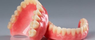
Crown of the tooth
The crown of a tooth (lat. corona dentis) is the part of the tooth protruding above the gum. The crown is covered with enamel - hard tissue, 95% consisting of inorganic substances and subject to the most powerful mechanical stress. There is a cavity in the crown of the tooth - dentin (hard tissue 2-6 mm thick) comes closer to the surface, then pulp, which fills both part of the crown and the root part of the tooth. The pulp contains the blood vessels and nerves of the tooth. Teeth cleaning and removal of dental deposits are carried out specifically from the crowns of the teeth.
Tooth neck
The neck of the tooth (lat. collum dentis) is the part of the tooth between the crown and root, covered by the gum.
Tooth roots
The root of the tooth (lat. radix dentis) is the part of the tooth located in the dental alveolus.
Fissure
On the chewing surface of the back teeth, between the cusps of the teeth there are grooves and grooves - fissures. The fissures can be narrow and very deep. The relief of the fissures is individual for each of us, but dental plaque gets stuck in the fissures of everyone. It is almost impossible to clean the fissures with a toothbrush. Bacteria in the oral cavity, processing plaque, form acid, which dissolves tooth tissue, forming caries. Even good oral hygiene is sometimes not enough. In this regard, fissure sealing has been successfully used throughout the world for 20 years.
Tooth enamel
Tooth enamel (or simply enamel, Latin enamelum) is the outer protective shell of the crown part of human teeth. Enamel is the hardest tissue in the human body, which is explained by the high content of inorganic substances - up to 97%. There is less water in tooth enamel than in other organs, 2-3%. Hardness reaches 397.6 kg/mm? (250-800 Vickers). The thickness of the enamel layer differs in different areas of the crown of the tooth and can reach 2.0 mm, and disappears at the neck of the tooth. Proper care of tooth enamel is one of the key aspects of human personal hygiene.
Dentine
Dentin (dentinum, LNH; lat. dens, dentis - tooth) is the hard tissue of the tooth, constituting its main part. The coronal part is covered with enamel, the root part of the dentin is covered with cement. Consists of 72% inorganic substances and 28% organic substances. Consists mainly of hydroxyapatite (70% by weight), organic material (20%) and water (10%), permeated with dentinal tubules and collagen fibers. Serves as the foundation of the tooth and supports tooth enamel. The thickness of the dentin layer ranges from 2 to 6 mm. Dentin hardness reaches 58.9 kgf/mm?. There are peripulpal (internal) and mantle (external) dentin. In peripulpal dentin, collagen fibers are located predominantly condensally and are called Ebner fibers. In mantle dentin, collagen fibers are arranged radially and are called Korff fibers. Dentin is divided into primary, secondary (replacement) and tertiary (irregular). Primary dentin is formed during the development of the tooth, before its eruption. Secondary (replacement) dentin is formed throughout a person’s life. It differs from the primary by a slower pace of development, a less systemic arrangement of dentinal tubules, a larger number of erythroglobular spaces, a larger amount of organic substances, higher permeability and less mineralization. Tertiary dentin (irregular) is formed during tooth trauma, tooth preparation, caries and other pathological processes, as a response to external irritation.
Dental pulp
Pulp (lat. pulpis dentis) is loose fibrous connective tissue that fills the tooth cavity, with a large number of nerve endings, blood and lymphatic vessels. Along the periphery of the pulp, odontoblasts are located in several layers, the processes of which are located in the dentinal tubules throughout the entire thickness of the dentin, performing a trophic function. The processes of odontoblasts include nerve formations that conduct pain sensations during mechanical, physical and chemical influences on dentin. Blood circulation and innervation of the pulp are carried out thanks to dental arterioles and venules, the nerve branches of the corresponding arteries and nerves of the jaws. Penetrating into the dental cavity through the apical opening of the tooth root canal, the neurovascular bundle breaks up into smaller branches of capillaries and nerves. The pulp helps stimulate regenerative processes, which manifest themselves in the formation of replacement dentin during the carious process. In addition, the pulp is a biological barrier that prevents the penetration of microorganisms from the carious cavity through the root canal beyond the tooth into the periodontium. The nerve formations of the pulp regulate the nutrition of the tooth, as well as the tooth’s perception of various irritations, including pain. The narrow apical opening and the abundance of vessels and nerve formations contribute to the rapid increase in inflammatory edema in acute pulpitis and compression of the nerve formations by the edema, which causes severe pain.
Tooth cavity
(lat. cavitas dentis) The space inside the tooth formed from the cavity of the crown and root canals. This cavity is filled with pulp.
Cavity of the tooth crown
(lat. cavitas coronae) Part of the tooth cavity, located under the crown and repeating its internal contours.
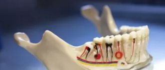
Tooth root canals
The root canal of a tooth (lat. canalis radicis dentis) is an anatomical space inside the root of a tooth. This natural space within the coronal part of the tooth consists of a pulp chamber, which is connected by one or more main canals, as well as more complex anatomical branches that can connect the root canals to each other or to the surface of the tooth root.
Nerves
(lat. nervae) Neuron processes passing through the apex of the tooth and filling its pulp. The nerves regulate the nutrition of the tooth and conduct pain impulses.
Arteries
(lat. arteriae) Blood vessels through which blood from the heart flows to all other organs, in this case - to the pulp of the tooth. Arteries nourish dental tissues.
Vienna
(lat. venae) Blood vessels through which blood returns from organs back to the heart. The veins enter the canals and penetrate the pulp of the tooth.
Cement
Cement (lat. - cementum) is a specific bone tissue that covers the root and neck of a human tooth, as well as the teeth of other mammals. Serves to firmly secure the tooth in the bone alveolus. Cement consists of 68-70% inorganic components and 30-32% organic substances. Cementum is divided into acellular (primary) and cellular (secondary). Primary cement is adjacent to the dentin and covers the lateral surfaces of the root. Secondary cement covers the apical third of the root and the bifurcation area of multi-rooted teeth.
Tops of tooth roots
(lat. apex radicis dentis) The lowest points of the teeth, located on their roots. At the tops there are openings through which nerve and vascular fibers pass to the tooth.
Apical foramina
(lat. foramen apices dentis) Places of entry of vascular and nerve plexuses into the dental canals. The apical foramina are located at the apex of the tooth roots.
Alveolus (alveolar socket)
(alveolar socket) (lat. alveolus dentalis) A notch in the jaw bone into which the roots of the tooth enter. The walls of the alveoli form strong bone plates impregnated with mineral salts and organic substances.
Alveolar neurovascular bundle
(lat. aa., vv. et nn alveolares) A plexus of blood vessels and nerve processes passing under the alveolus of the tooth. The alveolar neurovascular bundle is enclosed in an elastic tube.
Periodontium
Periodontium (lat. Periodontium) is a complex of tissues located in the slit-like space between the cementum of the tooth root and the alveolar plate. Its average width is 0.20-0.25 mm. The narrowest section of the periodontium is located in the middle part of the tooth root, and in the apical and marginal sections its width is slightly greater. The development of periodontal tissue is closely related to embryogenesis and teething. The process begins in parallel with the formation of the tooth root. The growth of periodontal fibers occurs both from the side of the root cement and from the side of the alveolar bone, towards each other. From the very beginning of their development, the fibers have an oblique course and are located at an angle to the tissues of the alveoli and cementum. The final development of the periodontal complex occurs after tooth eruption. At the same time, the periodontal tissues themselves are involved in this process. It should be noted that, despite the mesodermal origin of the constituent components of the periodontium, the ectodermal epithelial root sheath takes part in its normal formation.
Gingival grooves
(lat. sulcus gingivalis) Crevices that form where the crown of the tooth adheres to the gums. The gingival grooves run along the line between the free and attached parts of the gum.
Gum
Gums (lat. Gingiva) is a mucous membrane that covers the alveolar process of the upper jaw and the alveolar part of the lower jaw and covers the teeth in the cervical area. From a clinical and physiological point of view, the gums are divided into interdental (gingival) papilla, marginal gum or gingival margin (free part), alveolar gum (attached part), mobile gum. Histologically, the gum consists of stratified squamous epithelium and the lamina propria. There are oral epithelium, junctional epithelium, and sulcal epithelium. The epithelium of the interdental papillae and attached gingiva is thicker and can become keratinized. In this layer, there are basal, spinous, granular and stratum corneum. The basal layer consists of cylindrical cells, the spinous layer consists of polygonal cells, the granular layer consists of flattened cells, and the stratum corneum is represented by several rows of completely keratinized and nucleated cells that are constantly exfoliated.
Mucous papillae
(lat. papilla gingivalis) Fragments of gums located at their elevation in the area between adjacent teeth. The gingival papillae are in contact with the surface of the dental crowns.
Jaws
(lat. maxilla - upper jaw, mandibula - lower jaw) Bony structures that are the basis of the face and the largest bones of the skull. The jaws form the mouth opening and determine the shape of the face.
First lower molar
Average age of teething: 6 years
Average age of root formation: 9-10 years
Average length: 21.0mm
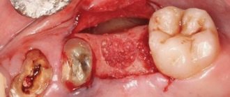
The mandibular first molar erupts earlier than other permanent teeth and most often requires endodontic treatment. It usually has two roots, but sometimes three roots are found, with two canals in the mesial root and one or two canals in the distal root.
The distal root is easily accessible for endodontic cavity preparation and mechanical treatment.
The doctor can directly see the opening(s) of the canal. The distal root canals are wider than those of the mesial root. Sometimes the mouth is wider in the buccolingual direction. This indicates the presence of two channels or a slit-like channel with a complex network-like configuration that can complicate cleaning and shaping.
The mesial roots are usually curved, with the greatest curve in the mesiobuccal canal. The orifices of the canals at the bottom of the pulp chamber are usually clearly separated from each other and located buccally and lingually relative to the upper tubercles.
The tooth often undergoes extensive filling. It almost always experiences a strong chewing load, so the coronal pulp cavity can be obliterated. It is easiest to identify the mouths of the distal canals.
Then the mouths of the mesial canals are found, which will be located in the above locations in the same horizontal plane.
Since the mouths of the mesial canals lie under the mesial tubercles, they may not be detected during normal cavity preparation. In this case, to determine their location, it is necessary to remove the hard tissue of the tubercle or filling. During access preparation, overhanging molar cusps need to be ground down [15]. Remember that this tooth, like all other lateral teeth, after endodontic treatment requires complete restoration of the entire occlusal surface area. Therefore, to identify anatomical landmarks and orifices, it is better to make a wider access cavity than to skip one or more channels for the sake of “sparing” preparation, which may cause failure.
Skidmore and Bjoradal [11] found that approximately one third of the mandibular first molars examined had four root canals. If there are two canals, “they either remain separate with separate apical foramina, or unite and form a common apical foramen, or communicate with one another by transverse anastomoses partially or completely... If the tooth, instead of the usual triangular shape, had a more rectangular shape, this would allow better vision and explore a possible fourth canal in the distal root.”
In the area of bifurcation of the roots of the lower molars there are several orifices of additional canals [9]. They are usually impossible to clean and shape, and are rarely visible except incidentally on radiographs when they are filled with root cement or heated gutta-percha during treatment. It would be correct to assume that if irrigation solutions tend to clean the canal from protein decomposition products, then the area of root bifurcation in the pulp chamber must be thoroughly cleaned (remove denticles, etc.) so that the solutions can reach the small mouths of the canals.
All infected dentin, leaking fillings, and pulp denticles must be removed before endodontic treatment begins. It is recommended to completely cover the cusps with a restorative structure after endodontic treatment.
Signs of distal bite
It is quite possible to identify occlusion independently, not only by the incorrect position of the teeth when closing, but also by other signs: by certain facial features, features of facial expressions, even by body position or behavior. There are so many nuances that they can be divided into 3 main types.
Intraoral
It is relatively easy to determine the anomaly by looking at the upper frontal incisors. Normally, they should overlap the lower ones by about one third. In case of violations, they are lowered more strongly, and even pushed forward - so much so that there is some distance between the rows, in scientific language it is called the sagittal gap.
External
You should think about how to correct a distal bite in an adult (or at least make an appointment with an orthodontist for a consultation) if:
- the facial profile is convex, “bird-like”;
- the chin is disproportionately small, sloping, with an impressive fold;
- the nose, small in itself, protrudes significantly;
- in a relaxed state, the lips do not close.
The problem is also reflected in posture: the body is always slightly tilted, the neck seems longer, the stomach sticks out, the shoulders are hunched.
Functional
If a person has this disorder, it is more difficult for him to breathe through his nose, chew food, swallow, even speak (often unclear diction, lisp). Although he usually does not notice these inconveniences, since the anomaly develops gradually, from the age of 4-6 years, and the body simply adapts to it.
Second lower molar
Average age of teething: 11-13 years
Average age of root formation: 14-15 years
Average length: 19.8 mm
The crown is somewhat smaller in size than the first molar, and the more symmetrical second lower molar is characterized by a close arrangement of roots. The roots gradually converge distally and have closely spaced apices.
Exposure is made at the mesial portion of the crown with access extending only slightly distal to the central sulcus. After trephination of the cavity with a fissure bur with a cutting apex, an elongated ball-shaped bur is used to expand it until free access is achieved. The distal root angles often allow for a smaller opening than the lower first molar.
Particular attention should be paid to the shape of the mouth of the distal canal. A narrow oval orifice indicates the presence of a slit-like lumen in the distal canal, which requires more careful treatment.
All carious tissue, leaking fillings and denticles should be removed and replaced with a suitable temporary filling prior to endodontic treatment.
The lower second molar is most susceptible to vertical fractures. After preparing the access, but before starting endodontic treatment, the clinician should examine the floor of the pulp cavity using fiber optics. For mesial-distal crown or root fractures, the prognosis is extremely unfavorable.
To avoid vertical fractures after endodontic treatment, it is necessary to completely cover the cusps with a restorative structure.