When the upper and lower jaws close, the dentition interacts with each other in a special way. This interaction is called "bite". How correctly the dentofacial apparatus functions depends on the nature of the closure of the dentition.
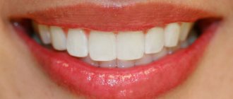
A normal or physiological bite ensures the high-quality functioning of the dental system. A person with a normal bite has clear diction, free breathing, and an attractive smile.
Pathological occlusion is a serious deviation in which the functioning of the masticatory muscles and the temporomandibular joint is impaired. As a result, the patient feels discomfort when chewing food, his teeth deteriorate, his appearance deteriorates, and his quality of life is disrupted.
In order to describe the types of disorders in the structure of the dentofacial apparatus, a classification of occlusions is carried out. There are three degrees in dentistry.
- The first degree assumes a normal or physiological bite. The main signs of a normal bite: there are no pronounced gaps between the closed upper and lower teeth, their contact occurs symmetrically, the central line of the jaws coincides with the midline of the face.
- The second and third degrees indicate the presence of deviations from the norm of varying intensity.
There are different types of normal and pathological occlusion.
Types of normal occlusion
According to statistics, no more than 30% of the population have a normal bite. There are several types of teeth arrangement that meet all the signs of a correct bite:
- Orthognathic. The teeth of the upper jaw overlap the lower teeth by a third of the crown size. When the jaws close, close contact occurs between the teeth.
- Straight. The cutting edges of the upper and lower teeth are in close contact. Correct occlusion of molars and premolars is mandatory.
- Biprognathic. The upper and lower incisors have a slight inclination towards the vestibule of the mouth. There is contact between the incisors, and the upper canines slightly overlap the lower ones.
- Progenic. The teeth of the lower jaw are pushed forward relative to the upper ones. The closure is maintained.
With any type of normal positioning of teeth, bite correction is not carried out, since the load on the teeth is carried out evenly. Food is chewed thoroughly, the temporomandibular joint is not overloaded.
Classification
In orthodontic practice, various assessment criteria are used to determine which dental bite is correct and how to correct existing anomalies. The ideal situation, in which nothing needs to be corrected, is characterized by the following indications:
- Semi-elliptical shape of the upper and parabolic shape of the lower arc.
- Small - no more than a third - overlap of the vestibular surface of the mandibular elements.
- Contact of antagonists during closure and absence of obvious gaps.
Compliance on all points is quite rare, but in most cases the deviations are minimal and do not require medical intervention. Often the cause of imbalance is not congenital defects, but bad habits, some of which are formed at an early age.
Types of correct bite
In accordance with the generally accepted classification, there are four main types of occlusal relationships, characterized by the absence of problems and not having a negative effect on the jaw region.
Orthognathic
The optimal, from an aesthetic point of view, is a condition in which all elements of the dentition have an even shape, are located in the correct manner, without gaps or deviations from the midline.
Straight
A common phenomenon, the main feature of which is the lack of overlap - the cutting edges of the incisors meet along a line, which causes a specific type of smile, but is not considered as a pathology.
Biprognathic
Another type of even dentition without threes and diastemas, the distinctive feature of which is a slight deviation of the vertical growth vector of the units. With this development, the crowns are slightly tilted forward.
Progenic
In a normal state it is not accompanied by abnormal manifestations. The identifying feature is a slight frontal protrusion of the lower units.
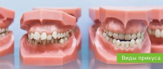
Types of abnormal bite
Any type of abnormal bite is characterized by the presence of gaps between the teeth of the upper and lower jaw. Lack of contact creates problems when chewing food, diction is impaired, and the oval of the face changes.
- Open is characterized by the presence of areas of non-closure in the anterior or lateral areas of the dentition. It is manifested by the presence of a gap between the teeth of the upper and lower jaw, non-closure of the lips, leading to disruption of the functions of chewing, breathing, and diction.
- Deep . The teeth of the upper dentition overlap the lower ones by more than 30% of the crown height. The most common type of abnormal bite. A strong degree of pathology leads to constant trauma to the mucous membrane of the gums, which is why this type of bite is called traumatic.
- Distal . It is characterized by protrusion of the upper dentition in relation to the lower one due to abnormal development of the upper jaw (or underdevelopment of the lower jaw). External signs are a sloping chin, shortening of the upper lip and retraction of the lower lip. The upper and lower incisors do not have contact, and the lateral ones do not close correctly.
- Cross is characterized by the crossing of the dentition when closing. Characteristic signs are facial asymmetry as a result of uneven development of the jaw areas. The patient complains of diction defects, accidental biting of the inner cheeks and pain when chewing.
- Mesial . It is characterized by the advancement of the lower dentition as a result of underdevelopment of the upper jaw or abnormal development of the lower jaw. Poor contact between the teeth of the lower and upper jaw leads to dysfunction of chewing and speech. Externally, the anomaly is manifested by the presence of a massive chin, a receding upper lip, and a concave face profile.
Dividing each type of abnormal bite into separate subcategories helps determine the degree and nature of the pathology.
If there are even minimal deviations, you should consult an orthodontist for advice. Pathology of the dentofacial apparatus has a tendency to progress and develop complications from the gastrointestinal tract.
Causes of pathological bite
The formation of occlusion is a long and complex process that begins at the stage of intrauterine development, continues in early childhood and ends after the end of puberty. Pathological occlusion is the result of long-term influence of a number of unfavorable external or internal factors at different periods of a person’s life:
- Genetic factor. A genetically determined anomaly is formed when there is a congenital discrepancy in the size of the teeth and dentofacial bones (crowding of teeth or the presence of spaces between teeth) or a hereditarily determined abnormal shape and size of the lower and upper jaws.
- The influence of negative factors on the body of a pregnant woman. The formation of the dental apparatus in the fetus can be influenced by bad habits, poor nutrition and diseases suffered by a woman during pregnancy.
- Birth injury. During childbirth, the baby may receive injuries leading to displacement of the temporomandibular joint.
- Negative factors affecting the baby’s body in the first years of life. The reason for the formation of a distal bite may be the incorrect selection of nipples or the late introduction of solid foods into the diet.
- Bad habits and behavioral factors in early childhood. The habit of sucking a pacifier or finger, or sleeping with the head thrown back can cause a child to develop a deep bite.
- Factors influencing the formation of anomalies in later life. These include carious lesions of the teeth, ENT pathologies, deviated nasal septum, and diseases of the skeletal system.
Treatment of abnormal bite should begin as early as possible. Early diagnosis and therapy make it possible to restore the normal functioning of the dentofacial apparatus more effectively. Doctors at the Consilium-Dent use specially developed diagnostic tests and tables. They are used both for direct examination of the oral cavity and for diagnosis using dental casts.
The need for a particular therapeutic measure is determined by a specialist based on the diagnostic results on an individual basis. You cannot resort to self-medication in this situation. This can lead to activation of the pathological process.
Eruption of canines in infancy
The order of formation of the primary occlusion provides for the appearance of fangs after the incisors and the first chewing units have erupted. The upper elements emerge at the age of 1.2-1.5 years, which is slightly earlier than the period of formation of the lower molars. Late eruption is determined by the deep position of the units in the structure of the jaw, which also determines the characteristic painful sensations that may occur during growth.
To relieve the discomfort experienced by the child, the following techniques are recommended:
- Massage of gum tissue - before the procedure, you need to thoroughly wash your hands, then use your index finger to massage the area above the canine for a couple of minutes. It is recommended to repeat the massage several times a day;
- The use of teethers that provide a cooling effect - a product filled with distilled water does not pose a danger to the baby’s health even if the baby manages to chew the shell;
- Applying anesthetic gels that relieve inflammation within a few minutes;
- For nasal congestion, use children's nasal medications that promote vasoconstriction;
- At elevated temperatures (more than 38 degrees) - take fever medications, available in the form of syrup or suppository.
It is worth emphasizing that the use of medications must be agreed with a doctor, who will determine the appropriate list, as well as the duration of the course.
Common types of malocclusions
The most common anomalies of the dental system include deep bite. A severe degree of pathology is characterized by a situation where the upper row of teeth completely overlaps the lower row. When the jaws are closed, the upper incisors can injure the mucous membrane of the lower gums. Therefore, this type of anomaly is called a traumatic bite.
A deep bite is accompanied by a violation of facial aesthetics, impaired diction, poor quality of chewing food, and increased abrasion of the enamel.
There are two degrees of development of pathology. They are caused by various provoking factors and are characterized by different forms of manifestation:
- Primary deep bite is often due to genetic reasons and is determined by the special structure of the facial bones. It manifests itself as pathological changes in the area of the dentition, alveolar process and basal part of the jaws. The movement of the jaws is blocked, and the patient experiences significant discomfort when chewing.
- Secondary deep bite is acquired in nature and is a consequence of a disease or injury. As a result of a decrease in the interalveolar distance and the lower third of the face, a deep traumatic closure of the incisors develops.
Another type of pathology classification is carried out depending on the nature of the interaction of the lateral teeth.
- A deep distal bite is characterized by the overlap of the lower row of teeth with the upper ones by more than one third of the length of the crown with a violation of the relationship of the lateral teeth. Characteristic disorders include difficulty biting and chewing, breathing problems, and pain in the temporomandibular joint.
- Deep neutral bite: when the overlap of the lower front teeth with the upper ones is disturbed, while the position of the lateral teeth is not disturbed.
If you experience even minor discomfort when chewing, you should immediately contact a specialist and follow his recommendations.
The presence of a malocclusion or a predisposition to it is determined by an orthodontist after an initial examination using special tests. Self-diagnosis does not provide an objective picture of the condition of the dentofacial apparatus, since different pathologies may have the same symptoms. Treatment in this case will be radically different.
If there are no problems with your bite, you should visit the orthodontist for a preventive examination approximately once every six months. The doctor will determine whether there is a tendency to a particular anomaly even before the problem is noticeable.
The importance of routine observations in normal occlusion
In children with an orthognathic or straight bite, the arrangement of dental units in a row may change due to untimely (early or late) change of primary teeth. During a preventive examination, the orthodontist will identify the presence of abnormalities in the rudiment itself. The following types of preventive measures are carried out:
- Clinical examination (allows us to identify existing anomalies and risk factors).
- Formation of groups of patients for further clinical observation.
- Drawing up a plan of preventive and therapeutic measures (pediatricians of all profiles participate).
In adult patients with normal dentition, malocclusion can develop under the influence of external and internal unfavorable factors. The main reasons that increase the risk of developing dental anomalies in adults are:
- carious process;
- loss of teeth;
- various types of prosthetics;
- prosthetics errors;
- bad habits.
During the examination, the dentist will identify the presence of predisposing factors, after which he will develop a scheme for carrying out preventive measures to maintain a normal bite. Selection is carried out on an individual basis, taking into account the condition of the dental system and the physiological characteristics of the patient’s body.
Treatment of malocclusions
Before prescribing treatment procedures, the orthodontist assesses the condition of the oral cavity, carefully examines the depth of the problem and the presence of concomitant pathologies, taking into account the patient’s age. Treatment of dental anomalies is carried out using conservative and radical methods:
- Braces are the most popular method of bite correction. This orthodontic method has the widest range of applications and is suitable for solving any problem of malocclusion. Treatment with this method is long and includes preparatory, main and retention (recovery) periods.
- Aligners. Removable structures are used to treat the initial stages of deviation. With their help, a gentle correction of the dentoalveolar apparatus is carried out, changing one set of mouthguards to another.
- Application of veneers, onlays, crowns. Weak areas of the dentition that are susceptible to caries are protected by using orthopedic structures. Chipped areas of enamel are restored using ceramic onlays and veneers.
- Surgical intervention is performed to treat late stages of dental anomalies (mainly of a hereditary nature). During the operation, the position of the jaws is changed, their size is reduced or increased.
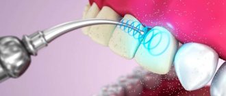
Laser therapy is performed as an auxiliary treatment to help speed up regeneration and reduce the risk of complications.
Types of prevention of malocclusions
Prevention of malocclusion in childhood is very important, as it determines whether the child will have problems in the future. Adults should monitor the eruption of permanent teeth in children and visit an orthodontist with the child for timely detection of anomalies and bite correction.
Prevention of malocclusions involves eliminating bad habits that cause one or another anomaly: lip biting, finger sucking in children. To do this, special plates are used that block the ability to use a bad habit. In adults, such methods help cure abnormalities in the early stages.
It is important to carry out sanitation of the oral cavity in a timely manner and periodically come for medical examinations to the dentist. Children from the age of six months should be taught to chew solid food.
A good method of prevention is specially designed therapeutic exercises. When performing exercises, the patient puts the correct load on the jaw muscles, which contributes to the harmonious development of the maxillofacial system.
International Viola system: a convenient diagram of the arrangement of a person’s teeth by numbers
The described method is very convenient and most common in dentistry. It has received international recognition and has been generally accepted among dentists since 1971, called the two-digit Viola system. The convenience of such a system lies primarily in the fact that there is no need to create a special map of a person’s teeth, the numbering is easily calculated in the mind and information about the condition of certain dental units of the patient can be easily conveyed in an oral conversation, by telephone or by e-mail.
However, many people, having read this far, may say: but our teeth were counted completely differently, and the map contains completely different designations! That’s right, because in addition to the Viola system, there are several other systems that can be used by dentists.
Bite formation in children
At the stage of bite formation in children, there are three most important periods:
- The milk bite begins to form at six months of age and continues until 3 years of age. This is the initial stage during which the foundation for the formation of a permanent bite is laid.
- The change to a permanent dentition lasts up to 9 years. During this period, the jaws actively develop and a permanent bite begins to form. During this period, when the dentition consists of baby and permanent teeth, children often develop malocclusions. Common causes are a violation of the pairing of teeth erupting with an unequal degree of development of the jaw sections.
- The formation of a permanent bite occurs from 12 to 15 years. The replacement of baby teeth ends, permanent teeth complete their growth and are installed in their permanent places.
Prevention of malocclusion in childhood is very important, as it determines whether the child will have problems in the future. Adults should monitor the eruption of permanent teeth in children and visit an orthodontist with the child for timely detection of anomalies and bite correction.
Histological structure of the tooth
The special hardness of teeth is due to their structure. The outside of the crown is covered with enamel, the basis of which is hydroxyapatite crystals. 96% of the enamel consists of inorganic substances, which is why it is so hard (which, however, does not mean that experiments can be carried out on teeth, testing their strength). But dentin, which is located under the enamel, has much less inorganic substances - about 70%. It is softer, and therefore is destroyed by caries more than enamel. Inside the tooth there is a cavity filled with a neurovascular bundle, or pulp. The entire rich spectrum of pain during the deepening of the carious process is associated with it. In addition to nerve fibers, the pulp also contains a large number of blood vessels, through which infection from the affected tooth spreads throughout the body. This is why it is so dangerous to leave dental diseases untreated.