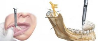
Tuberal anesthesia is performed to block the posterior superior and alveolar nerves involved in the formation of the dental plexuses. After such anesthesia, it becomes possible to carry out painless manipulations in the area of the upper molars.
In addition, this type of anesthesia can be used to provide local anesthesia to the area where surgery is planned. Tuberal anesthesia is also known as conduction or peripheral anesthesia.
Features and principle of action of conduction anesthesia
Conduction anesthesia is a local anesthesia and involves the introduction of a drug into the peripheral region of the nerve that ensures the functioning of this part of the body. Dentists in most cases use conduction anesthesia to treat teeth located in the lower jaw.
The technique is aimed at blocking impulses transmitted from nerves to the brain. As soon as the drug enters the nerve, sensitivity is reduced to a minimum level. The duration of action of the anesthetic depends on the substance used.
A number of features of conduction anesthesia can be distinguished:
- The injection is carried out with drugs, the concentration of which reaches 2%. Compared to other methods of pain relief, the content of active substances is at a high level.
- Loss of sensitivity does not occur at the site of drug administration, but in the nerve that is located in this area of the body.
- Conduction anesthesia has a minimal number of complications and side effects.
- The method allows you to control the duration of pain relief and the area of effect of the drug.
Tuberal anesthesia according to the method of P.M. Egorova (blockade of the posterior superior alveolar nerves)
Extraoral method The basis of the method of blocking the posterior superior alveolar nerves according to P.M. Egorov is to determine the individual anatomical landmarks of the injection site, the direction of insertion and the depth of immersion of the needle. To perform the method P.M. Egorov recommends using the extraoral route of needle insertion. With extraoral methods of local anesthesia, the surgical field is more accessible for review and selection of the injection site; there are all conditions for reliable antiseptic treatment of the injection site.
Indications for conduction anesthesia
- Implantation.
- Carrying out surgical intervention (removal of cysts, granulomas, ulcers and benign neoplasms).
- Therapy of dental diseases.
- Preparation before surgery.
- Removal of teeth or roots.
- Inflammation in the oral cavity.
- Carrying out preventive procedures to prevent periodontal diseases and caries.
- Inability to use general anesthesia.
Anesthesia at the incisive foramen
Such anesthesia is carried out by neutralizing the nasopalatine nerve in order to anesthetize the anterior region of the mucous membrane of the hard palate in the area of the anterior teeth.
The incisive foramen is located between the front incisors 7-8 mm from the edge of the gum at the intersection of the lines that connect the distal edges of the median palatal suture and the necks of the canines.
Anesthesia technique: the patient is in a chair, his head is thrown back, his mouth is open wide. The needle is inserted into the mucous membrane near the incisive opening to a depth of 3-4 mm. The anesthetic solution is released slowly. The process of inserting a needle into the papilla itself is very painful, so thin needles are used for such injections, and additional pain relief is performed. The anesthetic effect is achieved within a few minutes.
Contraindications to conduction anesthesia
- Poor blood clotting. This feature can lead to bleeding.
- Allergy to used painkillers.
- Disorders of the cardiovascular system (arrhythmia, ischemia, tachycardia, heart valve defects, etc.). These pathologies are not compatible with adrenaline, which can be used to constrict blood vessels.
- The presence of inflammation or ulcers at the injection sites of the anesthetic drug.
- Open wounds, infectious inflammations and contaminated wound surfaces on the patient’s extremities.
- Childhood.
- Mental disorders, unstable psycho-emotional background of the patient.
- Patient refusal to use this type of anesthesia.
Contraindications and side effects
Contraindications to dental anesthesia are:
- Allergic reactions to substances included in anesthetics;
- History of cardiovascular diseases;
- Diabetes;
- Pathology of the endocrine system organs;
- Some types of severe injuries of the maxillofacial area.
Side effects of anesthesia in dentistry
Whether the doctor is a professional in his field, complications with local anesthesia in dentistry are very unlikely. There are some things that worry patients after dental treatment and, in principle, are a variant of the norm: swelling of the lip or swelling of the cheek, pain in the gums or even a headache for several hours.
However, all these symptoms should go away within 1-3 days after treatment. If you see that the situation is not improving or even worsening, contact the dentist who performed the procedure.
Methods of administering conduction anesthesia
Methods of administering conduction anesthesia can be divided into 2 groups:
- Extraoral. Used in the presence of inflammatory foci in the oral cavity. These include:
- Submandibular. The pain medication blocks the inferior alveolar nerve.
- Podzykulova. The drug is injected under the edge of the cheekbone arch.
- Mandibular. The mandibular foramen loses sensitivity.
Once the anesthetic is injected into the treated area, the numbing effect occurs within 15 minutes. The duration of the anesthetic effect depends on the type of medication used.
- Intraoral. These include:
- Apodactylous. The alveolar nerve is blocked.
- Torusal. The anesthetic is injected into the mandibular ridge. Molars and premolars lose sensitivity.
- Tuberal. Anesthesia is injected into the area of the molars located on the upper jaw.
- Infraorbital. It is used to block sensitivity in the anterior wall of the maxillary bone, mucous membrane and alveolar process.
- Palatine. The palate and alveolar process are anesthetized.
- Incisive. The anesthetic is injected between the upper canine and incisor. The hard palate, nasopalatine nerve and soft tissues surrounding the tooth become numb.
When administered intraorally, the effect occurs 10 minutes after injection.
Preparation for conduction anesthesia
Before performing the operation under general anesthesia, it is necessary to undergo a standard examination. During diagnosis, special attention should be paid to the patient’s neurological condition, his psyche and behavior.
Before the procedure, the specialist will explain in simple, accessible words about the sequence and method of administering anesthesia, as well as the sensations that may accompany the patient during pain relief. In addition, the doctor will definitely check the patient’s reaction to the anesthetic drug to make sure there is no allergic reaction.
The anesthesiologist must follow the rules when administering conduction anesthesia:
- First of all, it is necessary to numb the skin using a gentle method.
- The needle must be fixed in a strictly defined place in order for the anesthetic to reach the desired area.
- Paresthesia must be present. It indicates that pain relief was carried out correctly and sensitivity has decreased.
- Drugs should be administered in portions with aspiration samples to prevent them from entering the bloodstream.
- During anesthesia, it is necessary to continuously monitor the patient's blood pressure and pulse.
- The room in which the procedure is performed must be equipped with all the necessary instruments for pain relief, as well as means of resuscitation and treatment of possible complications.
Stages of administration of conduction anesthesia
- A consultative appointment, during which a specialist determines the presence of chronic diseases and evaluates test results that may become contraindications to pain relief.
- Detection of nerve endings that need to be anesthetized.
- Disinfection of the skin puncture area with a special solution.
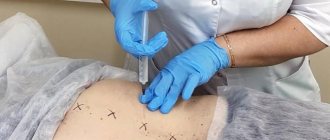
- Injection of anesthetic into certain areas. At the same time, the rate of administration of the anesthetic is observed and the volume of the injected substance is controlled.
- Monitoring the patient after pain relief.
- Before the start of surgery, the patient is asked control questions to determine the level of sensitivity.
The duration of the analgesic effect depends on the drug used and its dosage. Modern painkillers retain their effect for up to 40-60 minutes.
Area of influence
Tuberal anesthesia affects the area from the gums of the first molar to the first molar. The needle enters the space between the optic nerve and the fat pad. In some cases, it is possible to reduce or increase the working space.
The anesthetic is injected near the alveolar ending, located behind the fixed jaw bone. The action extends to adjacent mucous tissues, the posterior part of the alveolar process and directly to the molars.
Drugs used
The success of conduction anesthesia depends not only on the technique used, but also on the drugs used. The following medications are used for this pain relief technique:
- Solutions of the Articaine series. They have a long lifespan and high efficiency. These include:
- Ultracaine.
- Septanest.
- Ubistezin.
- Lidocaine. It is a highly effective anesthetic and at the same time contains a minimal amount of toxic substances.
- Melivacaine. The duration of action of this drug is about 40 minutes. Melivacaine has no contraindications for use.
- Novocaine. This is perhaps the most frequently used remedy. It is characterized by low toxicity and rapid hydrolysis in the body.
Intraoral access
Using the thumb and forefinger of the left hand, push the upper lip up and out, and with the middle finger hold the projection of the infraorbital foramen. With this type of access, it is located at the intersection of two axes. One of them, horizontal, runs 5-7 mm below the lower orbital margin, and the second, vertical, runs on the corresponding side along the axis of the second upper premolar. The needle must be inserted, retreating 5 mm from the upper edge of the attachment of the transitional fold between the lateral and middle incisors. Then it is pushed upward, forward and outward in the direction of the lower orbital foramen until it stops at the bone. And only there the anesthetic solution is released.
Advantages and disadvantages of conduction anesthesia
Advantages:
- The drug can be administered outside the surgical site.
- Anesthesia has a long-lasting effect. A specialist can monitor the duration of action of the medication.
- To achieve the desired effect, small doses of painkillers are sufficient.
- There is no deformation of soft tissues in the operated area.
- When exposed to conduction anesthesia, salivary activity decreases.
- The technique is safe, so it can be used from the age of 12, and also in some cases in pregnant and lactating women.
- The cost of the procedure is affordable.
Flaws:
- In order for anesthesia to pass without complications, the specialist must have certain skills and sufficient experience.
- There is a high probability of the anesthetic entering a blood vessel.
- Restricted use by age (only from 12 years old).
- Risk of injury to nerves or blood vessels.
Possible complications of local anesthesia
Local anesthetics are the most effective and safest drugs for pain control.
Dentists in the United States use more than 300 million local anesthetic carpules annually, but reports of serious complications are low. Complications are divided into local and systemic.
Local complications occur at the site of needle insertion or anesthetic application, while systemic complications involve the body as a whole. Local complications include needle breakage, paresthesia, trismus, hematoma, and facial nerve paresis; to systemic ones - a psychogenic reaction to the very fact of injection, allergies and drug overdose (toxic reaction). Below is a brief description of these potential complications. Local anesthesia is a primary method of pain control in dentistry. It is impossible to imagine a day in surgical practice without these wonderful drugs, which prevent harmful stimuli generated during surgery from entering the human brain, where they are interpreted as pain. Although other methods of pain control are available, including general anesthesia, hypnosis, acupuncture, and dental electroanesthesia, none are as reliable or safe as local anesthesia. It has been established that, despite the fact that, according to the most conservative estimates, more than 300 million capsules of local anesthetic drugs are used annually in the United States for dental procedures, the number of serious complications associated with their use is minimal. However, when prescribing drugs, problems can and do arise. Two types of problems can be distinguished: 1. Those arising locally, at the injection site. 2. Systemic reactions associated with the use of the drug. Local complications The etiological factor for most local complications is either trauma associated with the advancement of the needle through the soft tissue at the injection site, or the solution injected into this area. Most local complications associated with the use of local anesthetics are short-lived, although they cause inconvenience and concern for the patient. Some last only a few seconds (injection pain and burning), others last hours or even days (trismus, hematoma, infection, swelling, facial paresis), while paresthesia, which usually resolves within a few days, can in rare cases be permanent . Needle breakage When using modern disposable stainless steel dental needles, it is extremely rare to encounter a needle breakage during an intraoral injection. The most common cause of this is unexpected movement by the patient as the needle enters the muscle or contacts the periosteum. Smaller needles, such as 25 or 27, break more often than larger needles (such as 30). Previously bent needles break more often. It is clinically irrational to bend needles, except perhaps for intrapulpal or intraligamentary anesthesia. Broken needles that can be easily removed do not pose any danger. Only needles that have been inserted to the full length of the tissue may not be removable if they break. One of the basic rules when performing an injection is the following: do not insert the entire length of the needle, except in cases in which it is absolutely necessary for the successful implementation of this technique. Pain or burning when injecting These problems are almost always short-lived and can usually be prevented. Slow administration of the local anesthetic increases both the safety and comfort of the injection. The contents of a full carpule (1.8 or 2.2 ml) should be administered within approximately one minute. Solutions of local anesthetics containing a vasoconstrictor (eg, epinephrine) have a more acidic pH (about 3.5) compared to "pure" drugs (pH about 6). The introduction of a few drops of a “clean solution” before the drug with the addition of a vasoconstrictor provides greater comfort for the patient. Paresthesia (residual anesthesia) Most reported cases of paresthesia after dental care are associated with the intervention itself. Damage to the inferior alveolar and lingual nerves occurs as a result of surgical manipulations. Under certain circumstances, the frequency of this complication can be 22% of cases. Most needlestick injuries to nerves result in mild sensory loss that resolves spontaneously within a few weeks or months and is almost never accompanied by damage to the entire length of the nerve. Although local anesthetics are an extremely rare cause of paresthesia, it has been shown to occur more frequently with 4% anesthetic solutions (such as prilocaine and articaine) than with other less concentrated anesthetics. Trismus Trismus is a prolonged spasm of the masticatory muscles. Trismus results in the patient being unable to open his mouth more than a few millimeters. Although there are many causes, the most common associated with local anesthetic use is injury to the blood vessels or muscle in the infratemporal fossa. Other possible causes include contamination (eg, alcohol) of the local anesthetic solution, bleeding, and infection. It should also be added that all local anesthetic solutions have a slight myotoxic effect, while some tissue damage occurs with any needle insertion. Trismus usually develops of mild severity and resolves in most cases within 2-3 days. Hematoma This is the release of blood into extravascular spaces that develops when a sharp object (such as a needle) damages a blood vessel. Hematomas are most likely to occur in richly vascularized areas. More often, hematomas develop during conduction anesthesia on the lower jaw, but the most aesthetically noticeable hematoma, involving the side of the face from the temporomandibular joint to the lower edge of the chin, occurs after tuberal anesthesia. It is not always possible to prevent the occurrence of a hematoma, but the risk of its development can be minimized by following these recommendations: o never use a needle as a probe; o reduce the number of needle penetrations into tissue to a minimum; o change any anesthesia technique in accordance with the patient’s anatomy; o acquire knowledge of the normal anatomy of the proposed injection site. Infection With the introduction of disposable needles and carpules into dental practice, infection during local anesthesia has become an extremely rare occurrence. The main cause of post-injection infection is contamination of the needle before the local anesthetic is administered. The needles are already sterile before use and do not require “wiping” or other similar manipulations before insertion into the tissue. Another possible cause of post-injection infection is the injection of a local anesthetic solution into previously infected areas. When administered under pressure, such as intraligamentary anesthesia, the force with which the anesthetic is injected can push infected material into adjacent healthy tissue, causing bacteremia. Edema, tissue necrosis and soft tissue damage Edema following surgery is rarely associated with the use of local anesthetics. Angioedema caused by local anesthetics of the ester group (such as benzocaine) in allergy sufferers can affect the airway if the process involves the tongue, pharynx or larynx. Soft tissue necrosis, which is also quite rare, is most likely observed after the injection of local anesthetic solutions containing norepinephrine into the tissue of the palate. Norepinephrine causes severe and prolonged ischemia, which promotes the development of sterile abscesses on the palate. Soft tissue injury, usually caused by the patient biting the lip or tongue, is most often associated with the use of long-acting anesthetics. Self-harm of soft tissues is most often observed in children or mentally and physically disabled adults and children. Prevention of this complication consists of warning parents or accompanying persons about the possibility of developing such damage and the need to avoid eating, drinking hot liquids and biting the lips and tongue until the numbness passes; as well as in the choice of a local anesthetic with a duration of action appropriate to the planned intervention. Temporary paresis of the facial nerve The VII pair of cranial nerves, the facial nerve, is the motor nerve for the facial muscles. Paresis of this nerve results in clinical signs of muscle weakness in the anterior face, including the inability to close the eye (orbicularis oculi palsy) and drooping of the upper lip (levator labii palsy). When a local anesthetic solution is injected under the capsule of the parotid gland (located behind the ramus of the mandible), a block of the facial nerve may be observed. This will only happen if the tip of the needle does not touch the bone during the mandibular nerve block. The resulting paresis continues for the duration of the effect of the injected anesthetic on the soft tissue (for example, five hours for lidocaine with epinephrine). During this period of time, the protective reflex of the eyelid is absent (blinking and squinting of the eye becomes impossible) and the patient cannot close the eye. Contact lenses should be removed. An eye patch should be applied until the paresis stops. Systemic complications The most common reactions in dentistry to the use of local anesthetics are not related to the effect of these drugs, but are caused by the very fact of the administration of local anesthetics: these are psychogenic reactions. Two systemic reactions that are actually related to the use of the drug may also occur - allergy and drug overdose (toxic reaction). Psychogenic reaction The most common emergency condition seen in dentistry is loss of consciousness (syncope, vasodepressor syncope, vasovagal syncope). In a study of 4,309 dentists in North America, 53.9% of more than 30,000 emergency reports were cases of loss of consciousness. It should be added that hyperventilation (4.3%) and “reaction to adrenaline” (3%) are also psychogenic in origin. Analysis of the timing of emergencies showed that 54.9% of these reactions occurred either during or within 5 minutes after administration of the local anesthetic. Virtually all psychogenic reactions to local anesthetics can be prevented by: - placing all patients receiving local anesthesia in a supine position with the feet slightly elevated (thus preventing “fainting”); - identifying and controlling the patient’s fear of dental procedures (such as using sedatives). Medical management of loss of consciousness includes proper positioning of the patient and monitoring of the airway. MAIN POINTS OF MEDICAL ACTIONS IN ANY EMERGENCY CONDITIONS P - Patient position Unconscious - lying with feet slightly elevated Conscious - position comfortable for the patient B - Upper respiratory tract Unconscious - control and maintenance of patency Conscious - control of patency D - Breathing Unconscious - control and, if necessary, artificial ventilation Conscious - control of breathing K - Blood circulation Unconscious - control and, if necessary, chest compressions Conscious - control of blood circulation O - Certain manipulations Diagnosis Treatment Emergency medications and/or calling the emergency service. Allergies Actual, documented and recurrent allergic reactions to ester local anesthetics are fairly common, whereas allergies to amide local anesthetics (Table 1) are so rare as to be considered virtually unimportant. Table 1.
| Local anesthetics of the group of esters and amides | |
| Esters group | Amide anesthetics |
| Procaine (novocaine) Tetracaine (dicaine) 2-chloroprocaine Propoxycaine Cocaine | Lidocaine Mepivacaine Prilocaine Articaine Bupivacaine Etidocaine |
Very often the patient himself reports an allergy (for example: “Doctor, I am allergic to novocaine”). Suspecting an allergy to local anesthetics, the doctor should: - always trust the patient and not prescribe any local anesthetics, including topical ones; - find out what actually happened during the “allergic reaction”. Anamnesis should include the following questions: 1. Describe your reaction: itching, urticaria, rash, loss or impairment of consciousness, dizziness, perspiration. 2. What treatment was given? No adrenaline, antihistamines, corticosteroids, oxygen, ammonia, or treatment were required. 3. What position were you in during the reaction? Lying, sitting or reclining. Knowing the signs and symptoms of allergies will help your dentist quickly distinguish a true allergic reaction from the more common psychogenic one. If the patient or physician has any doubt, do not prescribe local anesthetics. Allergy tests performed by an anesthesiologist or allergist may be required to determine the true nature of the reaction. Although allergy to amide anesthetics is rare, a significant number of people are allergic to the antioxidant, sodium (meta)bisulfite, present in each capsule of a local anesthetic solution containing a vasoconstrictor (such as epinephrine, norepinephrine, felypressin). A significant number of patients with an allergic form of bronchial asthma are allergic to bisulfite. Sulfites are also found in dried fruits and wines. If there is a documented sulfite allergy, any “pure” local anesthetic solution (eg, 3% mepivacaine) can be used. Table 2.
| Volumes of local anesthetic solutions recommended for intraoral use | ||
| Anesthesia technique | Volume for adults (ml) | Volume for children (ml) |
| Infiltration (supraperiosteal) | 0,6 | 0,3 |
| Inferior alveolar nerve block | 1,5 | 0,9 |
| Mandibular anesthesia according to Gough-Gates | 1,8 | 0,9 |
| Mental anesthesia | 0,6 | 0,45 |
| Tuberal anesthesia | 0,9 | 0,45 |
| Infraorbital | 0,9 | 0,45 |
| Anesthesia at the greater palatine foramen | 0,45 | 0,2 |
| Incisal anesthesia | 0,2 | 0,2 |
| Block of the maxillary (2nd branch) nerve | 1,8 | 0,9 |
Overdose (toxic reaction) Overdose reactions occur when the serum level of a local anesthetic in the central nervous system or myocardium rises to a level at which the drug can have a potentially life-threatening effect. This reaction continues until the level of the drug in these target organs falls below toxic levels. There are several reasons why this too high level is achieved: - rapid intravascular administration; - use of too high doses; - rapid absorption from the injection site; - inability to undergo normal biotransformation of the drug; - inability to normal excrete the drug. The most common causes of overdose in dental practice are the first three. Overdose due to intravenous administration can be prevented by performing aspiration tests before and during each administration of local anesthesia. The speed of administration of the local anesthetic is also important. The ideal speed for drug administration is 1 ml/min. For dental interventions, a rate not exceeding one carpule (1.8 or 2.2 ml) per minute is recommended. The use of too much local anesthetic has become the most common cause of serious local anesthetic overdose in dentistry. Although most of the problems are associated with pediatric practice (when treated in an adult dental department rather than a pediatric one), there is also significant morbidity and mortality from high doses of local anesthetics in adult patients. Overdose of a local anesthetic due to exceeding its dose can be avoided by following a few simple rules: - use only the volume of the drug that is required for a given anesthesia technique (Table 2); - Always add a vasoconstrictor (for example, adrenaline) to the local anesthetic solution, unless there are serious reasons for its exclusion; - when treating patients with low weight (children or the elderly), do not exceed the recommended dose of local anesthetics, based on calculations in accordance with the patient’s body weight (Table 3). Almost all overdose reactions caused by local anesthetics are preventable if the dentist follows the simple recommendations outlined above. In those rare situations when an overdose reaction does develop, following the basic rules for providing emergency care leads to the successful removal of the patient from this state in almost all cases. Table 3.
| Maximum recommended dosages of local anesthetics | ||||||
| A drug | % | mg/ml | Mg/carpul (2.2ml) | mg/kg | mg/lb | Absolute maximum (mg) |
| Artikain | 4 | 40 | 88 | 7.0 - adults 5.0 - children | 3.2 - adult. 2.3 - children | 500 |
| Lidocaine | 2 | 20 | 44 | 4,4 | 2,0 | 300 |
| Mepivacaine | 2 | 20 | 44 | 4,4 | 2,0 | 300 |
| Mepivacaine | 3 | 30 | 66 | 4,4 | 2,0 | 300 |
| Prilocaine | 3 | 30 | 66 | 6,0 | 2,7 | 400 |
| Bupivacaine | 0,5 | 5 | 11 | 1,3 | 0,6 | 90 |
Conclusion Local anesthetics are the mainstay of dental pain control techniques used to reversibly block peripheral nerve transmission. When using medications, you should always remember that any medications can potentially have harmful effects on the body. All dentists authorized to handle local anesthetics should be aware of the possibility of these problems and be prepared to deal with them quickly and effectively. DISCUSS IN THE FORUM
Source: Magazine “Clinical Dentistry” No. 1, 2000
Register and log in to the site to be able to leave comments
JComments
Possible complications
In the practice of specialists at the World of Dentistry clinic, severe complications after conduction anesthesia occur in extremely rare cases. Negative effects include neuropathy and an inadequate response of the body to the administered drug. Complications that are normal and go away on their own include:
- Muscle weakness.
- Goosebumps effect.
- Partial loss of sensitivity.
The above symptoms disappear without a trace a maximum of a month after the procedure. Damaged nerves are completely restored.
Anesthesia technique.
We feel the surgical field on both the right and left sides with the left hand, and always carry out the injection with the right hand. When performing extraoral tubercular conduction anesthesia on the right side, we turn the patient’s head to the left, and when operating on the left side, to the right.
When injecting on the right side, we place the index and thumb of the left hand from top to bottom on the tissues covering the zygomatic-alveolar ridge so that the index finger probes the anterior surface of the said ridge, and the thumb - the angle formed by the lower edge of the zygomatic bone and the zygomatic-alveolar ridge, and posterior surface of the ridge.
With the same fingers we try to pull down the soft tissues and fix them (especially with the thumb) as far as possible to the surface of the upper jaw, behind the zygomatic-alveolar ridge. We take a strong needle, with a diameter of 0.75 mm and a length of 4-5 cm. We make an injection at the named angle immediately to the bone (the back surface of the ridge). We immediately release a little anesthetic solution and from here direct the needle into the tissues, not too obliquely upward, inward and backward by 2-2.5 cm.
When injecting on the left side, place the thumb on the anterior surface of the zygomatic-alveolar ridge and the index finger on the (zygomatic-alveolar) angle and posterior surface of the ridge (Fig. 84). With these fingers, we pull the soft tissues of the cheek down and press this time (mainly with the index finger) to the surface of the jaw behind the ridge. Then everything goes the same way as with anesthesia on the right side.