Dental granuloma is an inflammatory formation at the apex of the root. It is a proliferation of granulation tissue. Granuloma is formed as a result of the action of protective mechanisms in which the body localizes the source of infection and seeks to isolate it from other tissues. According to ICD-10, the disease was assigned code K04.5.
Typically, a granuloma is formed against the background of inflammation of the neurovascular bundle - the pulp. If pulpitis is not treated, its root part becomes inflamed, and the infection spreads beyond the boundaries of the tooth, into the peri-root tissue. As a result, a kind of pouch is formed, filled with decay products of dead cells.
A granuloma is considered to be a formation up to 0.5 cm in size, but it can grow, and as it grows, it transforms into a cystogranuloma, the size of which reaches 1 cm. With a diameter of more than 10 mm, we are talking about a tooth root cyst. There is no cavity in a granuloma; it is an area of tissue surrounded by a capsule. Due to the latter, the granuloma is firmly attached to the apex of the tooth root.
Reasons for the development of pathology
There are two reasons for the development of granulomas on the root of a tooth.
1. Untreated pulpitis. The development of caries leads to the appearance of a deep cavity in the tooth. Pathogenic microorganisms enter the pulp, it becomes inflamed, and severe pain appears. Lack of medical care leads to the gradual death of the pulp. Bacteria penetrate beyond the tooth through root canals. A focus of inflammation appears at the apex of the root. We are talking about periodontitis.
A deep carious cavity in this case is not always observed. Inflammation can develop internally when secondary caries appears under the filling.
2. Poor quality endodontic treatment. Granuloma can develop at the root of a tooth in which root canal filling was previously performed. Usually there is underfilling: the doctor has not completely filled the canals with material. In the remaining voids, pathogenic bacteria develop, and the tissues surrounding the root react with inflammation.
These causes cause most cases of granuloma formation. But there are others, less common:
- poor quality orthodontic treatment;
- previous dental trauma;
- other inflammatory diseases - tonsillitis, abscess, etc.
In the latter case, the infection enters the tissues through the blood or lymph flow.
Ask a Question
Pathogenesis
Typically, pyogenic granuloma, also called telangiectatic granuloma, occurs at the site of a minor skin injury, such as a scratch or abrasion. The formation process is quite fast - it begins 1-2 weeks after a minor scratch and is benign; it can be tumor-like, granulomatous, rising above the skin and forming a structure the size of a lentil grain, cherry bone or nut.
The formation of pyogenic granuloma occurs in several stages:
- phagocytes accumulate in the area of minor injury ;
- then the transformation of cellular structures into macrophages and degeneration into granuloma occurs;
- Subsequently, an epithelioid pyogenic tumor is formed due to the transformation of macrophages and phagocytes into epithelioids, which additionally gives it the properties of a granuloma.
The structure is usually represented by granulation tissue in the form of an atypical proliferation of numerous small capillaries ( angioblastoma ), inflammatory infiltrate and endothelium; in later stages it can become lobulated. Then a regression stage is possible due to extensive fibrotic processes.
In most cases, botryomycoma occurs at the site of minor injuries, however, it can also occur without previous trauma; in this case, elevated tumor-like formations of a bright red or bluish color develop, which contain a large number of superficially located vessels, which gives it the features of a lobulated capillary hemangioma. Ulceration and damage to the tumor entail an increased risk of secondary infections and recurrent inflammation. It is important to remember that any benign neoplasm can degenerate into malignant.
Granuloma symptoms and complications
Symptoms of dental granuloma are nonspecific. Often the patient is unaware of the disease, since there may be no signs at all. Usually the tooth does not bother you, but moderate pain occasionally occurs when biting, drinking hot drinks or eating food. Such symptoms are characteristic of all forms of periodontitis.
It is worth noting that from time to time the disease may worsen. For example, in case of hypothermia, an infectious disease, surgery - in all cases when the body's defenses are reduced. During an exacerbation, the following symptoms appear:
- sharp pain, intensifying when biting, tightly closing the jaws;
- swelling of the gums in the projection of the root apex;
- pain in the gums when touched.
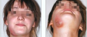
An exacerbation can go away on its own, and the disease returns to its chronic form. But sometimes inflammation develops before the appearance of purulent contents in the tissues - periostitis or gumboil.
Inflammation can cause resorption or dissolution of a section of jaw bone tissue. The appearance of purulent complications is dangerous due to its consequences: from tooth loss and damage to surrounding units to tissue melting and sepsis. Therefore, it is important to get timely help from a doctor. Treatment of dental granulomas is carried out by a dental therapist, and if removal is required, you need to contact a dental surgeon.
Clinical case
Based on Lam S. et al, Eosinophilic granuloma/Langerhans cell histiocytosis: Pediatric neurosurgery update. 2015
A 17-year-old young man was hospitalized due to an increasing scalp lesion over the past 6 weeks. The formation is painful on palpation and periodically bleeds due to ulceration, but no neurological deficit has been identified. CT and MRI revealed a large lesion in the frontal bone on the right, compressing the superior sagittal sinus. A total resection of the formation was performed, and the diagnosis of Langerhans cell histiocytosis of the skull bone was confirmed. At the outpatient stage, cytostatic therapy was carried out.
Figure 6. (a) CT examination without contrast - frontal scan (upper and middle part) and 3D reconstruction of the skull (lower part). (b) MRI scan. T1-weighted image in the coronal plane (top) and T2-weighted image in the sagittal plane
Sources
- Coppes-Zantinga A., Egeler RM The Langerhans cell histiocytosis X files revealed. Br J Haematol, 2002. - V. 116 - N. 1 - P. 3–9.
- Lam et al., Management of adult patients with Langerhans cell histiocytosis: recommendations from an expert panel on behalf of Euro-Histio-Net. Orphanet J Rare Dis, 2013 - V. 8. - N 72.
- Lam S., Reddy GD, Mayer R., Lin Y., Jea A. Eosinophilic granuloma/Langerhans cell histiocytosis: Pediatric neurosurgery update. Surg Neurol Int, 2015. - N. 6 (Suppl 17): S435 - S439.
- Langerhans' Cell Histiocytosis (Histiocytosis X). What is it? Harvard Medical School. Harvard Health Publishing. October, 2014. www.health.harvard.edu
- Sharma R., Singh R. et al. Langerhans cell histiocytosis (skeletal manifestations). Radiopaedia. https://radiopaedia.org/articles/langerhans-cell-histiocytosis-skeletal-manifestations-1
- Shea C. R, James W. D. et al. Langerhans Cell Histiocytosis. Medscape, 2022. https://emedicine.medscape.com/article/1100579‑overview
- Churilov L.P. Death on takeoff, or Who are you, Doctor Taratynov? Health is the basis of human potential: problems and ways to solve them, 2014. - T. 9 - No. 2 - P. 919–929.
- Yusupova L. A., Yunusova E. I., Garayeva Z. Sh., Mavlyutova G. I. Histiocytosis X. Practical Medicine, 2014 - T. 08 - No. 14.
Conservative treatment
For granulomas, conservative treatment is more often used. It consists of mechanical treatment of the root canals. After this, they are filled with temporary healing material - pastes based on calcium hydroxide. After 2-3 weeks, a control image can be taken, and if the inflammation is eliminated, the canals are filled with a permanent material - gutta-percha. A new permanent filling is placed on the crown of the tooth.
There are two treatment tactics depending on the initial condition of the tooth.
1. Treatment of granuloma of a tooth in which the root canals are not filled. In this case, treatment involves the following steps:
- removal of carious tissues, old filling on the crown, if any;
- mechanical treatment of canals - with the help of special tools they expand, the walls are smoothed;
- antiseptic treatment of canals.
Further actions depend on the size of the granuloma. If it is small, up to 3 mm, simultaneous filling is allowed. If the formation is more than 3 mm, then the root canals are filled with temporary paste. It helps the granuloma shrink or disappear completely.
You will have to wear temporary material for no more than 3 weeks. At the end of the period, the doctor will order a repeat x-ray, and if he sees positive dynamics, he will fill the root canals with permanent material. The restoration of the tooth crown is also carried out.
2. Treatment of a tooth in which the root canals have already been filled. In this case, the doctor will first remove the old material. If a tooth has a crown, it must be removed. The root canals must be resealed, and the treatment tactics correspond to those described above: sometimes the installation of a temporary therapeutic filling is required.
Is it possible to put a crown on a granuloma on the root of a tooth?
Dental prosthetics imposes special requirements on the doctor and the patient. If we are talking about installing a crown on a tooth of which no more than 1/3 remains, that is, only the root, then it must be carefully prepared: the canals are sealed, the infection is eliminated. A granuloma under a tooth is an indicator that an inflammatory process is occurring in the tooth, and before dental prosthetics it is necessary to get rid of it, or sooner or later the granuloma will still have to be treated, since it can become inflamed under the crown. So why not do this right away, before installing a denture?
Surgery
Surgical treatment of dental granuloma may be required only in a few cases:
- obstruction of the root canals - complex, tortuous structure, too thin, narrow canals;
- impossibility of unsealing channels;
- the presence of a pin in the root canal - attempts to remove it may cause injury;
- patient's reluctance to remove the crown.
Many patients prefer granuloma removal because they do not want to resort to long-term treatment and remove a good crown. In this case, an apical resection operation is performed - part of the root is removed along with the granuloma through a small incision in the gums. Less commonly used is hemisection - removal of one root of a multi-rooted tooth along with part of the crown. In this case, further restoration of the crown of the tooth with a prosthesis will be required.
In rare cases, it is not advisable to preserve a tooth with granuloma. For example, if the crown is severely damaged and cannot be restored. In this case, when removing a tooth, the doctor must remove the granuloma from the socket in order to prevent the development of inflammation.
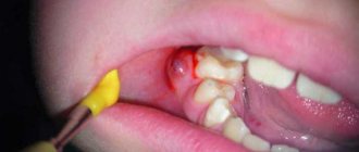
If purulent complications develop against the background of a granuloma, it is important to get help from a doctor immediately. The specialist will provide first aid: relieve acute pain by opening the tooth. Previously sealed canals are opened, and purulent contents are subsequently removed through them. In this case, relief comes instantly.
If severe swelling of the gums or cheeks appears, this may be due to the release of inflammatory contents under the periosteum or oral mucosa. In this case, a small incision is made to drain the pus. Further treatment is possible only after relief of acute symptoms. Drug therapy will also be required - the doctor will prescribe a course of antibiotics. You should not take them on your own. Moreover, it makes no sense to be treated only with antibiotics in the hope that the inflammation will go away - they are not able to eliminate the source of the disease or even reduce it, it is important to take local measures to eliminate the inflammatory process.
General information
Pyogenic granuloma is conditionally classified as a group of pyoderma , because previously it was believed that the provoking factor was a staphylococcal infection , but now it is defined as benign inflammatory hyperplasia of granulation tissue.
The pathology is usually localized on the skin of the feet, hands, red lip border, on the abdomen, oral mucosa, periungual ridges, in places of cuts, injections, burns, splinters, ingrown nails , in extremely severe cases in the gastrointestinal tract and is most often single.
Features of prevention
The main condition for the prevention of dental granulomas is timely assistance from a dentist when caries occurs. You should not allow severe tooth decay or the development of pulpitis. The peri-root tissues are healthy until the pulp becomes inflamed. Therefore, if symptoms of caries or pulpitis appear, it is important to immediately consult a doctor.
Endodontic treatment also increases the likelihood of developing periodontitis. Therefore, it is better to eliminate caries in the early stages and avoid the need for root canal filling. If this is unavoidable, it is important to carefully choose a dental clinic - the professionalism of a specialist will help eliminate possible mistakes and prevent complications.