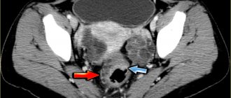
Local swelling of the root zone, compactions of various sizes, throbbing pain, sensitivity to temperature when eating, enlarged lymph nodes may indicate an acute inflammatory process of infectious etiology. In dentistry, there are several types of diseases, among them periapical abscess. It can be caused by advanced caries, chipping of an element of the dental system, overheating of the pulp due to tooth preparation and various pathologies. This can also happen due to poor-quality root canal treatment.
A periapical tooth abscess is an accumulation of pus between the molar and the gums. Purulent exudate can accumulate both in the inner and outer surfaces of the alveolar process of the lower or upper jaw. It most often begins in a tooth with a non-viable pulp, when the nerve dies, causing infection and progressive inflammation. The infection may gradually spread to surrounding tissue. Clinical symptoms are in many ways similar to acute periodontitis. The patient experiences increased sensitivity, acute pain, the mucous membrane may have swelling, there is active hyperemia, and overflow of blood in the vessels of the circulatory system.
Symptoms
- Intense dull throbbing pain.
- Discomfort when chewing food.
- Swelling in this area.
- Painful sensations when swallowing.
- Enlarged lymph nodes.
- Unpleasant sensations when opening the mouth.
- Change in molar color.
Other symptoms also include malaise, fever, weakness, and bleeding. It should be noted that in some cases the inflammatory process can be practically asymptomatic; activation can occur periodically, for example in the case of hypothermia. In this case, immediate assistance from a dentist is required so that he can quickly and accurately diagnose and establish a treatment regimen for effective recovery.
Causes of abscess
Complications associated with the inflammatory-destructive process can occur due to the following reasons:
- Poor quality endodontic treatment.
- Carious formations.
- Injuries to elements of the dentition.
- Unqualified prosthetics.
- Infectious diseases in the maxillofacial area, such as sinusitis.
Treatment of a periapical abscess without a fistula may be required in case of violation of the endodontic therapy protocol or incomplete obturation of the canal system. Provoking factors can be serious infectious diseases, hypothermia, and a reduced level of the body's immune defense. Leukocyte infiltration also often causes the formation of an abscess and microabscess.
Reasons for development
The appearance of a purulent neoplasm in the oral cavity can be provoked by:
- failure to consult a doctor in a timely manner for the treatment of caries or periodontitis,
- penetration of filling material beyond the root canal during dental treatment,
- jaw injury,
- necrosis of the neurovascular bundle,
- inflammation of soft tissues in the oral cavity.
The risk of tissue suppuration is increased by weak immunity, frequent infectious diseases, vitamin deficiency and other factors that weaken the body as a whole.
Kinds
Based on the nature of the localized accumulation of pus in the elements of the dentition and nearby structures, two groups of abscess are distinguished.
- Periapical abscess without fistula. In this case, transcanal infection of periodontal tissue is observed. The adjacent gum may have swelling, hyperemia is observed, pain is felt when biting on a molar, and the appearance of purulent exudate is recorded. The disease has a pronounced clinical picture.
- Periapical abscess with fistula. With this phenomenon, the inflammatory destructive process spreads to the bone marrow and mucous membrane. The fistula most often opens in the projection of the apex of the root element of the dentition. There may be hyperemia on the mucous membrane, a scar from a fistula, and the gums may be quite sensitive to palpation.
In case of periapical abscess without a cavity and with a cavity, complaints of intense dull throbbing pain, deterioration of general condition, and constant pain are recorded.
Diagnostics
Diagnosis of an acute inflammatory process of infectious etiology with localization in the hilar region includes a visual examination and radiographic examination. In some cases, thermography may be performed. The dentist assesses the condition of the mucous membrane, hyperemia of the tissue, and changes in tooth color. X-ray examination helps to assess the location of the pus. At the diagnostic stage, the specialist determines the viability of the pulp.
A clinical study allows us to determine the degree of pathology and select the correct endodontic treatment or surgical intervention. If necessary, additional examination methods may be required to determine the condition of the periapical tissues. In each specific case, the treatment method is selected individually depending on the history and clinical examination. The patient’s age, local symptoms, and the presence of general diseases are also taken into account. Additionally, microbiological diagnostics can be performed to identify various types of microbial pathogens.
Periapical pathologies: how to treat?
What is the main determining factor for a dentist when choosing an intervention algorithm during endodontic treatment: existing knowledge, experience or the results of CBCT analysis? This article describes a clinical case of a 10-year-old patient with periapical pathology, which confirms the idea that endodontically affected teeth should not always be extracted. Even in cases of roots with very curved morphology, successful treatment results can be achieved using special nickel-titanium files.
Although clinical dentistry is rich in evidence in favor of various treatment algorithms, in most cases such evidence is of very low quality. In the end, doctors rely more on their own experience. Even a simple clinical case can be treated using different methods with a fairly good prognosis. Discussion of such topics is very common on social networks. A study recently conducted at Ghent University in Belgium found that dentists are often biased and make mistakes when making necessary clinical decisions. For example, in cases of persistent and chronic asymptomatic apical lesions, the endodontist will likely opt for repeat endodontic treatment or apical surgery rather than extraction followed by implantation. Orthopedists and surgeons, on the contrary, will lean towards the latest intervention algorithm. In fact, the choice of treatment method should take into account the wishes of the patient after he has signed an informed consent that he is fully aware of all possible rehabilitation options. In this article we will discuss the best way to select a treatment algorithm in the presence of a specific apical lesion.
Endodontic treatment
Recent data from Ghent University and other epidemiological studies show that in 22% of cases, doctors choose extraction as a treatment method for periapical lesions larger than 1 cm. If the size of the pathology exceeded 1 cm, more than 50% of doctors were inclined to remove it. In principle, the choice of such intervention tactics is logical: doctors do not know the true histological picture of the lesion (whether it is a cyst or a periapical granuloma), therefore, the treatment algorithm is based on the obtained radiological data. Naïr points out that it is simply impossible to differentiate between cystic and non-cystic lesions based on radiographs alone. Similarly, during dynamic monitoring it is difficult to say whether the lesions are enlarging or shrinking after treatment.
Moreover, the limitations of 2-D radiographs must be taken into account during clinical intervention planning. Wu et al showed that targeted images often indicate a healthy apical region or extensive healing, when in fact the CBCT results show significant signs of apical periodontitis (Figures 1 and 2).
Photo 1. X-ray showing endodontically treated tooth No. 11 and cold-sensitive tooth No. 12.
Figure 2: CBCT findings from the same region of interest showing the presence of a large apical defect.
Although CBCT allows a more accurate determination of the presence and size of the apical lesion, there has not yet been enough research to support the need for CBCT scanning as a standard diagnostic method in endodontic practice. Morris et al concluded that endodontic treatment of teeth followed by direct and indirect restorations provides fairly predictable rehabilitation results, which, in turn, justifies endotherapy as the first choice.
Size matters?
In general, literature data suggest that the larger the periapical lesion, the less favorable conditions for its healing. In principle, everything is logical: the larger the pathological focus, the longer it took for it to grow. The longer a bacteriological association has developed, the more different bacteria are included in its composition, which means the more difficult it will be to cure such a pathology. Of course, a number of additional studies still need to be conducted, but in general, an increase in the focus of periapical lesions by every millimeter reduces the prognosis of successful treatment by 14 percent compared to clinical cases of endotherapy in which there are no radiological signs of periapical pathology at all. A similar trend is observed in periapical surgery: there is a negative correlation between the size of the apical lesion and the possibility of complete healing of the intervention area. During orthograde endodontic intervention, apical lesion size greater than 2 mm is categorized as a risk factor for the need for future endodontic retreatment. However, it must be remembered that the size of the formation on periapical images largely depends on their quality and positioning angle. Although it was previously believed that the larger the periapical lesion, the more likely its cystic structure, however, such conclusions are unsubstantiated without additional histological studies. Large periapical formations are characterized by a tendency to increase with the possibility of involving adjacent anatomical structures in the process. This does not mean that treatment of such lesions is impossible, but its predictability becomes much less predictable.
Clinical case: a young patient with a chronic apical abscess
A 10-year-old patient was referred for endodontic treatment of a mandibular right molar. In the area of tooth No. 46, the presence of a fistulous tract on the palatal side was noted. The patient and his parents were informed that the success rate of future endodontic intervention was reduced given the enormous size of the periapical lesion. However, they still did not want to remove the problematic tooth. After clinical examination and radiological control, a diagnosis of pulp necrosis and chronic apical abscess was made (Figure 3-4).
Photo 3. X-ray of the area of tooth No. 46 with visualization of the periapical area of the lesion.
Photo 4. Clinical appearance of the affected area.
Probing of the fistula tract confirmed its direction towards the apex. The choice of endodontic treatment was also justified by the need for corrective orthodontic and orthopedic interventions in cases of tooth extraction at such a young age. During the first visit, the tooth was isolated using a rubber dam, after which the doctor cleaned out all existing caries. To process the canals, NiTi files (HyFlex EDM) from the Swiss manufacturer COLTENE were used (photo 5). The canals were processed up to sizes 40/04. This system was used during the treatment because it is characterized by high flexibility of the instruments, which was extremely important in the patient’s existing canals with a curved morphology. In addition, only a few instruments were required to process the entire endospace. Chemical treatment of the canals was carried out using a 5.25% sodium hypochlorite solution and ultrasonic activation. After placement of an interim endodontic dressing with unsaturated calcium hydroxide, the tooth was subsequently restored using a composite restoration. To minimize the effect of microleakage, the tooth was covered with Teflon tape between visits.
Photo 5. Sequence of HyFlex EDM files.
Development of channels up to file size 60/02
During the second visit, it was noted that the patient continued to have a fistulous tract, but the swelling had almost completely subsided (Figure 6). The tooth was again isolated using a rubber dam, after which the medial canal system was treated to 50/03, and the distal canal system to 60/02. Medicinal treatment of the canal was performed with sodium hypochlorite and citric acid (40%). Since no discharge from the endospace was noted after drying, the mesial canals were obturated with bioceramics. The distal canal was filled with a plug made of MTA. The top of the tooth was covered with a composite restoration (photo 7). After three months, no fistula tract was noted, and radiographs confirmed healing of the periapical area (Figures 8-9).
Photo 6. Healing of the fistula tract.
Photo 7. Quality control of obturation.
Photo 8. X-ray after 3 months.
Photo 9. Clinical appearance of the tooth after 3 months.
At the same time, the doctor should always remember the main parameters that justify the advisability of extraction instead of endodontic treatment:
- doubts regarding the success of endotherapy on the part of the dentist who referred the patient;
- a large area of the lesion on the resulting x-ray;
- no signs of healing of the fistula after installing a calcium hydroxide bandage;
- compromised initial periodontal status;
- taking into account age-associated features of the clinical algorithm of endodontic treatment.
Is radiographic success everything?
Considering the studies mentioned in this article, the presented case shows that prejudice and subjective diagnosis should not prevent us from performing endodontic treatment even in cases of large apical lesions. With informed consent and proper motivation on the part of the patient, root canal therapy should always be the first choice of iatrogenic intervention. Although conventional 2-D imaging, which is used for early diagnosis, may hide the presence of prolonged apical periodontitis, CBCT allows us to objectify all the necessary parameters of the periapical lesion. At the same time, neither two-dimensional nor three-dimensional diagnostic methods make it possible to differentiate the differences between scar tissue in the area of the “healing” apex and the infectious process in the area of the affected apex in the absence of corresponding clinical symptoms. Based on this, success on a radiograph is not always clinical success.
Conclusion
Combining several imaging tools helps endodontists make the correct diagnosis and select the most appropriate treatment algorithm for each individual clinical case. With modern NiTi file systems, root canals with very curved morphology can be effectively treated. Ultimately, all this contributes to the fact that those teeth that were categorized as unsuccessful and planned for removal, it turns out, can still be treated if you approach the diagnostic and planning stages correctly.
Author: Dr Christophe Verbanck (Belgium)
Treatment of periapical abscess
Depending on the stage and form of the pathology, the specialist selects a therapeutic or surgical method of treatment. At the initial stage, the canals are usually cleaned, in particular the elimination of pathogenic microflora between the teeth and gums, and the cavity is treated with antibacterial components. Endodontic therapy may include the installation of a temporary filling and the application of an antiseptic dressing with antimicrobial properties in the root canal. Antibiotics may be prescribed to stop the inflammatory process.
High-quality endodontic therapy allows you to eliminate the infectious focus. As inflammation increases, an element of the dental system can be removed. In some cases, the specialist performs appropriate tooth-preserving procedures, for example, hemisection, that is, removal of one of the molar roots.
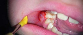
The opening of an abscess is always performed using anesthesia; discomfort and pain are excluded during high-quality endodontic treatment. After the abscess removal procedure, drug therapy and surface treatment with special bactericidal compounds may be prescribed. The final step is to take an x-ray to ensure complete recovery.
The specialists of the AlphaDent clinic are always ready to help if you have complaints about a periapical abscess without a fistula or with a fistula. Dentists have the necessary knowledge and sufficient experience to provide high-quality endodontic treatment.
Qualified treatment will eliminate the recurrence of the infection that caused the abscess. Timely dental care will help keep your teeth and mouth in good condition. It is recommended to make visits to the dentist for preventive examinations in order to exclude complex endodontic treatment associated with the removal of an abscess.
Treatment
Treatment tactics for periodontal and periapical abscess are different.
Treatment of periodontal abscess
The primary goal of treating a periodontal abscess is to eliminate acute inflammation. To do this, periodontal pockets are washed with antiseptic solutions. It may be necessary to open the abscess, in which case the doctor will first perform anesthesia.
After this, curettage of periodontal pockets is carried out - scraping out granulations, dental plaque, epithelial particles, and necrotic tissue. Finally, the doctor applies stitches. In most cases, the patient is prescribed to wear a periodontal bandage. This is the application of medicinal substances to the area of periodontal pockets.
Treatment of periodontal abscess must be comprehensive. The patient is also prescribed rinsing the mouth with antiseptic solutions and taking broad-spectrum antibiotics. As a rule, the inflammatory process can be stopped.
Treatment of periapical abscess
To ensure the drainage of pus, the doctor opens the tooth cavity and creates access to the root canals. Also, drainage of pus can be carried out through an incision in the gum tissue, which does not eliminate the need for endodontic treatment (canal treatment).
After opening the tooth, the pulp or filling material is removed, if the pulp was removed earlier, and root canal treatment is performed.
It is important to completely remove purulent masses and infected dentin tissues and rinse the canals with disinfecting solutions. Then an anti-inflammatory medicine is placed in them and a temporary filling is installed. The chances of saving a tooth depend on the quality of endodontic treatment. Broad-spectrum antibiotics are prescribed for oral administration. After the inflammatory process has been completely eliminated, the canals can be filled with permanent filling material and the crown of the tooth can be restored. If treatment is ineffective and the inflammatory process increases, the tooth will have to be removed. Both types of abscesses are usually accompanied by swelling of the soft tissues, and swelling may also persist after tooth extraction. Physiotherapy helps speed up recovery: UHF therapy, magnetic therapy, laser therapy.