Flux is a common, non-medical name for a whole group of diseases that require immediate, and in some cases, emergency care. One of the main manifestations of flux is a violation of the shape of the face associated with a complication in an inflamed tooth.
The main groups of diseases that are commonly called flux: periodontitis, periostitis, osteomyelitis, abscess and phlegmon, lymphadenitis, odontogenic sinusitis.
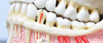
Periodontitis
- inflammation of the tissues of the apical part of the tooth.
Periostitis
– inflammation of the periosteum in the area of the teeth.
Osteomyelitis
– inflammation of the jaw bone, which necessarily ends with necrosis (death) of the affected area of the bone.
Abscess
- This is a limited purulent inflammation of fatty tissue.
Phlegmon
- This is a diffuse purulent inflammation of fatty tissue.
Under the skin, under the mucous membrane, under and between the muscles there is fatty tissue. With an exacerbation of odontogenic infection, pus spreads under the periosteum, axillary and intermuscular spaces, causing inflammatory and edematous-infiltrative changes in the soft tissues of the face and neck.
Inflammatory diseases of the maxillofacial area (flux, abscess, phlegmon) and neck are divided into two main groups according to localization. The first is abscesses and phlegmons located near the upper jaw. The second is abscesses and phlegmons located near the lower jaw.
Lymphadenitis
– inflammation of the lymph node (group of lymph nodes) adjacent to the odontogenic (dental) focus of infection. Odontogenic sinusitis is often an inflammation of the maxillary (maxillary) sinus (sinusitis), caused by the anatomical features of the position of the roots of the maxillary teeth involved in the pathological process, in which the roots of the teeth either stand in the lumen of the maxillary sinus, or are located close to the bottom of the maxillary sinus. In this case, infection of the mucous membrane of the maxillary sinus occurs with the subsequent development of sinusitis.
Causes
Inflammatory (infectious-inflammatory) diseases of the maxillofacial area are caused by microbial microorganisms, which are usually part of the normal microflora of the oral cavity. Microbial pathogens are staphylococci, streptococci, enterococci, diplococci, gram-positive and gram-negative bacilli (intestinal, protea, etc.). In addition, during flux, fungi, mycoplasmas, treponema, and protozoa from the Trichomonas family are found in foci of odontogenic infection. According to various authors who analyzed the microbial flora in foci of odontogenic infection (flux), the microflora is represented by a monoculture of staphylococcus (aurus and epidermal) or streptococcus of groups D, F and G. Associations of the above microorganisms are often identified.
The question arises of how non-pathogenic or opportunistic microorganisms trigger the initiation of an infectious-inflammatory process (flux). For the occurrence of a disease, the presence of only non-pathogenic or pathogenic microorganisms is not enough. The answer to this question is given by the so-called Arthus-Sakharov phenomenon. According to the infectious-allergic theory, its essence boils down to the following: under the influence of a foreign serum protein, which has antigenic properties, antibodies are produced, which leads to sensitization of the body. Sensitization of the body is the acquisition by the body of increased sensitivity to foreign substances (proteins) - allergens. Against this background, repeated introduction of the protein into the vascular bed causes the formation of antigen + antibody complexes, which are fixed on the membranes of blood vessels and turn into target cells. Then the cell membrane is damaged, enzymes are released, mediators (from the Latin mediator - mediator) of inflammation are released. This is accompanied by activation of the third platelet factor and can cause intravascular coagulation, which leads to impaired microcirculation and necrosis (death) of tissue. This immunopathological reaction is also involved in odontogenic infection, leading to the occurrence of gumboil. The waste products of microbes participate in the role of antigen. This explains why in many patients non-pathogenic microorganisms are found at the site of inflammation.
The dynamic balance between the focus of chronic odontogenic infection and the body is ensured by the connective tissue capsule surrounding the focus. It limits the penetration of microbes into the tissues and vascular bed adjacent to the lesion, and limits the effect of immune factors on the infectious focus. The balance is also maintained by the fact that part of the waste products of microorganisms and tissue decay through the root canal of the tooth, fistula or periodontal fissure is released from the infectious focus into the oral cavity.
An imbalance can be caused by a disruption in the outflow of exudate from the lesion through the root canal due to food masses entering the carious cavity or when filling a carious cavity by a dentist. In the infectious focus, the concentration of microbes, their toxins, and waste products, which penetrate through the connective tissue capsule into the surrounding tissues, increases. Here their direct damaging effect on tissue can occur, and upon penetration into the vascular bed, the mechanism of the Arthus-Sakharov phenomenon is triggered. Clinically, this is manifested by an exacerbation of a chronic focus of odontogenic infection and the occurrence of gumboil.
Another mechanism of imbalance between the focus of chronic infection and the human body is associated with damage to the connective tissue capsule itself. This can happen when a tooth is removed, when there is increased load on the tooth while chewing hard food, etc. Damage to the capsule leads to the spread of microorganisms (their toxins, waste products) beyond the infectious focus, which in a sensitized organism leads to the occurrence of the Arthus-Sakharov phenomenon. These are the reasons and the main mechanism of exacerbation of odontogenic infection, leading to the development of gumboil.
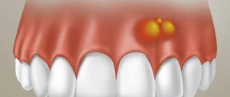
Abscess on the gum due to periodontitis
With this disease, ulcers often form. The drugs only temporarily relieve discomfort, but the pathological process continues to develop, gradually destroying the bone tissue of the jaw.
Timely intervention by the dentist allows you to remove the source of infection and achieve 100% cure.
Antibiotics, rinses, physiotherapy - all this is included in the treatment of purulent infection. It is important not to let the process take its course, because complicated purulent infections are extremely difficult to treat.
What are the consequences of negligence?
As a rule, aching pain speeds up the patient, and few people delay a visit to the doctor. And this is correct, because even if there are no painful symptoms, swelling of the gums is quite enough to urgently consult a dentist for consultation, diagnosis and treatment (after which the swelling soon goes away).
In the absence of qualified and timely assistance, the patient may develop very serious complications. Thus, a slight inflammation can quickly affect the entire gum and even the jaw bone. And this will require a long and serious treatment with antibiotics, in addition, the prospect of inevitable surgical treatment of teeth in a dream looms before him - to eliminate the source of infection.
Symptoms
The main manifestations of gumboil disease are pain in the area of the causative tooth and the appearance of swelling in the soft tissues of the face. If at the beginning of the disease the swelling is mild, then as the process progresses the swelling increases and can spread to the eye area, scalp or neck. As the symptoms of flux increase, pus from under the periosteum can spread through the fatty spaces closer to the skin - this is manifested by the fact that the skin turns red, becomes dense and hot to the touch. Further, the skin in the center of the flux softens, becomes thinner, and a fistula with purulent discharge may appear. In this case, pain may decrease somewhat, which is associated with a decrease in pressure in the flux cavity during the formation of a fistula tract.
In addition to pain, symptoms such as limited mouth opening or inability to open the mouth, and pain when swallowing may occur. If the flux is located deeply, facial swelling may be mild or not expressed at all, and the main manifestations will be limitations in mouth opening, pain in the flux area and pain when swallowing.
Inflammation: where does it come from?
The most common cause of swollen gums is an inflamed tooth. In the vast majority of cases, swelling occurs as a result of chronic inflammation of the dental tissues under the crown and/or filling, lasting more than one day or even more than one week, against the background of progressive complicated untreated caries and/or after mechanical trauma to the mucous membrane.
Unfilled canals, improper treatment of caries - all this ultimately leads to gum swelling. Voids in the dental canals, perforation of the tooth root against the background of poor quality treatment involuntarily help the infection to spread further.
In this case, the swelling of the gums also affects the area of the diseased tooth, accompanied by a feeling of aching pain. For some people, the pain may be intermittent and only occur when biting.
The usual swelling of the gums can also hide such serious problems as a dental cyst or an abscess on the gum.
Diagnostics
To diagnose flux, methods such as questioning, examination, and additional research methods are used: radiography, ultrasound, computed tomography, magnetic resonance imaging, blood test data, culture of exudate, etc. An experienced maxillofacial surgeon who treats patients with flux, can, based on the patient’s complaints and examination, carry out an accurate topical diagnosis of the disease (localization of pus) and select the optimal surgical approach to ensure sanitation of the purulent focus and adequate drainage.
What is a dental cyst?
The inflammatory process - a dental cyst (from the Greek “kystis” - bubble) - is formed in the form of a small inflammatory ball that swells as part of a response to injury or infection. The inflamed area looks like a pus-filled bubble, sometimes reaching several centimeters in size. The body’s active work to restore order and remove slagged cells occurs constantly. At some point, a lot of them accumulate, and our excretory system selects the weakest area of the body through which they can be removed. In this case, the soft tissue around the tooth becomes the target, where a purulent neoplasm appears. An emerging problem can be diagnosed in the early stages using an orthopantomogram.
Treatment
Treatment for flux can be boiled down to a catchphrase in Latin: “Ubi pus, ibi incision,” which translates as “Where there is pus, there is an incision.” Treatment of flux begins with eliminating the cause that caused the flux. That is, from the removal of the causative tooth. Next, they begin to drain (empty) the purulent focus by opening (incision) in the area of the flux. The abscess is emptied, the abscess cavity is washed with antiseptic solutions, and drainage is installed through which pus will be released. Regardless of the type of flux, the principle of treatment remains the same: opening the lesion, drainage.
In the postoperative period, daily antiseptic treatment (washing) with antiseptic solutions is carried out. There is a peculiarity in the treatment of these diseases (flux, periostitis, phlegmon, etc.), which is that immediately after surgery and up to 1-2 days, facial swelling (flux) may increase. This should not be taken as a worsening of the disease. The fact is that any surgical intervention is perceived by the body as an injury, and the body responds to this by increasing tissue swelling in the area of injury. This may be accompanied by increased swelling of the eyelids, cheeks, etc. Painful sensations decrease and subsequently decline as acute inflammatory phenomena subside. As a rule, antibacterial drugs, antihistamines and painkillers are prescribed after surgery.
If there is an advanced case of flux, which is accompanied by a violation of the function of external respiration (the patient may suffocate), then hormonal drugs are prescribed, and even an operation is performed - a tracheotomy. The operation consists of making an incision in the area of the tracheal rings and installing a tube. As the swelling of the upper respiratory tract (pharynx, larynx) decreases, when the threat of asphyxia (suffocation) has passed, the tube is removed from the trachea, and the patient breathes through the natural airways. The duration of prescription of drugs is determined by the attending physician. The drainage in the wound is periodically changed, and the wound is removed when cleansed. The duration of drainage in the wound is also determined by the attending physician, based on the clinical picture of the disease. The drainage can remain in the wound for an average of 3 to 10 days.
If the incision for flux was made through the oral mucosa, then additional suturing of the wound is not required in the future. The wound in the oral cavity will heal on its own (by secondary intention). If the incisions were on the skin side, then after the acute inflammatory processes have subsided, the wound can be sutured. After suturing the wound (and this can be 2-4 weeks after surgery), the patient is also prescribed antibacterial drugs and painkillers, and the wound is drained.
With inadequate treatment, as well as in advanced cases of flux, complications such as suffocation, penetration of infection into the cranial cavity, onto the membranes of the brain, into the neck and into the mediastinum (part of the chest cavity where vital organs are located), penetration into the orbital cavity with damage to the optic nerve and the occurrence of a systemic inflammatory reaction (sepsis), which leads to multiple organ failure and even death.
Soft tissue incisions, which are used in the surgical treatment of flux, must ensure complete sanitation of the purulent focus and adequate drainage. Surgical treatment should not be replaced by the prescription of strong antibacterial drugs. Waiting tactics, delays in performing surgery, and small incisions that do not provide adequate drainage of the purulent focus are unacceptable. We must not forget that surgery for a purulent process in the maxillofacial area and neck is often an operation to save the patient’s life. The occurrence of complications is more likely when the patient has concomitant chronic diseases: diabetes mellitus, coronary heart disease, anemia, diabetes mellitus and others. Elderly and senile age are also factors that can aggravate the course of this disease.
Other provocateurs
In addition to diseases and pathological changes that cause swelling of the cheek from the tooth, there are a number of other causes.
If an infection enters the patient’s body, medications that have an antibacterial effect should be taken. Thanks to this, you will get rid of the main cause, that is, infection, and the consequence - swelling. With inflammation of the lymph nodes in children, this situation often occurs.
The presence of a sebaceous gland cyst provokes changes in the facial part. In this case, surgical intervention will be required.
The cause is identified as neurological diseases. In addition to swelling of the cheek, the patient may suffer from congestion in the ear canal and discomfort in the throat area. To find out the reasons, you need to consult a neurologist.
A similar situation occurs with injuries or bruises. You can reduce the size of swelling by using a cold compress.
With a pathological change in the condition of the internal organs, swelling of the cheek is observed. To find out the reasons, you should consult your doctor.
It is important to know what to do if your cheek is swollen. Here are some tips to help reduce unpleasant symptoms:
- Often, after you have a wisdom tooth pulled out, you may experience swelling. To get rid of this, you should rinse your mouth with chamomile or sage. Chlorhexidine is perfect for these purposes.
- A fairly effective remedy is a saline (soda) solution. Rinsing with these products relieves pain and produces an antiseptic effect.
- While a wisdom tooth is coming out, it is worth using the help of special ointments and creams. Their use helps to minimize unpleasant symptoms.
- If your cheek is swollen after a wisdom tooth has been pulled out, you should use Kalanchoe or aloe juice. To do this, soak a cotton pad in the medicine, then apply it to the painful area.
- This manifestation is also possible with an insect bite. In this case, a decoction of chamomile, combined with aloe, helps.
Experts advise taking vitamin complexes daily to help improve the protective function of the immune system. It is better to reconsider your diet, in particular this concerns reducing the consumption of sweets. You can perform a light gum massage for preventive purposes.
By following basic rules of personal hygiene and regularly visiting the dentist, you will prevent complications and the development of pathological conditions.
Dental disease and its treatment is an unpleasant phenomenon that many adult men and women try to avoid or delay until an emergency. But the most dangerous thing about far-fetched fear is that while a person waits for the disease to become unbearable, destruction of teeth and gums occurs, which in the initial stages could be treated with fairly simple, almost painless and inexpensive methods. In this article we will talk about gumboil, how to get rid of it, and how to help the patient at home.
What is the difference between a cyst and a granuloma?
There are other diseases in the oral cavity that are symptomatically similar to a cyst. Specifically, a granuloma is an outwardly swollen round projection in the root zone of a tooth. The doctor must correctly diagnose the disease, since the sequence of exposure, removal techniques, and pharmacology have significant differences. Granuloma is eliminated with the help of medications, as well as accompanying mouth rinsing with phytocomponents. The cyst has deeper roots and does not go away without surgery.
What to do with a dental cyst?
Features of the development of purulent inflammation of various forms and localizations show that the formation occurs at a slow pace. When pronounced symptoms appear, the cyst emerges on the surface of the mouth, gum or root, inflamed and filled with pus. Patients complain of severe malaise, “fever,” experience pain and discomfort when chewing food.
A decade ago, the only solution to getting rid of the problem was the ability to remove the source of pain. Modern equipment, special instruments, and medications (painkillers, anti-inflammatory drugs) make it possible to save a tooth by treating a serious tumor. Depending on the stage of development, surgical or therapeutic methods are used.
The latter option involves mechanical cleaning of the canals, careful processing and filling. The surgical procedure involves removing the damaged surface while preserving the tooth. The missing root tissue is replaced with a specific material. Complete removal is considered only in the case of a wisdom tooth or when damaged tissues occupy most of the tooth and root. An exceptional situation when there is no need to treat a dental cyst is if it appears next to a baby tooth and develops slowly, and there is only a short time left before replacing a molar one.