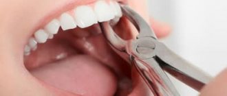
Modern dentistry is constantly developing and improving methods of dental treatment, trying to achieve maximum restoration of the aesthetic appearance and functionality of the dentofacial apparatus. Until recently, in Russia, any tooth that could not be filled was simply removed, after which an implantation operation was performed according to one of the selected types. However, in some cases, such a radical solution to the problem is not always justified and it is possible to solve the problem without removing the tooth. In many dental clinics, dentists realized that for some categories of patients, due to psychological barriers, even implantation cannot be a full replacement for a natural tooth and have developed a number of special surgical interventions to save teeth.
Tooth-preserving operations are dental treatment methods performed with the aim of preserving natural teeth that were previously simply removed. It is important to save the tooth, but to stop the worsening of the inflammatory process, the development of infection and the atrophic phenomena of the jaw, which will inevitably progress if it is removed, you will have to work hard. Also in dentistry, there are clinical cases in which amputation of the root of a problematic tooth does not eliminate the disease and inflammation further develops independently, and tooth-preserving surgery is a more effective therapeutic technique.
In what cases is it possible to save a tooth?
To carry out such procedures, it is important to clearly determine the degree of success of tooth-preserving therapy.
!Important: This is not a panacea and, unfortunately, this is not possible under all conditions and not for all patients; tooth-preserving surgery is not always the ideal solution to the situation; sometimes you still have to remove a tooth and replace it with an implant, removable or fixed prosthesis. The main indication for such operations is the ineffectiveness of conservative treatment, which almost always begins therapy and the first stage of such treatment.
Indications for tooth-saving surgery:
- Perforation of the tooth root (most often a consequence of an incorrect method of filling the root canal or filling the tooth);
- Rapid progression of inflammatory dental diseases with the threat of surrounding bone resorption, with the risk of infection of neighboring teeth;
- Destruction of preapical tissues due to ineffective treatment of teeth that are located under the crowns, a cyst at the root of the tooth;
- The presence of crowns or a bridge, the removal of a tooth under which threatens to weaken the structure and total removal, is not advisable as a temporary delay in more radical treatment;
- The presence of a deep pocket at the border of the tooth neck and gums or between the teeth;
- Lack of adequate filling of root canals and low patency of the root canal due to which has led to the fact that a cyst or granuloma has now appeared on the root;
- A supernumerary or dystopic tooth in the position of fusion of the roots, with a risk for neighboring teeth, as well as tooth root caries;
- Excessive atrophy of bone tissue in a large area of the root, with the prospect of atrophy transferring to the main jaw bone;
- Pathological foci in the area of bifurcation (connection) of the roots of multi-rooted teeth.
The doctor makes a decision to perform tooth-preserving surgery on an individual basis, taking into account the characteristics of a particular patient, comparing the benefits and risks. If a dentist has the opportunity to save a tooth, he will definitely take advantage of it.
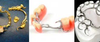
Risks of implantation
The survival rate of implants today is a record 99%. But no one will ever give 100%; it is impossible to eliminate all risks. Someone will definitely fall into this 1%. Complications during implantation are very rare, but they do occur. During implantation of an artificial root, the following may happen:
- Inflammation of the tissue around the implant (peri-implantitis).
The doctor will rule out the cause of the inflammation and treat the root with special solutions. In case of relapse, the implant will have to be removed and the bone tissue will have to be restored. - Implant rejection.
It happens extremely rarely. The artificial root is removed. - The implant is unscrewed together with the plug.
This may occur during abutment placement. In the absence of inflammation, the titanium root is put in place. - The implant is pushed into the maxillary sinus.
In such cases, only removal of the titanium root will help. - Exposing the upper part of the implant.
A fairly common complication that affects aesthetics more than health.
Contraindications to tooth preservation
During the initial examination and examination of the patient, much attention is paid to eliminating contraindications to tooth-preserving operations. This type of surgery is not performed if the patient has:
- That the operation takes place in cases of severe somatic pathologies in the decompensation stage (cardiovascular problems, diabetes, bronchial asthma, and so on);
- Mental disorders in the acute stage;
- Any problems with blood clotting (congenital and acquired coagulopathies without correction with drugs);
- Decompensated dysfunction of the immune system (any uncompensated immunodeficiency, including HIV infection);
- Old age of patients (poor tolerance of surgical interventions, increased all possible risks, weakened body);
- Poor quality of oral hygiene (the problem can be solved by professional cleaning);
- The presence of oncological tumors of any location and stage.
- During the examination, the dentist’s task is to exclude the listed contraindications, the omission of which can lead to disastrous consequences.
!Important: Special attention should be paid to the tolerability of both local and general anesthesia (depending on the choice made), this includes the allergic component and the selection of the drug for older patients with hypertension, preventing the occurrence of a crisis.
The dentist should be aware of the frequent allergic reactions that occur in patients to local anesthetics of the novocaine series. Despite the fact that modern drugs are quite safe, you need to make sure every time that the body does not hyperreact.
Signs and symptoms of oral cancer
The following changes in and around the mouth may be signs of cancer. They can also be caused by other factors. You should contact your healthcare provider if you have any of the following:
- a non-healing sore, nodule, or lump (an area that feels thicker or harder) on the lip or mouth;
- white or red spots on the gums, tongue, or lining of the mouth;
- an infection in the mouth that cannot be cured within 1 month;
- loose teeth or dentures that no longer fit;
- chronic (long-term) pain in the mouth or throat;
- bleeding or numbness of the lips or mouth.
to come back to the beginning
Types of tooth-preserving operations
There are many types of tooth-preserving operations, each of which has its own advantages and disadvantages, and is also performed for certain dental diseases. Modern dentistry distinguishes the following methods of tooth preservation:
Excision and correction of the apical portion of the root
The operation is carried out in cases where there is any pathological focus in one of the areas in contact with the root. In order to eliminate the problem, a small incision is made through which the cyst and inflammation are removed and the damaged root is partially removed. The cavity formed as a result of surgery is filled with natural, synthetic or osteoplastic material. This promotes rapid tissue healing and preservation of the full functioning of the natural tooth. The most common reasons for resection of the root apex are the presence of cysts and granulomas, accompanied by inflammation and not amenable to conservative treatment methods.
Complete root amputation
Human teeth do not always have only one root, which makes tooth-preserving surgery such as complete amputation of a dead root possible. It is carried out on those teeth where there are two or three roots, while tissues not involved in the pathological process remain in place. The dentist’s task is to rid the patient not of the tooth, but only of the diseased area of the multi-rooted tooth.
No changes in the functioning of the tooth occur, since the mechanical function of the tooth does not change, the remaining roots completely compensate for the loss of one area and distribute the load among themselves.
Hemisection of tooth root
This surgical intervention involves the removal of one of the affected tooth roots with partial amputation of the crown, usually performed on molars or premolars. During the surgical procedure, the area involved in the pathological process is carefully cut out and removed, after which the resulting space is filled with osteoplastic material. Hemisection of the tooth root is a sparing corono-radicular separation and gives very good results.
Increasing the length of the coronal area of the tooth
This type of surgical intervention is required in cases where the remaining tooth tissue is too weakened, it is necessary to improve the appearance of the tooth, and also when the natural tooth is too short or is quickly worn away. As a rule, such a problem affects several areas of the dentition at once and is caused by an incorrect bite or excessive load on the tissue. There is also a congenital predisposition to increased tooth wear, which also requires lengthening of the crown part. The procedure is non-traumatic, very fast and cosmetically always very successful.
Manipulations in pathological processes in the periodontium
Periodontal soft tissue diseases pose a great danger of losing the patient's entire dentition. In addition, their progression is accompanied by the addition of secondary infections, which aggravates the risk of loss of natural teeth. Treatment of such diseases must be timely and as radical as possible with the removal of all compromised tissue.
The use of tooth-preserving surgery for periodontal diseases is performed not only on damaged teeth, but also to strengthen healthy teeth. This method of treatment is almost the only solution for periodontal damage, which involves preserving teeth. They try to treat diseases conservatively, but very often the desired effect is not achieved. During the surgical procedure, infected soft tissues and formations in them are removed. Microsurgical access is carried out through the mucous membrane of the gums.
There are two main types of operations for periodontal pathologies:
- Flap tooth-saving operations. This surgical intervention is the main solution to the problem of periodontal pathologies. There are several of its varieties: reconstruction and correction of bone pockets, gingivoplasty, the use of the method of directed regeneration for periodontal tissues, the use of an allogeneic graft to strengthen teeth. Thanks to the operation, the infectious process is transferred to the stage of remission, and subsequently, the teeth become less mobile, signs of bacterial damage to the oral cavity disappear (unpleasant odor, discharge of pus, and so on).
- Curettage. This operation involves the delicate removal of affected areas from periodontal pockets without incisions. If the gums around the crown are pulled back, the curettage is called open curettage. It allows you to better eliminate infected areas and achieve a positive treatment result. Bone material is often introduced into the surgical area, but sometimes this is impossible due to severe infection, then the resulting cavities are gradually filled with dense scar connective tissue, which is not sensitive to external irritants, and itself strengthens the tooth. If the empty spaces are too large, they are filled with osteoplastic material in a second stage after several months in clean conditions. Closed curettage allows you to deal with small foci of the infectious process (up to 5 mm) in a less traumatic way.
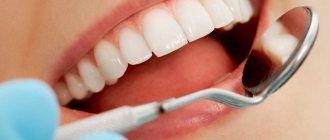
Preparatory stage
Before prescribing an operation, the dental surgeon conducts a thorough diagnosis using visual and hardware methods. X-ray and (or) computed tomography make it possible to assess the condition of tissues, identify foci of inflammation, and develop a surgical plan. If indicated, a complete sanitation of the oral cavity is carried out: treatment of teeth and gums, a comprehensive professional cleansing procedure. If the elements of the dentition are unsteady, splinting is performed (fixing a splint on loose teeth to stabilize their position).
Retrograde filling
This atypical filling of teeth from the side of the tooth root is not available in all cases in the treatment of cysts and cystogranulomas, but it is effective under certain conditions. The attending physician will determine when such manipulation is required. Previously, if a cyst was detected and in similar situations, the tooth was immediately removed, but now a retrograde filling method is used to sterilize the area where the cyst was removed and the tooth lasts for a very long time. The essence of the surgical intervention is that access to the affected area is through the alveolar process of the jaw, usually through the fistulous tract. The operation allows you to save teeth that cannot be treated conservatively for a number of reasons:
- Curvature of channels;
- Lack of channel patency;
- The presence of fragments of a surgical instrument in the canal;
- The presence of metal-ceramic crowns, as well as structures with core inlays or pins.
Using retrograde filling without filling the canal, the affected apical surface is hermetically sealed, the cyst is removed and the spread of the pathological process to surrounding tissues and healthy teeth is prevented.
Diagnosis of oral cancer
Methods for diagnosing oral cancer are described below.
Biopsy
A biopsy takes a small piece of tissue from an area that may be affected by cancer. The tissue is sent to a pathologist (a doctor who examines body tissue to diagnose diseases). A pathologist will examine it under a microscope to look for cancer cells. Typically, biopsy results are ready no earlier than after 5 days.
If you have not yet had a biopsy, it may be done at your first appointment with your doctor at MSK. If you have already had a biopsy, a pathologist at MSK will review tissue samples taken during the biopsy to confirm the diagnosis.
Medical imaging
You may also have medical imaging tests, such as computed tomography (CT), magnetic resonance imaging (MRI), or panoramic X-rays of your upper and lower jaws. A panoramic x-ray shows the entire upper and lower jaw, including the sinuses. These images provide detailed information about the lesion (an area of diseased or damaged tissue). This can show how deep the tumor has gone and whether it has spread to other organs.
to come back to the beginning
Recovery period after surgery
Modern approaches to surgical intervention in dentistry can eliminate almost all risks and adverse outcomes. The rehabilitation period passes quite quickly and ends with a full restoration of the functioning of the dentofacial apparatus.
In the first days after surgery, the following may persist: pain of moderate intensity, swelling, and the release of pinkish saliva. Gradually, clinical manifestations should disappear, completely stopping to bother the patient within a week after surgery.
!Important: If in the postoperative period you notice any questionable symptoms, you should immediately contact +74957752001 for consultation and determine the causes of atypical manifestations; at night and on Sunday you will have access to a 24-hour support telephone number, it will be included in the package with medications and a brochure with postoperative recommendations.
Basic recommendations for the postoperative period:
- Careful oral hygiene without using hard toothbrushes during the first week after surgery;
- Periodic use of antiseptic solutions to disinfect the oral cavity;
- Using healing ointments and taking antibiotics (the doctor can individually prescribe different medications that must be taken strictly according to the instructions);
- Eating only well-chopped food at room temperature without chemical irritants for three days;
- Avoid intense physical activity for a week after surgery.
The patient must strictly adhere to the dentist's instructions, as they are all aimed at minimizing the risk of infection and the development of other complications. Correct implementation of the recommendations contributes to the speedy healing of postoperative injuries and a return to a normal lifestyle.
About the oral cavity
Figure 1. Oral cavity
The oral cavity (or simply the mouth) is used for speech, chewing, swallowing, and breathing. The oral cavity consists of:
- lips;
- anterior two-thirds of the tongue;
- palate (hard and soft);
- floor of the mouth (under the tongue);
- lining of the inner surface of the cheeks (buccal mucosa);
- gums;
- a small area behind the wisdom teeth on the lower jaw (retromolar triangle).
to come back to the beginning
Cost of tooth-preserving operations in Moscow
| Name of service | Price |
| Removal of a dystopic tooth | 7000 rub. |
| Removal of an impacted tooth | 13500 rub. |
| Applying and removing sutures | included in deletion |
| Anesthesia, sedation | 12,000 rub./hour |
It is impossible to talk about the cost of the operation with certainty, since it depends on many components. These include:
- Type of surgical intervention;
- Consumables and use of medications, in particular anesthesia;
- Qualification and experience of the doctor;
- Modern equipment;
- The need for additional consultations;
- Additional examinations if necessary and other individual factors.
Carrying out tooth-preserving surgery in Moscow, in any case, will be cheaper than tooth extraction followed by prosthetics. The main thing is to choose a qualified specialist who will perform the treatment correctly and avoid complications.
Example: The cost of tooth-preserving surgery to remove a cyst is 18,000 rubles, there is no way to indicate the cost of bone materials, since often infection does not allow the simultaneous installation of a sterile regeneration stimulator at the time of surgery, and the second stage is carried out in a defect that has already actually healed, so the cost of CT no more than 10,000 rubles.
Sincerely, Levin D.V., chief physician
Which implants to choose - basal or classic?
So, on the one hand, the classic two-stage procedure is the most common and internationally recognized option for dental restoration. On the other hand, in some clinical situations it is more appropriate to perform basal implantation.
Among modern implantation methods, it is impossible to single out clearly good or bad. Only a doctor can select technologies that are suitable for a specific patient and his clinical case. Experienced dentists-implantologists of the “Full Order” clinics will select the appropriate method for restoring teeth, even in difficult cases.
Don't put off visiting your doctor! Just a few visits to the clinic can restore your health and the ability to smile freely.
Historical reference
The first doctors of human civilization “treated” diseased teeth in the only way - without finding out the reasons, they removed them. But over time, doctors in different parts of the world made attempts to treat teeth, including surgically, as opposed to removing them. This is confirmed, for example, by a fragment of a jaw found by archaeologists, dated approximately 2680-2563. BC, with traces of a successful intravital surgical operation to remove pus1. In the Middle East, the famous surgeon Abu al-Qasim Khalif ibn al-Abbas al Zarawi (936-1013), who lived in the Cordoba Caliphate, advised his followers not to rush into removing teeth, calling them “noble organs.” The most famous surgeon of the 16th century. During the Renaissance, the Frenchman Ambroise Pare (1517–1590), developed surgical methods for excision of epulis and replantation (transplantation) of teeth. On the territory of our country, dentistry, including surgical dentistry, was forced to develop – “thanks” to wars. Surgical dentistry was established on a scientific basis only in 1758, when the very first medical faculty in the history of our country was opened at Moscow University, where they taught, among other things, the skills of surgical dentistry. In pre-revolutionary Russia, the dental system was considered as an autonomous organ, for the treatment of which “handicraft” medical craft was sufficient. Progressive figures in medicine, such as Professor N.V., opposed this point of view. Sklifosovsky, who considered dentistry an undeservedly forgotten branch of surgery. In 1896, the operation of resection of the root apex for chronic apical cementitis entered dental practice. Another significant event in the field of oral surgery was the operation of cystotomy and cystectomy, developed in 1892 by K. Parch.
Surgical dentistry became an independent field of medical science after the First World War - faculties and departments of dentistry and odontology began to open throughout the country, in all 15 republics2.
Thanks to the intensive development of surgical dentistry in the 20th and early 21st centuries, today there are a sufficient number of methods of surgical treatment of teeth for the purpose of preserving them. As a result of the generalization of such surgical interventions, the concept of “tooth-preserving surgery” appeared.