Reason for breakage
The canals in our teeth have a very complex structure; they are curved, very thin and branched in different directions. To clean the canal to the very end, the doctor uses special devices, which are also very thin and flexible. Unfortunately, it is impossible to completely protect yourself from item damage. Even the most experienced professional cannot be 100% insured against such an incident.
The process of removing a foreign object is quite difficult, so not every master can cope with the task. The final decision on the need to remove a part should be made only after an adequate assessment of all the pros and cons.
There are many nuances, for example, if part of the instrument is lost in a living tooth and at the final stage of treatment, no complications should arise in the future. The patient will not encounter an allergic reaction, inflammatory processes or increased sensitivity to various irritants in the future.
Introduction
A broken instrument in the tooth and one untreated canal is the background to this clinical case, which was encountered in the practice of the German Implantology Center.
A direct participant in the events, the doctor who saved the patient’s tooth, endodontist of the German Implantology Center Melikov Azer Fuadovich, tells about this interesting case, when it was necessary to remove an instrument previously forgotten by one of the doctors from the root of a tooth:
The patient was referred to me by a colleague from another clinic after obturation of 2 root canals of a tooth, to remove a broken instrument during the work. A broken and forgotten instrument in a canal is not good. During the initial CBCT diagnosis, we identified the fact that the broken instrument was located in the root canal.
The fact that an instrument was left
and in this form it was sealed - this is an extremely negative point. But the situation was complicated by the undertreatment of the tooth as a whole, since during the examination a missed 4th root canal was discovered:
The patient was sent to me with a temporary filling, which I immediately removed. Here is a view of the tooth cavity after removing the temporary filling and monitoring the location of the fragment of the broken instrument in the root canal:
It is necessary to evaluate the situation with an open, unfilled canal missed during treatment for the first time. All work of this level is carried out exclusively using a microscope
, about which there will be a short video at the end of the article. In the following image you can see the appearance of the tooth cavity after removing the temporary filling and checking the missed canal before starting work:
Possible complications
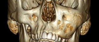
It is worth noting that a piece of the instrument itself does not pose any threat to the patient’s health. This is explained by the fact that such items are made from hypoallergenic materials. However, the part may prevent complete cleaning of the dental canal, which will negatively affect the outcome of the entire treatment. As a result, inflammation may develop, severe pain will occur, and in rare cases, tooth extraction may be necessary.
Parts remaining in the canal can negatively affect recovery after treatment. In rare cases, the patient may develop an allergy; sometimes it is accompanied by a strong tooth, swelling and redness of the soft tissues and mucous membrane located next to the diseased tooth.
The final prognosis of endodontic treatment will depend on many factors, for example, on the stage at which the working instrument broke, as well as on the degree of preparation and sufficient treatment of the canal with medications at this stage.
Price for removing a broken instrument from a dental canal in Partner-Med
| Dental service | Price* for cash in rubles |
| Extraction of 1 foreign body, or 1 pin, or 1 broken instrument from the root using a microscope (Promotion) | 5 500 ₽ |
| Extraction of 1 foreign body, or 1 pin, or 1 broken instrument from the root using a microscope | 8 690 ₽ |
*The indicated cost of the service is valid for DECEMBER 2020 for patients visiting our clinic for the first time. Please check the current cost with individual prices available for you by calling + 7
Just pick up the phone and call us!
8
We will definitely make you an offer that you cannot refuse!
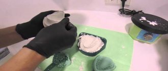
The process of removing debris from the canal
Even if the fragment does not affect the outcome of treatment in any way, a conscientious doctor will definitely inform his patient about the problem. This is necessary so that if the slightest discomfort is detected, a person immediately goes to the clinic, and does not treat the tooth with folk remedies at home. If the instrument breaks at the initial stage of the intervention before the infection is eliminated, the dentist must try to remove the part from the canal.
This will require a special dental microscope; without it, it will be simply impossible to detect such a small detail in a thin canal. At the first stage of the procedure, the doctor must create direct access to the foreign particle; for this he can use a dental bur.
Next, ultrasonic instruments with different tips come into play; it is with their help that the fragment can be removed from the dentin with minimal consequences. After this, the canal is thoroughly cleaned and sealed again, and medications are placed inside if necessary. A temporary filling is first placed on the tooth to ensure that there is no inflammation. After some time, you can put a permanent filling.
previous post
The dangers of teeth whitening at home
next entry
Treatment begins with removing the metal fragment
The instrument, which broke and remained in the tooth canal, actually acted as a pin that had to be removed, since inflammation could develop in the tooth canal at any moment. The consequences of such forgetfulness could be the most dire.
The following image shows the restoration of the mesial wall of the tooth, removal of the broken instrument:
After removing the instrument from the tooth canal, we place the fragment on an endodontic ruler to document the size. The length of the metal fragment was as much as 7 millimeters
!
A little about X-ray monitoring during treatment
Such control is mandatory. Especially when treating root canals.
First stage of control
. In this clinical case, we monitor the passage of root canals (with a broken instrument and the previously missed 4th canal):
Monitoring the patency of the root canals after filling and determining the length is carried out using an apex locator - a special electronic device that determines the position of the apical narrowing and allows you to determine the length of the root canal of the tooth.
What does work look like under a microscope
you can see using an apex locator in this short video:
Causes and symptoms of the appearance of a foreign object in the dental canal
Our clinic’s specialists conduct therapy carefully, following the protocol. However, even dentists with extensive experience have cases when unforeseen situations arise. Most often they occur during the treatment of too narrow and curved canals, as well as when removing dead tooth roots.
The main reasons for tool failure include:
- Defects in the anatomical structure of the dental canal.
- Low quality of the material from which the tool is made.
- An old tool in which the metal has become thin.
Thus, the doctor must monitor the frequency of use of dental instruments and change them on time.
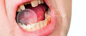
When a foreign body enters the dental canal, the patient does not feel anything. Symptoms of the presence of a foreign object appear a little later. There are several warning signs of pathology:
- The appearance of pain when biting and chewing food.
- Swelling of the gum tissue around a recently treated tooth.
- Formation of a fistula in the oral cavity.
- Development of a purulent focus.
If you have the above symptoms, you should immediately contact your dentist. The doctor will order an x-ray and determine the presence of possible foreign objects in the dental canal.
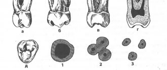
Types of pins and their main features
The pins are either fiberglass or metal. Fiberglass is used when installing tile crowns. Such an instrument does not show through the crown. It is quite easy to remove it if necessary. However, metal knitting needles are stronger, which is why they are used more often. Sometimes they are also called anchor.
Anchor pins are made from materials such as titanium, palladium or stainless steel. There are also pins made of brass and precious metals. They are used in the most difficult cases. Anchor pins are very popular because they make it possible to restore an almost completely destroyed tooth. It is only important that its root is intact.
The pins are fixed in the canal with phosphate cement. The use of ultrasound can help destroy it. This has to be done when repeated caries begins around the pin. Excessive strength of the material often causes displacement of the pin when chewing food, which can lead to damage to the prosthesis. In this case, the entire structure will need replacement or correction. Most often, the violation is detected already at the stage when it is already quite difficult to cure the tooth. In this case, you have to remove the pin along with the tooth. In some cases, the tooth can be saved. In this case, a small part of the dental tissue is removed around the pin using a drill, and then the pin is carefully unscrewed. After removal, re-treatment is carried out, the canals are properly processed and fillings are placed again.
Parapulpal pin wires are non-metallic. They are made from gold or special stainless steel, and then they are necessarily coated with polymer. Such knitting needles are used for retention of fillings and their reinforcement. A special feature of parapulp pins is that they are installed in hard tissue without touching the pulp. The absence of actions inside the tooth cavity ensures the complete elimination of the likelihood of contracting any infection or the development of inflammatory processes in the oral cavity. The disadvantage of such tooth strengthening means is the limited possibilities of use. The fact is that these products are located very close to the surface. They often break teeth and need to be reinstalled.
Dentists divide pin instruments into two groups: standard and special. The first ones are used to eliminate minor damage, and are available in the form of cylinders or cones. Special devices are made in a variety of shapes. Pin devices are also distinguished by fastening methods. Among screw materials, the main drawback is the danger of the tooth part breaking off during screwing.
Removing file debris from root canals.
Our patients' understanding of the importance of preserving their own natural teeth has increased significantly over the past few decades. The emergence of aesthetic restorations in combination with advanced materials, techniques and technologies in all areas of dentistry has led to increased demands on conservative dentistry. One segment of dentistry whose popularity has increased significantly is endodontics.
How does this happen
Although endofile technology has advanced rapidly over the past few years, despite the best efforts of dentists, misunderstandings still occur. Files break inside the canals, which may be caused by root shape, metal fatigue, excessive force, or a manufacturing defect.
As tools for internal canal processing, files shape the canal according to pre-planning. This common routine process involves the use of files of increasing size to create the required anatomy. The file is a segment of twisted wire with sharpened blades that perform the shaping.
Under ideal conditions, cavity cleaning runs smoothly. However, the instrument may become accidentally jammed in the canal or be bent beyond its normal tolerance. Simply attempting to remove or straighten a file inside a canal can cause it to accidentally break. This is more likely to happen with smaller, thinner files, but can also happen with larger files.
The file fragment is always located deep in the canal and can be difficult to grasp or remove with a conventional instrument. The biggest problem is the limited space available in the root canal. Not only are most tools virtually impossible to enter within narrow access boundaries, but even when they are introduced, manipulation of these tools is extremely limited.
Regular extras
These cases usually pose a lot of stress for the entire team.
The available selection is limited:
* The patient may be referred to an endodontist.
* The dentist may try to remove the broken file; insufficient access and visibility, and the lack of special instruments create a serious risk of damage to the canal walls and possible perforation of the surrounding periodontium.
* The dentist can use instruments and then obturate the broken file.
*Treatment of the tooth can be continued through semi-excision, apex resection or other surgical interventions.
* The tooth may be removed.
Another solution
Each of these opportunities is associated with a large investment of time and certain difficulties. It would be more practical to have an effective extractor for broken fragments directly in the office. The ideal design parameters for this tool are:
* Quick and effective removal of file debris and silver pins from endodontic canals.
* Preservation of healthy tooth structure.
* Applicability for both frontal and lateral teeth.
* Quick learning.
* Predictable success.
Meisinger recently introduced the Meitrac instrument system, which provides a treatment process and multiple solutions to the problem. Teeth that were considered non-viable can now be completely restored in terms of form and function.
Meitrac I and II instruments are made of surgical stainless steel, are autoclavable and can be reused multiple times. Their size and shape ensure a process with minimal intervention and allow the preservation of healthy tooth tissue. This is a very important aspect when treating the inner part of the root, which is a relatively delicate structure of the tooth. The size of the pulp and the extent of caries can significantly affect the reduction in the amount of remaining dentin, so every effort to minimize the loss of healthy teeth is important.
Meitrac I and II are designed to remove broken file fragments with minimal loss of remaining root dentin. The function of the instruments is to preserve the trephine around the file fragment, providing sufficient space for the extractor to grasp the fragment and remove it. The trephine is similar to a typical circular bur used at low speeds. The main difference is that it is hollow throughout its entire length. This means that when it is used for carving, it functions more as a fruit pitter. In the case of a broken file (or silver post), the trephine cuts the tooth structure immediately adjacent to the metal fragment, releasing it so that it can be captured by the extractor.
Clinical process
The trephine is inserted into the canal entrance until it reaches the broken file (Fig. 4). This is done slowly, carefully, since there is almost no visibility. The endodontic entrance to the canal must be wide enough to allow the insertion of an appropriately sized instrument.
The trephine is then inserted further into the canal to a position where the file fragment enters the hollow center of the trephine. This is not a difficult step, since both the end of the broken file and the trephine cavity are centered in the endodontic canal. In some cases, when this did not happen immediately after insertion, a slight movement of the trephine at an angle will allow the broken file to be captured. The trephine is then used at very low speeds to maintain complete control during the release process.
The shape and movement of the trephine ensures both minimal intervention and proper direction. Since the inside of the trephine is not cutting, the fragment of the file serves as a guide along which the trephine is processed along the length of the fragment. This also aims to limit the trephine treatment to the center of the root where the pulp is located, avoiding lateral perforation. The scope of trephine processing is limited by the narrowness of the instrument and the circular cutting tip.
When the trephine has opened the fragment to a height of 1 - 2 mm, an appropriate extractor can be inserted into the canal, which is located around the file. There are 4 different extractor sizes that match the diameter of the broken file. The extractor consists of two components - a small hollow tube attached to a clamping device button at one end and long vertical slots cut into its active end. These vertical slits allow the small tube to be compressed, reducing the internal volume of the tube. The small tube is pressed tightly into a larger hollow tube that also has a clamping button at one end and a slightly compressed volume at the other end.
Since the smaller extractor component is inserted into the larger component, it is supported in the neutral position by a spring. The extractor is carefully inserted into the entrance to the canal, which was prepared with a hollow trephine. Based on the inner part of the canal prepared with a trephine, the fragment of the file is centered in the extractor and the inclined inner walls of the larger tube insert the file into position inside the smaller hollow tube, as can be seen in the illustration in section and inside the canal.
The extractor is activated by pushing the smaller tube further into the larger component against spring resistance. As the smaller tube exits through the larger one, the reduced lumen at the active end of the larger tube compresses the smaller tube, reducing the internal lumen. By this reduction of the internal lumen of the smaller tube, the metal fragment is firmly captured in the slotted sections of the inner tube, as seen in the cross-sectional illustration and inside the endodontic canal. The pressure on the spring is maintained, ensuring that the internal hollow tube, which firmly grasps the file fragment, is removed from the endodontic canal.
Due to the combination of diameters, the file fragment and the inner hollow tube are firmly held within the larger outer tube and the entire system can be carefully removed from the entrance to the canal. If the file has been compressed in the canal, apply a light counterclockwise rotational force to the extractor.
Once the file fragment (or silver post) has been removed, normal endodontic treatment can be continued immediately.
Conclusion
A problematic situation with a broken file deep inside the canal can disrupt the treatment, lead to tooth loss and, in any case, cause serious stress for the doctor and his assistants. The advent of the Meitrac systems (Meitrac I for small files and Meitrac II for larger files and silver pins), a set of tools specifically designed to remove broken files and silver pins in hard-to-reach places, makes a file failure inside the canal a momentary misunderstanding rather than a permanent disaster. .
Article provided by the magazine “Dental Doctor”