It is difficult to find a person who does not dream of maintaining healthy, strong teeth until old age. After all, healthy white teeth mean a beautiful smile in the photo, self-confidence, and absence of pain. Healthy teeth are also a good help in the proper functioning of many systems of our body.
Doctors say that healthy, strong teeth are always a pressing issue in dentistry. In fact, all their activities are aimed at keeping people’s teeth healthy.
And in the same way, in order for healthy white teeth to delight us in old age, we need to take care of them all our lives, using all means of disease prevention, including folk ones.
Teeth of ancient people
The dentofacial apparatus of prehistoric and modern humans differs significantly. Ancient people had more than 36 teeth, protruding fangs and a massive jaw. This was explained by the need to chew rough food and raw meat. With the addition of thermally processed foods to the diet, the dentition began to change. The canines were the first to transform, becoming aligned with the bite line. Then the jaw arch narrowed, the interdental spaces disappeared, and the teeth themselves decreased in size. Currently, 32 teeth in humans are the norm, but third molars are considered to be an atavism.
Interesting fact!
The teeth of ancient man cannot be called aesthetic, but they were healthy. According to scientists, cavemen never suffered from caries and other oral diseases.
Treatment for missing teeth
The specificity of the anatomical structure of the jaw apparatus is that even complete edentia does not guarantee the formation of correct occlusion after implantation. The result of prosthetics is largely determined by the degree of development of defects and anomalies that formed even before the loss or removal of elements. If the row ratio was within acceptable limits, it is enough for the doctor to take into account the necessary adjustments when developing a replacement structure, but the presence of problems with bone tissue will require the use of more complex restoration techniques.
How to check and find out if the bite is correct when making a jaw prosthesis without teeth? For this purpose, special wax rollers are used, placed in the oral cavity. The measuring instruments are a ruler arc and an intraoral plate. After collecting the necessary readings, first a plaster prototype is created, and then the final structure.
Name of human teeth
Depending on the location and structure, dental units have their own functional characteristics and are called differently.
- Incisors.
On both jaws there are four front teeth in humans - medial and lateral incisors, which are used for biting food. - Fangs.
Sharp teeth designed for chewing hard foods. - Premolars.
"Fours" and "fives" on the left and right sides of each jaw arch grind soft or small pieces of food. - Molars.
Three large outer teeth in each row are aimed at grinding coarse substances. - The canines
and incisors are part of the anterior group, or the “smile zone,” and the human molars are part of the chewing segment.
In addition, teeth are divided into temporary and permanent. In the first case, we are talking about dairy products that appear in children from the fifth month of life to three years. The second refers to the final bite, which is formed between six and thirteen years of age. Milk teeth differ from permanent teeth only in size, but in structure they are identical.
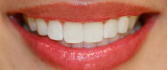
Healthy teeth, photos of patients. Photo of healthy teeth
We often see the dazzling smiles of celebrities in photographs in glossy magazines. And many people believe that this is what healthy teeth look like. Photos in such press, however, do not always reflect the truth.
After all, photographs in magazines can be processed in special programs, and in order to recognize the signs of an impending problem in time or simply make sure that you have healthy teeth, it is advisable to look at the photos provided by a specialist.
On dental websites, almost any material about the prevention of oral diseases is accompanied by at least one photo of healthy teeth. At the same time, photos are often presented from different angles, including their inner surface. Photos of healthy teeth can also be viewed in the clinic, in special catalogs. Therefore, today it is not difficult to see how strong and white teeth look in the photo.
How many teeth does a person have?
The number of teeth a person has depends on age and anatomical features. The child has a set of 20 primary teeth, which are replaced by a permanent bite of 28 teeth. Third molars erupt, as a rule, after twenty years or do not grow at all, which is not a pathology.
In dentistry, a single numbering of human teeth is adopted. Doctors classify teeth as lower and upper and distinguish the right and left segments of the jaws. Each of them includes two incisors, a canine, two premolars and three molars. The countdown starts from the first front tooth and ends, accordingly, with a figure eight. Sometimes a number is added to the serial number indicating the location zone. For example, the right canine of the top row is numbered 13. This order in the schematic representation is called the formula of human teeth.
Polyodontia
In rare cases, an anomaly such as polyodontia is observed - supernumerary, or extra teeth in a person. Dental units can appear in the primary and permanent dentition anywhere in the jaw, separate from or fused with the main teeth. The defect affects not only the aesthetics of the smile, but also leads to the formation of incorrect occlusion, impairs the quality of chewing food and diction. Most often, supernumerary teeth are removed in childhood or built into the dentition.
Edentia
There is also a deviation of the opposite meaning called edentia - congenital or acquired absence of dental units. The causes of the phenomenon include heredity or improper development of the embryo in the womb. People without teeth cannot fully eat and speak, have a deformed facial contour and weakened immunity.
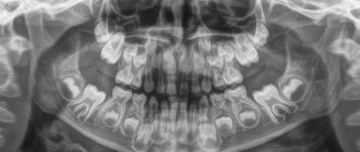
Panoramic dental x-ray
If pulpitis or another disease is suspected, the dentist sends the patient to take a photo of the diseased tooth. But sometimes it is difficult to determine exactly which tooth is the source of the problem. If the gums are inflamed, a person simply cannot understand where exactly it hurts.
Previously, in such cases it was necessary to take x-rays of nearby teeth. Today, the doctor will definitely refer you for an orthopantomogram - a panoramic image. This method allows you to get a “picture” of the entire oral cavity at one time. Moreover, from such an image it is possible to determine not only caries or periodontal destruction. A panoramic x-ray of the teeth will reflect the condition of the alveoli, sinuses and even maxillofacial bones.
Experts also advise periodically taking panoramic dental x-rays for prevention. This diagnostic method will allow you to identify inflammatory processes at the tops of the roots of the teeth even before toothache begins to torment you. And having seen the general picture of the state of your dental system, the dentist will prescribe a targeted photo of the questionable tooth in case of any deviation.
After the therapy, especially if it took place in several stages, you may need to repeat dental x-rays. This will allow you to notice and eliminate defects in the canal filling in a timely manner.
If, according to indications, you are offered to remove a wisdom tooth, a panoramic photograph will also be useful. The surgeon needs to see the entire area around the problem area - the area of the upcoming operation.
If you decide to have prosthetics, install braces or implant an implant, be sure to take a panoramic photo. Such an X-ray of the teeth in this case will allow the doctor to fully assess the condition of your oral cavity and decide on a treatment regimen.
So, if you are determined to correct your bite, the orthodontist will ask you to do an orthopantomogram in order to use it to analyze at what angle the roots of the teeth are, and whether there are teeth that are not visible, but they are located inside the jaw bone - the so-called impacted teeth. This information will allow you to perform bite correction as successfully as possible.
If you are going to have dental prosthetics, it is necessary to exclude hidden problems - a dental cyst, granuloma or tumor. The size and boundaries of these neoplasms are quite clearly visible on a panoramic photograph of the teeth. With a traditional x-ray, when only 1-2 teeth are reflected in the image, the doctor risks missing the threat.
When placing an implant in the upper jaw, the implantologist will use this image to estimate the distance to the maxillary sinuses. This is necessary so that during the procedure the implant does not enter the maxillary sinus. If you need to place an implant in the lower jaw, it is important to make sure that it does not touch the nerve.
How is a panoramic dental x-ray performed?
Everything is elementary simple: you just need to stand at a special installation for a few seconds. And within a couple of minutes the photo is in your hands. Before starting the procedure, be sure to remove all metal objects from yourself. They interfere with the passage of the X-ray beam, which means they can cause image distortion. The most modern devices are digital, they allow you to store your pictures in the computer's memory. After all, they can be useful to you at an appointment with a variety of doctors - dental therapists, maxillofacial surgeons, orthodontists, implantologists, and so on.
Another plus is the price. A panoramic dental x-ray will cost you much less than taking pictures of several problem areas. In addition, modern technologies make this procedure as safe as possible, even for young patients. After all, the radiation dose is reduced to a minimum. But do not forget that for some chronic diseases, as well as during pregnancy, any x-ray is contraindicated. Therefore, before the procedure, be sure to discuss all the nuances with your doctor.
Dimensions of human teeth
The upper central incisors are twice as wide as their antagonists. The remaining dental units of the same name have approximately equal parameters. The size is determined using special tables with the optimal size and permissible deviations. Experienced doctors calculate proportions by dividing the length of a person’s teeth by the width. A result of about 0.75 millimeters is considered close to ideal. For more detailed diagnostics, other professional formulas and techniques are used.
Size deviations from the norm occur due to improper formation of the jaw, fusion of tooth buds, or genetic predisposition. Teeth that are too large are called macrodentia, and abnormally small teeth are called microdentia. Pathologies are accompanied by problems with bite and chewing functions, but can be successfully corrected by a dentist.
Interesting fact!
The longest tooth in the world belongs to an Indian teenager. The size of its crown is almost four centimeters. About a year ago, the tooth was removed, and the young man was included in the Guinness Book of Records.
Causes of tooth enamel destruction
There are a number of factors that exacerbate the rate of enamel damage. These include:
- the anatomical structure of the tooth, when a large amount of food accumulates in the dental cavities and spaces between the teeth;
- insufficient saturation of enamel with fluoride;
- genetic predisposition;
- weak immunity, various diseases of the body;
- composition of salivary fluid - a small amount of saliva, viscous in composition, causes the accumulation of bacteria on the surface of the tooth and in the dental spaces, and consequently, the formation of dental plaque;
- insufficient oral hygiene, improper brushing of teeth;
- eating mostly soft food (soft food weakens the protective functions of enamel, while hard food helps to naturally strengthen teeth);
- lack of vitamins and beneficial microelements in the human body;
- The bristles of a toothbrush used for oral hygiene are too hard.
Restoration of tooth enamel is required for those who eat large amounts of citrus fruits (oranges, lemons, grapefruits), which significantly weaken tooth enamel. The acid contained in these fruits tends to dissolve the protective surface of the tooth. Therefore, after eating citrus fruits, be sure to rinse your mouth with water.
The structure of the human tooth
Anatomy
From an anatomical point of view, a human tooth consists of three parts.
- Crown.
The visible part protruding above the gum. It has four sides: the occlusal, or cutting edge, in contact with the antagonist teeth; contact wall adjacent to adjacent dental units; vestibular and lingual surfaces facing the lips and tongue, respectively. - Root.
Fixed in the socket by connective tissue, located in the recess of the jaw. As a rule, premolars have two roots, and molars have three, four or even five. The remaining dental units have one root canal. - Neck.
It is located between the coronal part and the root of a human tooth, surrounded by periodontium.
Histology
What are human teeth made of? Let's look at the cross-section of the structure of a human tooth.
- Enamel.
A transparent protective coating of the crown, almost entirely consisting of inorganic microelements. - Dentine.
The hard base of the tooth, containing 80% mineral components and 20% organic substances. The shade of dentin is responsible for the color of dental units, as it shines through the enamel. - Cement.
The bone tissue covering the tooth root. Plays the role of a fastening element connecting the tooth to the alveolus. - Pulp.
Soft tissue filled with bundles of nerves and capillaries. Painful sensations during caries are explained precisely by the presence of nerve endings.
Myths about tooth enamel
Here are some common myths and misconceptions about tooth enamel.
The first myth is that enamel is destroyed very quickly. Actually this is not true. The process of enamel destruction is gradual and can last ten years. This is why proper prevention of tooth enamel destruction is so important.
The second myth is that tooth enamel is destroyed immediately on most of the teeth. This is also not true. It happens that one or more teeth suffer from enamel destruction more than the rest. This is a sign that inflammatory processes have begun inside the tooth and it is necessary to urgently contact a dental clinic.
The third myth is that enamel destruction is not a disease. In fact, damaged enamel is not only a cosmetic defect, as many people think. This is a disease, the progression of which can lead to complete destruction of all tooth tissues and subsequent loss.
The fourth myth is that enamel destruction can be diagnosed only by external signs (darkening of the tooth surface, the formation of pigment spots). This is not true. Symptoms such as an acute reaction of teeth to external irritants (cold, hot and cold food), as well as the presence of toothache (cutting or aching), indicate that your tooth enamel has been destroyed.
Human wisdom teeth
A “wisdom tooth” is the third outer molar with three to five roots. In structure it is no different from its “neighbors”. To the question “How many wisdom teeth does a person have?” cannot be answered unambiguously. They erupt around the age of twenty, one on each side of both jaws. However, there are people without wisdom teeth. This is a variant of the norm, since in the process of human evolution the need for the “eight” disappeared, and the structure of the jaws underwent corresponding changes. Today, third molars are considered a vestigial organ.
Why is tooth enamel destroyed?
The destruction of tooth enamel may be a natural consequence of the patient’s age. People in the age group of fifty years and older very often complain of a decrease in the protective functions of enamel. This is a consequence of age-related changes in the body.
Enamel destruction can also be observed in pregnant women. In this case, this is a consequence of hormonal changes occurring in a woman’s body.
There are also slightly curious reasons for the destruction of the tooth surface. For example, when a person tries to crack a too-hard nut with his teeth (special tools are available for this) or demonstrate to friends how he can open glass bottles with his teeth. Such events are very dangerous because they can result not only in the destruction of the enamel, but also in the fact that the tooth can break or chip off.
Photos of dental implantation before and after
Photo of dental implantation on a beam
All on 4 implantation photo
All on four implantation photo
Installation of 4 implants of the Superline Dentium system using All on 4 technology and installation of a fixed denture. Orthopedist Shagin L.V., surgeon-implantologist Kurbatov V.O.
All on Four implantation photo
Photo of one-stage dental implantation
Tooth extraction and photo of one-stage implantation
Simultaneous removal and implantation in the area of 25 and 26 teeth. Without bone grafting, but using a centrifuge.
Gum disease: symptoms and treatment
Gum diseases in adults and children occur in the same way. Below we will analyze each disease - with a description of its characteristic symptoms and photographs of the patients’ teeth and gums. The information will allow you to easily distinguish one disease from another, and understand how much professional treatment you will need in different situations.
The author of the article has more than 10 years of experience as a periodontist, and therefore our recommendations really work (state-issued documents on advanced training in the Periodontology program can be viewed in the editorial section).
How does the procedure work?
Panoramic photography of teeth is the specialty of a dental radiologist. It is carried out using an orthopantomograph - a modern device that exposes the body to a minimal amount of x-ray radiation.
To take a photo, you need to stand or sit near the device. Next, the patient must wear a protective apron that protects the chest from radiation. When the patient is ready, the doctor starts the orthopantomograph. The entire procedure lasts no more than 15 minutes and is completely painless.
During the process you must remain stationary. After the panoramic photo is taken, you need to wait a few more minutes. During this time, the specialist will prepare the image and provide it to the patient. The entire procedure lasts no more than 15 minutes, including the time required for preparation and waiting.
Types and degrees of curvature
The method of restoring crooked teeth depends on the degree of curvature and the age of the patient. On the Internet you can find many photos of celebrities with crooked teeth before and after restoration. How does the correction process work?
First, let's look at the degrees of curvature:
- First degree: refers to individual teeth that differ from the rest in shape, size or location.
- Crooked teeth 2 degrees: incorrectly formed dentition due to the simultaneous eruption of several teeth at once.
- Third degree: curvature at the level of the jaws, which leads to improper closure of the upper and lower dentition.
In addition, the front teeth, incisors or wisdom teeth may be crooked. The front ones, as a rule, change shape or location due to external factors: injuries, the habit of chewing a pen or seeds.
Orthodontist Anastasia Sergeevna Arkhangelskaya from the 32 Dent clinic explains why teeth grow crooked in adults: “In adulthood, people often have to have their teeth removed. If an implant or other prosthesis is not installed in their place, the dentition may become crooked. In addition, the development of dental anomalies is facilitated by the eruption of wisdom teeth. This is especially true for people with a narrow jaw, which simply does not have enough space for large wisdom teeth.”
If you have a problem similar to that described in this article, be sure to contact our specialists. Don't diagnose yourself!
Why you should call us now:
- We will answer all your questions in 3 minutes
- Free consultation
- The average work experience of doctors is 12 years
- Convenient location of clinics
Single contact phone number: +7
Make an appointment
Photos of veneer installation before and after
The color and shape of teeth are changed by veneers. The total cost of installing veneers was RUB 320,000. Orthopedic doctor Yuri Anatolyevich Lychko
Photo of veneer installation
Photo of installing veneers on front teeth
The shape and color of teeth are changed through the use of veneers. The cost of installing veneers is RUB 370,000. Orthopedic doctor Yuri Anatolyevich Lychko
Photo of installation of veneers on the upper jaw
The shape and color of teeth are changed using veneers. The cost of installing veneers is 180,000 rubles. Orthopedic doctor Yuri Anatolyevich Lychko
Photo of veneer installation on one tooth
Trauma to the central incisor. The tooth was restored with a veneer. Installation price - 34,000 rubles. Orthopedic doctor Yuri Anatolyevich Lychko
Installation of a metal-ceramic crown
Installation of a mandibular bridge on the right
Photo of installation of a zirconium dioxide crown
Photos of a zirconium crown before and after
Installation of a zirconium crown on a Nobele Biocare implant, screw fixation. Orthopedic doctor Leonid Vladimirovich Shagin.
Photo of installing a zirconium crown on an implant
Zirconium crown on the implant, installed using screw fixation. Orthopedic doctor Leonid Vladimirovich Shagin.
What causes malocclusion to form?
Defects appear for various reasons:
- Genetic predisposition. In most cases, mesial and distal bites are inherited. Therefore, parents who have this problem should take their child to an orthodontist in early childhood.
- Anomalies of fetal development. A child’s teeth are formed while still in the womb. During a complicated pregnancy or the influence of negative factors on its course, anomalies of the dental system can form.
- Birth injury. Mesial bite in some cases is a consequence of dislocation of the baby’s lower jaw during childbirth.
- Bad habits. Constant thumb or pacifier sucking, improper latching of the nipple, incorrect sucking during artificial feeding - all this provokes the development of malocclusion pathologies.
- Inferiority of the dentition. You can resort to removing baby teeth if there is less than a year left before the change. Otherwise, the dentition becomes asymmetrical, the midline shifts, and the chewing movements of the jaw are blocked. The formation of a permanent bite ends at approximately 15 years of age. For adults, after tooth loss, it is important to undergo prosthetics in a timely manner.
- Displacement of the jaws as a result of hypertonicity of the masticatory muscles, which can be observed due to stress.
Jaw injuries, as well as improper prosthetics, can provoke the development of malocclusions.
What is an ideal smile?
Probably, every person has his own idea of a beautiful smile. But in dentistry this concept has clear parameters. When a dentist makes a Hollywood smile, he strives for these criteria:
- Geometry. On a person’s face, you can draw several conventional lines that form the eyes, lips, edges of the incisors and fangs. For a beautiful smile, the parallelism of these lines to each other is important.
- Teeth are physiologically correct size. Ideal teeth don't just have different sizes. The largest teeth are the incisors. Then the size of the teeth gradually decreases. With this size, the smile looks attractive.
- Correct bite. Antagonist teeth must touch each other at physiologically correct points.
- Upper lip. When smiling, it only exposes the teeth, but not the gum line.
It is customary to call a Hollywood smile a smile that meets all these criteria. She's not just beautiful. It makes you more attractive, younger.
Correct bite: what is it like?
Bite is the way the teeth of both jaws interact when they are completely closed. If it is correct, there are no difficulties when chewing and pronouncing sounds, the aesthetics of the smile is not impaired, but minor defects are still possible. What does a bite that does not require orthodontic treatment look like? Its main characteristics:
- Presence of 28–32 teeth. Not all people develop “eights”, which is determined by genetic factors. Their presence (if they have grown correctly) or absence does not affect the closure of the jaws. Moreover, in some cases, wisdom teeth removal is indicated during orthodontic treatment. Then there is more free space in the mouth.
- Symmetry in the closure of the dentition.
- The incisors of the upper jaw overlap the lower ones by one third.
- No interdental gaps, cracks or chips.
- Correspondence of molars and premolars of both dentitions to each other. Their closure is tight.
- Ease of chewing food, even hard ones. Opening the mouth is easy and there is no pain.
Correct bite does not always look aesthetically ideal. But the face is harmonious.
How to find out what kind of bite you have
Patients are often interested in how to determine whether their bite is correct. Usually the presence of abnormalities can be identified by facial features. It is also worth taking into account the criteria for correct bite described above. Go to the mirror, open your mouth and clench your teeth. In some cases, violations are immediately visible. Just because your smile looks aesthetically pleasing does not guarantee that everything is fine.
Even if you know what a correct bite should look like, it is impossible to make a correct diagnosis on your own. Only a specialist can do this. In addition to a visual examination and medical history, the doctor diagnoses the problem based on the results of a CT scan (computed tomography). Such an examination allows you to comprehensively assess the condition of the oral cavity. Disturbances in the placement of teeth are also identified by an impression made from an alginate mass. The orthodontist also needs photographs of the person’s face taken from several angles. Using them, you can evaluate the occlusal relationships of the dentition.
How to straighten crooked teeth?
The selection of a means for straightening crooked teeth is carried out according to the type of anomaly and taking into account the patient’s age.
To correct malocclusion in children, removable plates or trainers are used. They perform several functions:
- align the position of teeth and jaws;
- normalize muscle function;
- relieve excess pressure from the jaws that the tongue and cheeks exert during conversation or eating.
The use of removable corrective devices is effective only up to 10-12 years. After this, the child develops a permanent bite, which can only be corrected with special mouth guards or braces.
Modern dentistry provides a huge selection of braces. Most often they are installed on the outer surface of the teeth. Unfortunately, they are noticeable during conversation and spoil the aesthetic appearance of a smile. If the aesthetic factor plays a key role, you can install special lingual braces. They are placed on the inside, so the adjustment occurs almost unnoticed by others.
Prevention of tooth enamel destruction
As you know, prevention is always better than cure. This also applies to cases of tooth enamel destruction. Keeping your enamel strong and healthy is not difficult if you follow a number of recommendations.
Use a brush with soft or medium bristles to brush your teeth. Choose toothpastes that contain calcium and fluoride. Do not be lazy to maintain complete oral hygiene, that is, brush your teeth twice a day.
Use dental floss after eating - it perfectly cleanses the dental spaces of food deposits (and it is the cracks between the teeth, not even visible to the eye, that are potential spreaders of bacteria). If possible, use a mouth rinse after each meal - most of them contain microelements that serve to strengthen the tooth surface.
Eat foods fortified with calcium. These are fish, dairy products, cheeses, eggs. Eat more foods that are a source of vitamin C (pepper, rose hips, black currants, cabbage, garlic), as well as vitamin D (salmon, tuna, fish oil, potatoes, oatmeal).
When you brush your teeth, try to leave the paste on the tooth surface for at least three minutes. During this time, the medicinal substances included in the composition penetrate into the structure of the teeth, nourishing the enamel.
Types of correct bite
The above describes the orthognathic bite, which is the reference. It occurs in only 5% of people. But there are other types of teeth closure in which violations are not detected. What does each one look like? Experts identify the following bites:
- Biprognathic. Both jaws are slightly pushed forward, and the teeth are slightly directed towards the lips.
- Opisthognathic. The opposite of the previous type. The teeth are directed inward. Opisthognathic occlusion is rare. Its features are noticeable when viewed in profile.
- Progenic. The lower jaw protrudes slightly, but this feature is not pathological. The functioning of the temporomandibular joint (TMJ) and the functionality of the teeth are not impaired.
- Straight. The upper row of teeth is parallel to the lower one. The incisors touch their cutting edges. This feature is not a deviation, but it threatens rapid wear of the teeth due to increased load.
Sometimes, if you have one of these malocclusions, minor adjustments may be needed. But the treatment in this case will be easy.